

Genomic analysis reveals few genetic alterations in pediatric acute myeloid leukemia
- PMID: 19651601
- PMCID: PMC2716382
- DOI: 10.1073/pnas.0903142106
Genomic analysis reveals few genetic alterations in pediatric acute myeloid leukemia
Abstract
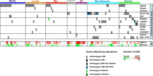
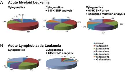
Pediatric de novo acute myeloid leukemia (AML) is an aggressive malignancy with current therapy resulting in cure rates of only 60%. To better understand the cause of the marked heterogeneity in therapeutic response and to identify new prognostic markers and therapeutic targets a comprehensive list of the genetic mutations that underlie the pathogenesis of AML is needed. To approach this goal, we examined diagnostic leukemic samples from a cohort of 111 children with de novo AML using single-nucleotide-polymorphism microarrays and candidate gene resequencing. Our data demonstrate that, in contrast to pediatric acute lymphoblastic leukemia (ALL), de novo AML is characterized by a very low burden of genomic alterations, with a mean of only 2.38 somatic copy-number alterations per leukemia, and less than 1 nonsynonymous point mutation per leukemia in the 25 genes analyzed. Even more surprising was the observation that 34% of the leukemias lacked any identifiable copy-number alterations, and 28% of the leukemias with recurrent translocations lacked any identifiable sequence or numerical abnormalities. The only exception to the presence of few mutations was acute megakaryocytic leukemias, with the majority of these leukemias being characterized by a high number of copy-number alterations but rare point mutations. Despite the low overall number of lesions across the patient cohort, novel recurring regions of genetic alteration were identified that harbor known, and potential new cancer genes. These data reflect a remarkably low burden of genomic alterations within pediatric de novo AML, which is in stark contrast to most other human malignancies.
Conflict of interest statement
The authors declare no conflict of interest.
Figures

Similar articles
-
Acquired copy number alterations in adult acute myeloid leukemia genomes.Proc Natl Acad Sci U S A. 2009 Aug 4;106(31):12950-5. doi: 10.1073/pnas.0903091106. Epub 2009 Jul 27. Proc Natl Acad Sci U S A. 2009. PMID: 19651600 Free PMC article.
-
Broad copy neutral-loss of heterozygosity regions and rare recurring copy number abnormalities in normal karyotype-acute myeloid leukemia genomes.Genes Chromosomes Cancer. 2010 Nov;49(11):1014-23. doi: 10.1002/gcc.20810. Genes Chromosomes Cancer. 2010. PMID: 20725993
-
RAS mutations in pediatric leukemias with MLL gene rearrangements.Genes Chromosomes Cancer. 1998 Mar;21(3):270-5. Genes Chromosomes Cancer. 1998. PMID: 9523205
-
Familial mutations of the transcription factor RUNX1 (AML1, CBFA2) predispose to acute myeloid leukemia.Leuk Lymphoma. 2004 Jan;45(1):1-10. doi: 10.1080/1042819031000139611. Leuk Lymphoma. 2004. PMID: 15061191 Review.
-
Genomic analysis of acute myeloid leukemia: potential for new prognostic indicators.Curr Opin Hematol. 2010 Mar;17(2):75-8. doi: 10.1097/MOH.0b013e3283366c43. Curr Opin Hematol. 2010. PMID: 20075726 Free PMC article. Review.
Cited by
-
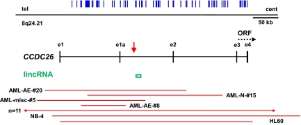
Long noncoding RNA, CCDC26, controls myeloid leukemia cell growth through regulation of KIT expression.Mol Cancer. 2015 Apr 19;14:90. doi: 10.1186/s12943-015-0364-7. Mol Cancer. 2015. PMID: 25928165 Free PMC article.
-
Long Noncoding RNAs in Acute Myeloid Leukemia: Functional Characterization and Clinical Relevance.Cancers (Basel). 2019 Oct 24;11(11):1638. doi: 10.3390/cancers11111638. Cancers (Basel). 2019. PMID: 31653018 Free PMC article. Review.
-
Acquired genomic copy number aberrations and survival in adult acute myelogenous leukemia.Blood. 2010 Dec 2;116(23):4958-67. doi: 10.1182/blood-2010-01-266999. Epub 2010 Aug 20. Blood. 2010. PMID: 20729466 Free PMC article.
-
SNP array analysis in hematologic malignancies: avoiding false discoveries.Blood. 2010 May 27;115(21):4157-61. doi: 10.1182/blood-2009-11-203182. Epub 2010 Mar 19. Blood. 2010. PMID: 20304806 Free PMC article. Review.
-
RUNX1 and its understudied role in breast cancer.Cell Cycle. 2011 Oct 15;10(20):3461-5. doi: 10.4161/cc.10.20.18029. Epub 2011 Oct 15. Cell Cycle. 2011. PMID: 22024923 Free PMC article.
References
-
- Kelly LM, Gilliland DG. Genetics of myeloid leukemias. Annu Rev Genomics Hum Genet. 2002;3:179–198. - PubMed
-
- Reya T, Morrison SJ, Clarke MF, Weissman IL. Stem cells, cancer, and cancer stem cells. Nature. 2001;414:105–111. - PubMed
-
- Schimmer AD. Induction of apoptosis in lymphoid and myeloid leukemia. Curr Oncol Rep. 2006;8:430–436. - PubMed
-
- Mullighan CG, et al. Genome-wide analysis of genetic alterations in acute lymphoblastic leukaemia. Nature. 2007;446:758–764. - PubMed
-
- Mullighan CG, et al. BCR-ABL1 lymphoblastic leukaemia is characterized by the deletion of Ikaros. Nature. 2008;453:110–114. - PubMed
Publication types
MeSH terms
Substances
Grants and funding
LinkOut - more resources
Full Text Sources
Other Literature Sources
Medical
Molecular Biology Databases
