Review
. 2009:88:63-101.
doi: 10.1016/S0070-2153(09)88003-4.

Hox specificity unique roles for cofactors and collaborators

Affiliations
Review

Hox specificity unique roles for cofactors and collaborators

Richard S Mann et al. Curr Top Dev Biol. 2009.

Abstract

Hox proteins are well known for executing highly specific functions in vivo, but our understanding of the molecular mechanisms underlying gene regulation by these fascinating proteins has lagged behind. The premise of this review is that an understanding of gene regulation-by any transcription factor-requires the dissection of the cis-regulatory elements that they act upon. With this goal in mind, we review the concepts and ideas regarding gene regulation by Hox proteins and apply them to a curated list of directly regulated Hox cis-regulatory elements that have been validated in the literature. Our analysis of the Hox-binding sites within these elements suggests several emerging generalizations. We distinguish between Hox cofactors, proteins that bind DNA cooperatively with Hox proteins and thereby help with DNA-binding site selection, and Hox collaborators, proteins that bind in parallel to Hox-targeted cis-regulatory elements and dictate the sign and strength of gene regulation. Finally, we summarize insights that come from examining five X-ray crystal structures of Hox-cofactor-DNA complexes. Together, these analyses reveal an enormous amount of flexibility into how Hox proteins function to regulate gene expression, perhaps providing an explanation for why these factors have been central players in the evolution of morphological diversity in the animal kingdom.


Figures
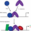
Figure 3.1
Figure 3.1
Two contributing steps in Hox specificity. In principle, Hox specificity can be broken down into two separate steps. The first step is DNA binding by Hox proteins, which can occur either with or without cooperatively binding cofactors. The second step involves the recruitment of additional factors, Hox collaborators, to the cis-regulatory element. The recruitment of these factors may depend on contacts between them and the DNA and/or protein-protein contacts between them and the Hox-cofactor complex. It is the recruitment of these collaborators, which we suggest depends on the architecture of the entire cis-regulatory element (including the details of the Hox-binding site) that ultimately determines the sign of the transcriptional regulation.
Figure 3.2
Figure 3.2
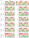
Comparison of in vitro and in vivo Hox-binding site preferences. Shown are LOGO diagrams summarizing Hox-binding site preferences for most paralogs. The column on the left lists the LOGOs generated using the binding sites identified by the bacterial 1-hybrid (B1H) method (Noyes et al., 2008). The column on the right lists the LOGOs generated using the in vivo binding sites in Table 3.1. To generate these LOGOs, we used CONSENSUS (as part of Target Explorer; http://luna.bioc.columbia.edu/Target_Explorer/) to generate position weight matrices (PWMs). PWMs that maximized alignment of an “AT” sequence were converted to Transfac format using the phiSITE conversion server (http://www.phisite.org/main/index.php?nav=home). enoLOGOS (http://chianti.ucsd.edu/cgi-bin/enologos/enologos.cgi/) was then used to generate the LOGOs using nucleotide frequency for the Y-axis. The number of binding sites used to generate each LOGO was as follows: Labial: 31 (B1H), 17 (in vivo); Dfd: 24 (B1H), 17 (in vivo); Scr: 34 (B1H), 12 (in vivo); Antp: 19 (B1H), 16 (in vivo); Ubx: 20 (B1H), 57 (in vivo; the resulting LOGO was only subtly affected if the 30 sites from the Antp-P2 element were omitted); Abd-A: 23 (B1H), 39 (in vivo); and AbdB: 21 (B1H), 49 (in vivo).
Figure 3.3
Figure 3.3
Three types of Hox target genes. “Paralog-specific” Hox target genes are those that are uniquely regulated by only a single Hox paralog, such as the activation of fkh by Scr. “Semi-paralog-specific” Hox target genes are those that are shared by a small subset of Hox paralogs, such as the repression of Dll by the abdominal Hox proteins Ubx, Abd-A, and Abd-B (schematized here is the Dll304 embryonic enhancer element). “General” Hox target genes are those that are regulated by most, or perhaps all, Hox paralogs, such as the control of optix in Drosophila. Ideally, this classification should apply to individual cis-regulatory elements, not entire genes, to allow for the scenario that the same gene may fall into more than one of these categories (in two different tissues or times of development). For fkh and Dll, specific cis-regulatory elements that fit the “paralog-specific” and “semi-paralog-specific” criteria have been identified. In contrast, a single cis-regulatory element that is a “general” Hox target has not yet been identified and therefore remains hypothetical.
Figure 3.4
Figure 3.4
Common and unique features of PBC-Hox-DNA complexes. (A) Consensus PBC-Hox-binding sites have a PBC half-site (typically TGAT or AGAT, blue) and a Hox half-site (typically NNATNN, red). Minor groove (Arg5) and major groove (Asn51) contacts observed in all five of the PBC-Hox crystal structures are indicated. N2-3 reflects the observation that the PBC and Hox Asn51-contacted “AT” are usually separated by 2 bp, but 3 bp spacings have also been observed. (B)-(F) Overviews of the five existing PBC-Hox-DNA crystal structures. In all examples, the PBC protein (for most examples, just its homeodomain) is shown as a blue surface. The Hox proteins, which include the YPWM motif (which is FDWM in Hoxb1 and ANWL in Hoxa9), linker, and homeodomain, are color-coded as indicated. Only side chains around the YPWM, linker, and N-terminal arm are shown; homeodomain helices and loops are shown in cartoon format. The Trp (W) in the YPWM motif is colored red in all cases to indicate its conserved interaction with the TALE motif in the PBC homeodomain. In all cases, Arg5 of the Hox N-terminal arm is observed in the minor groove (black arrows). In only two cases (C; Exd-Scr bound to fkh250 and F; Pbx-Hoxa9 bound to a consensus sequence) are additional N-terminal arm and linker regions observed; these regions are disordered in the other three structures. The DNA sequences present in these structures are shown below the structure, with the PBC and Hox half-sites color-coded. These images were generated using PyMol; the PDB accession numbers for these structures are (B) 1B72, (C) 2R5Z, (D) 2R5Y, (E) 1B8I, and (F) 1PUF.
Figure 3.5
Figure 3.5
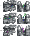
Interactions between Hox proteins and the DNA minor groove. Shown are images from X-ray crystal structures of PBC-Hox-DNA complexes, focused only on the interaction between the minor groove (shown as the gray surfaces) and the amino acid side chains of N-terminal arm/linker residues. The left-hand images (A, C, E) look into the minor groove from the top; the right-hand images (B, D, F) look along the axis of the minor groove. (A, B) Exd-Scr bound to the fkh250con consensus-binding site. Only Arg5 from the N-terminal is observed in the minor groove. (C, D) Exd-Scr bound to the fkh250 in vivo binding site. In contrast to the fkh250con structure, Arg3 (from the N-terminal arm) and His-12 (from the linker) are observed in the minor groove, in addition to Arg5. Note also that the minor groove in the fkh250 structure appears narrower than in the fkh250con structure (compare B with D). See Joshi et al. (2007) for details. (E, F). In the Pbx-Hoxa9 structure, one additional N-terminal arm residue, Arg2, is observed, together with Arg5. The Hoxa9 linker is unusually short (four residues), and none of them are seen inserting into the minor groove. See LeRonde-LeBlanc and Wolberger (2003) for details.
Figure 3.6
Figure 3.6
Two tiers of Hox-DNA-binding specificity. Hox proteins bind DNA using two levels of protein-DNA contacts. DNA contacts made by Arg5 (in the N-terminal arm) and Ile47, Gln50, Asn51, Met54 (in the third helix) are used by all Hox proteins to bind “AT”-rich DNA sequences (“general” Hox-DNA contacts), but are not good at distinguishing between Hox paralogs. With the help of cofactors (such as PBC proteins), paralog-specific DNA contacts are mediated by linker and N-terminal arm residues. “General” DNA contacts make hydrogen bonds in the DNA major groove. “Paralog-specific” DNA contacts may read a DNA structure, such as the narrow minor groove seen in the Exd-Scr-fkh250 structure.

Similar articles

Cited by

References

    1. Affolter M, Percival-Smith A, Müller M, Leupin W, Gehring WJ. DNA binding properties of the purified Antennapedia homeodomain. Proc. Natl. Acad. Sci. USA. 1990;87:4093–4097. - PMC - PubMed
    1. Affolter M, Slattery M, Mann RS. A lexicon for homeodomain-DNA recognition. Cell. 2008;133:1133–1135. - PubMed
    1. Akam M. Hox and HOM: homologous gene clusters in insects and vertebrates. Cell. 1989;57:347–349. - PubMed
    1. Appel B, Sakonju S. Cell-type-specific mechanisms of transcriptional repression by the homeotic gene products UBX and ABD-A in Drosophila embryos. EMBO J. 1993;12:1099–1109. - PMC - PubMed
    1. Arata Y, Kouike H, Zhang Y, Herman MA, Okano H, Sawa H. Wnt signaling and a Hox protein cooperatively regulate psa-3/Meis to determine daughter cell fate after asymmetric cell division in C. elegans. Dev. Cell. 2006;11:105–115. - PubMed

Publication types

MeSH terms

LinkOut - more resources