. 2009 Jul 30;63(2):230-43.
doi: 10.1016/j.neuron.2009.06.022.

GABA(B) receptor activation inhibits neuronal excitability and spatial learning in the entorhinal cortex by activating TREK-2 K+ channels

Affiliations

GABA(B) receptor activation inhibits neuronal excitability and spatial learning in the entorhinal cortex by activating TREK-2 K+ channels

Pan-Yue Deng et al. Neuron. .

Abstract

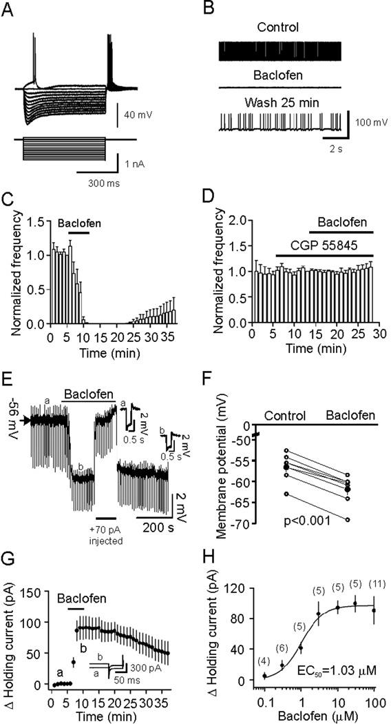
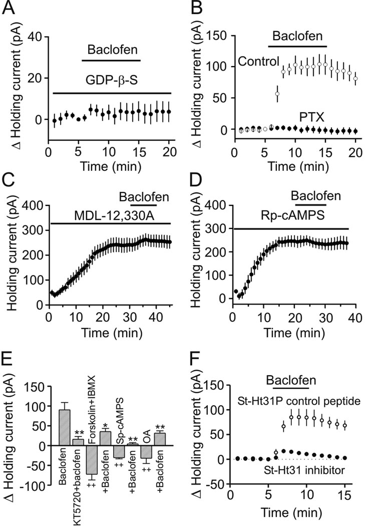
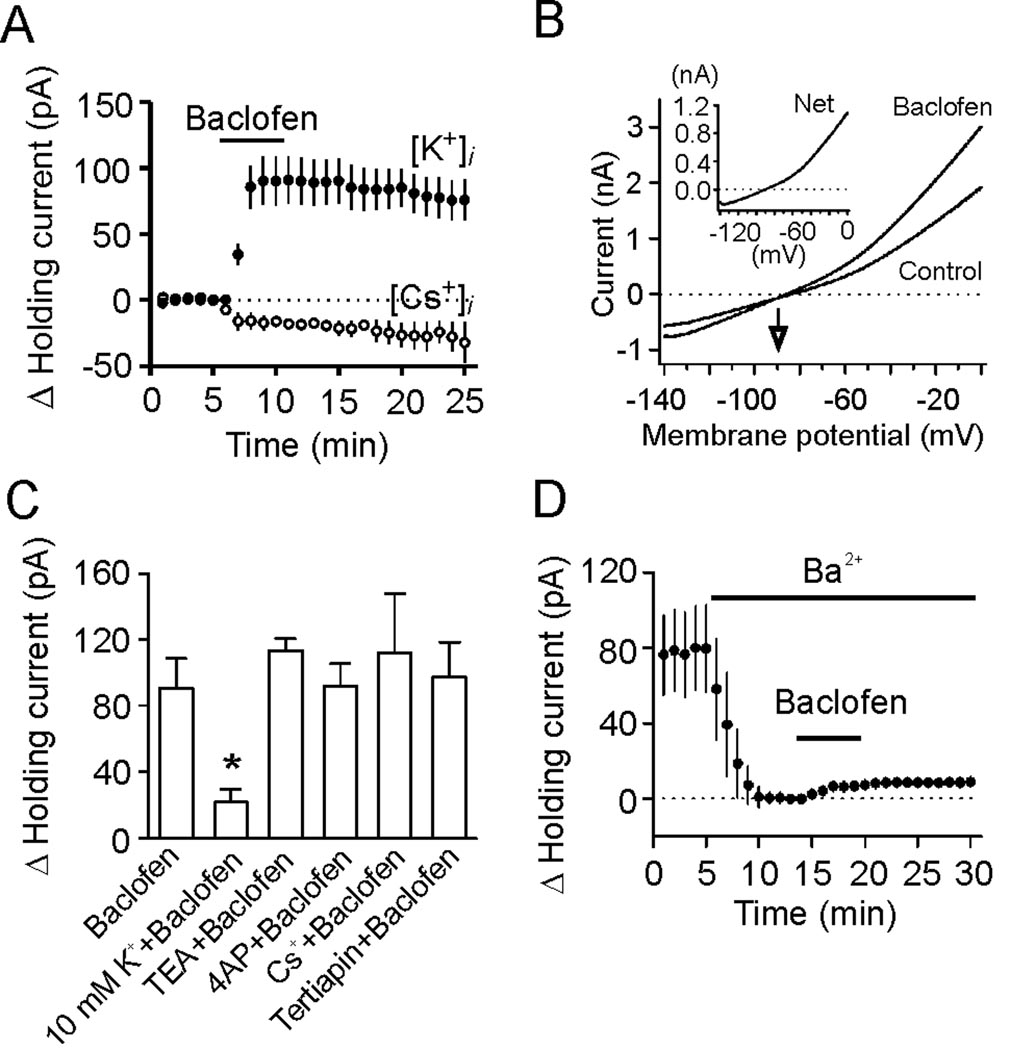
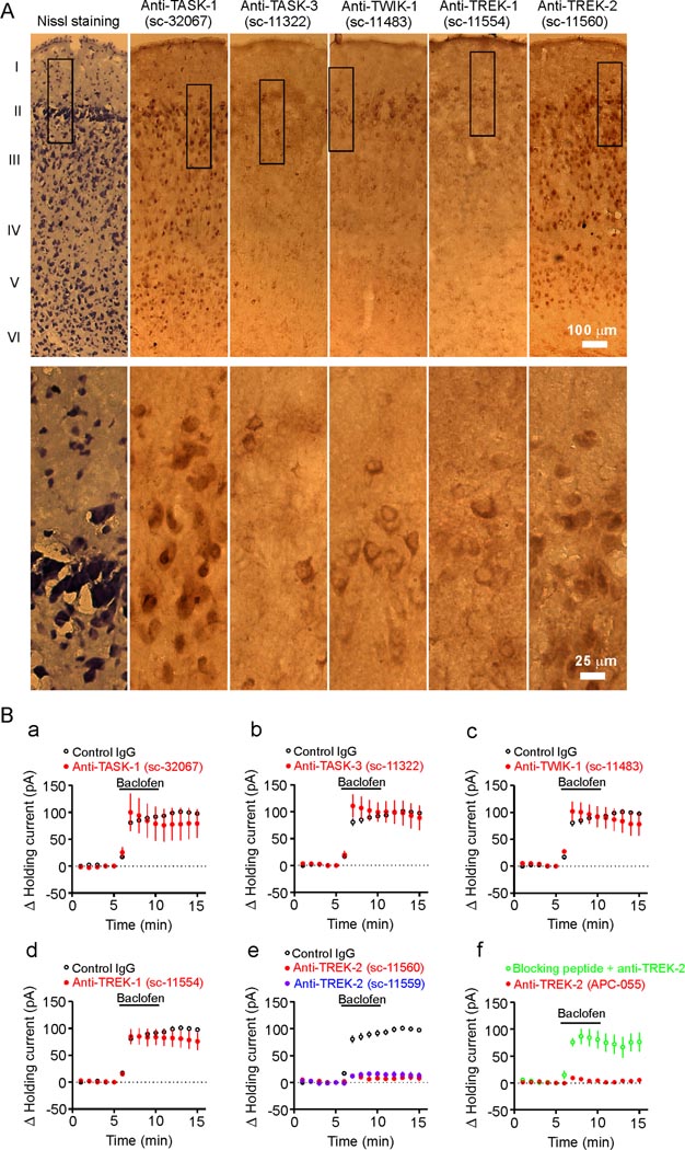
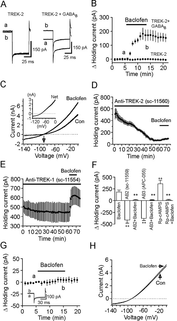
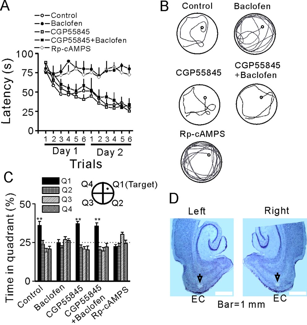
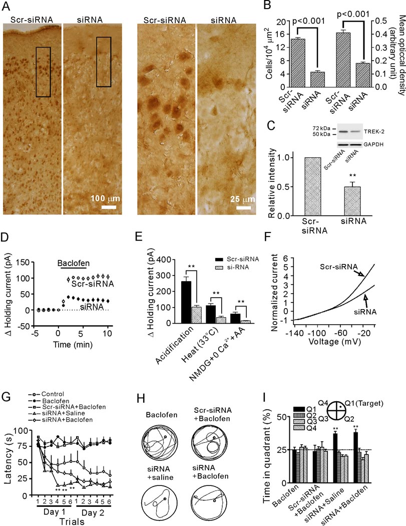
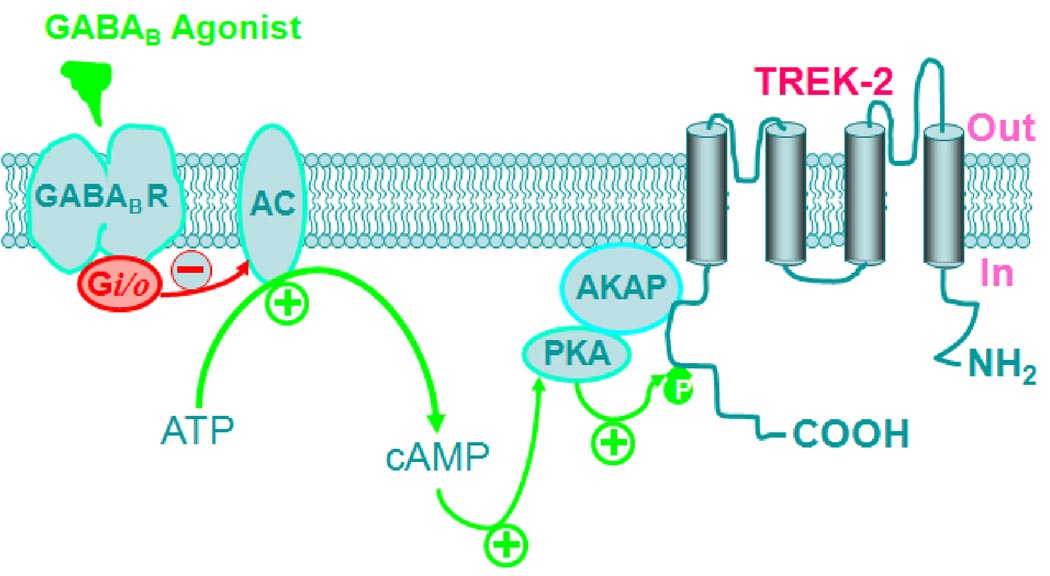
The entorhinal cortex (EC) is regarded as the gateway to the hippocampus and thus is essential for learning and memory. Whereas the EC expresses a high density of GABA(B) receptors, the functions of these receptors in this region remain unexplored. Here, we examined the effects of GABA(B) receptor activation on neuronal excitability in the EC and spatial learning. Application of baclofen, a specific GABA(B) receptor agonist, inhibited significantly neuronal excitability in the EC. GABA(B) receptor-mediated inhibition in the EC was mediated via activating TREK-2, a type of two-pore domain K(+) channels, and required the functions of inhibitory G proteins and protein kinase A pathway. Depression of neuronal excitability in the EC underlies GABA(B) receptor-mediated inhibition of spatial learning as assessed by Morris water maze. Our study indicates that GABA(B) receptors exert a tight control over spatial learning by modulating neuronal excitability in the EC.


Figures

Figure 1
Figure 1
Baclofen reduces the excitability of stellate neurons by generating membrane hyperpolarization. A, Voltage responses (upper panel) generated by current injection from +0.1 nA to −1 nA at an interval of −0.1 nA (lower panel) recorded from a stellate neuron in layer II. Note the depolarizing voltage sags in response to hyperpolarizing current pulses. B, AP firing recorded prior to, during and after application of baclofen from the stellate neuron in A. C, Pooled time course of AP firing frequency before, during and after application of baclofen. D, Baclofen-mediated inhibition of AP firing was blocked in the presence of the GABABR blocker, CGP 55845. E, Baclofen generated membrane hyperpolarization and reduced input resistance. A negative current (−50 pA for 500 ms) was injected every 5 s to assess the changes of input resistance. Insets are the voltage traces taken before (a) and during (b) the application of baclofen. Note that baclofen induced membrane hyperpolarization and reduced the voltage responses induced by the negative current injections suggesting a reduction in input resistance. To exclude the influence of baclofen-induced membrane hyperpolarization on input resistance, a constant positive current (+70 pA indicated by the horizontal bar) was injected briefly to elevate the membrane potential to the initial level. Under these conditions, the voltage responses induced by the negative current injections (−50 pA) were still smaller compared with control suggesting that baclofen-induced reduction in input resistance is not secondary to its effect on membrane hyperpolarization. F, Summarized data for baclofen-induced changes in RMPs. Filled circles denote the averaged values. G, Baclofen induced an outward HC. HCs were averaged per min and zeroed to the level just prior to the application of baclofen. Inset shows the averaged HCs recorded at the time points denoted in the figure. A −5 mV hyperpolarizing voltage step was used at the end of each trace to monitor potential changes of series resistance during recordings. H, Concentration-response curve for baclofen-induced changes in HCs. Numbers in the parenthesis were number of cells recorded.
Figure 2
Figure 2
GABABR-mediated inhibition requires the functions of inhibitory G proteins, AC, PKA and AKAPs. A, Intracellular application of GDP-β-S via the recording pipettes blocked baclofen-induced increases in outward HCs. B, Treatment of slices with PTX abolished the effects of baclofen on HCs, whereas treatment of the slices in the same fashion without PTX failed to alter the effect of baclofen. C, Intracellular dialysis of MDL-12,330A induced an outward HC and significantly inhibited baclofen-induced increases in outward HCs. HCs at −60 mV were recorded immediately after the formation of whole-cell configuration and the actual HCs were used to plot the figure. D, Application of Rp-cAMPS in the recording pipettes induced an outward HC and blocked baclofen-induced increases in outward HCs. The actual HCs were used to plot the figure. E, Baclofen-induced increases in outward HCs were significantly reduced by co-application of KT5720, forskolin plus IBMX, Sp-cAMPS or okadaic acid (OA) (*p<0.05, **p<0.01 vs. baclofen alone; ++p<0.01 vs. baseline=0). F, Inclusion of St-Ht31, the AKAP inhibitory peptide, in the recording pipettes significantly inhibited baclofen-induced increases in outward HCs whereas application of St-Ht31P, the control peptide, had no effects.
Figure 3
Figure 3
GABABR-mediated increases in outward HCs are mediated by activation of K2P channels. A, Bath application of baclofen induced a small inward current when Cs+-gluconate-containing intracellular solution was used in contrast to baclofen-induced outward currents when the intracellular solution contained K+-gluconate. B, Voltage-current relationship induced by a ramp protocol from −140 mV to 0 mV at a speed of 0.1 mV/ms prior to and during the application of baclofen. Subtraction of the current prior to the application of baclofen generated a net current of outward rectification (inset). The traces were averages from 9 cells. Note that the reversal potential was ~ −90.1 mV, close to the calculated K+ reversal potential (−92.2 mV). C, Baclofen-induced increases in outward HCs were insensitive to extracellular application of TEA, 4-AP, Cs+ and tertiapin, but reduced when the extracellular K+ concentration was increased to 10 mM (* p=0.02). D, Inclusion of Ba2+ in the extracellular solution induced an inward HC, but significantly reduced baclofen-induced increases in outward HCs.
Figure 4
Figure 4
TREK-2 channels are involved in GABABR-mediated hyperpolarization. A, Immunocytochemical staining of K2P channels in the EC (layer I–VI). Upper panels: low magnification, Low panels: high magnification of the regions marked in the upper panels. The catalog numbers of the ABs were labeled on the top. B, a–d, Intracellular infusion of ABs to TASK-1 (a), TASK-3 (b), TWIK-1 (c) and TREK-1 (d,) at 40 µg/ml failed to significantly change baclofen-induced increases in outward HCs. e, Intracellular dialysis of two TREK-2 ABs (40 µg/ml) drastically reduced baclofen-induced increases in outward HCs. f, Intracellular application of the third TREK-2 AB (APC-055, Alomone Labs, 40 µg/ml) significantly reduced the effect of baclofen whereas application of the TREK-2 AB preabsorbed with the corresponding blocking peptide via the recording pipettes significantly reduced the inhibitory effect of TREK-2 AB.
Figure 5
Figure 5
GABABR-induced hyperpolarization is dependent on PKA-mediated phosphorylation site on TREK-2 channels. A, Left panel: HCs recorded at −60 mV from a HEK293 cell transfected with TREK-2 alone before (a) and during (b) the application of baclofen (100 µM). Right panel: HCs recorded at −60 mV from a HEK293 cell cotransfected with GABABRs and TREK-2 channels before (a) and during (b) the application of baclofen. B, Summarized data from 6 HEK293 cells transfected with TREK-2 channels alone and 7 HEK293 cells cotransfected with GABABRs and TREK-2 channels. C, Baclofen-induced currents had a reversal potential (−89.2±2.2 mV, n=6) close to the reversal potential of K+ (−96.1 mV). Inset, net current induced by baclofen. D, Intracellular application of the first TREK-2 AB (40 µg/ml) to the HEK293 cells co-expressing GABABRs and TREK-2 channels generated an inward HC per se and significantly reduced baclofen-induced increases in outward HCs. HCs at −60 mV were recorded immediately after the formation of whole-cell configuration and the actual HCs were used to plot the figure. E, Intracellular application of TREK-1 AB (40 µg/ml) had little effects on HCs and did not significantly change baclofen-induced increases in outward HCs. HCs at −60 mV were recorded immediately after the formation of whole-cell configuration and the actual HCs were used to plot the figure. F, Intracellular dialysis of the second TREK-2 AB (AB2, sc-11559, 40 µg/ml) into the HEK293 cells co-transfected with GABABRs and TREK-2 channels generated an inward holding current (n=6, ++p<0.01 vs. baseline=0) and significantly inhibited baclofen-induced increases in outward holding currents (n=6, **p<0.01 vs. baclofen alone). Intracellular perfusion of the third TREK-2 AB (AB3, APC-055, Alomone labs, 40 µg/ml) induced an inward holding current (n=6, ++p<0.001 vs. baseline=0) and significantly inhibited the effect of baclofen (n=6, **p<0.01 vs. baclofen alone). Intracellular application of Rp-cAMPS (1 mM) into the HEK293 cells co-transfected with GABABRs and TREK-2 channels induced an outward holding current (n=7, ++p<0.01 vs. baseline=0) and significantly reduced the effect of baclofen (n=7, **p<0.01 vs. baclofen alone). G, Application of baclofen did not change the HCs significantly in HEK293 cells co-transfected with S359A mutant TREK-2 channels and GABABRs. Inset shows the holding currents before and during the application of baclofen. H, Application of baclofen did not induce significant changes in the voltage-current relationship recorded from HEK293 cells co-expressing GABABRs and S359A mutant TREK-2 channels.
Figure 6
Figure 6
Activation of GABABRs impairs spatial learning in Morris water maze. A, Mean latencies to the platform from the acquisition trials were presented by groups. Rats were microinjected with normal saline (control), baclofen, Rp-cAMPS, CGP55845 or CGP55845 followed by baclofen. B, Representative swimming traces from the last trial of day 2. Note that baclofen markedly lengthened the swimming path, which was mimicked by Rp-cAMPS. C, Probe trial performance of each group as presented by the proportion of total time spent in each quadrant of the Morris water maze (**p<0.01 vs. 25% chance in each quadrant). D, Horizontal brain sections showing the microinjection sites indicated by the arrows.
Figure 7
Figure 7
Knockdown of TREK-2 channels by siRNA annuls baclofen-induced impairment of spatial learning. A, TREK-2 channels were significantly knocked down after delivery of TREK-2 siRNAs to the EC. Left two panels: immunoreactivity of TREK-2 channels in a region of the EC adjacent to the injection site from rats treated with Scr-siRNA or siRNA in low magnification. Right two panels: high magnification of the regions denoted in the left two panels. B, siRNA-treatment significantly deceased the number of TREK-2-positive cells (left) and the mean optical density of TREK-2-positive cells (right) in layer II of the EC. C, Western blot showed that siRNA-treatment significantly decreased the level of TREK-2 channel proteins in the EC (**p<0.001). A band that had a molecular mass of ~60 kDa corresponding to the reported molecular mass of TREK-2 channels (Kang et al., 2007b; Simkin et al., 2008) was detected in the lysates of the EC. D, Baclofen-induced increases in outward HCs were significantly reduced in slices cut from rats treated with siRNA. E, siRNA treatment significantly reduced the increases in outward HCs induced by intracellular acidification (262.1±28.4 pA, n=10 slices from 3 Scr-siRNA treated rats vs. 101.8±11.5 pA, n=12 slices from 4 siRNA treated rats), heat (111.1±13.2 pA, n=10 slices from 3 Scr-siRNA treated rats vs. 37.5±7.4 pA, n=11 slices from 3 siRNA treated rats) and AA (in the presence of NMDG and 0 Ca2+; 59.9±10.6 pA, n=10 slices from 3 Scr-siRNA treated rats vs. 16.5±3.0 pA, n=12 slices from 4 siRNA treated rats) (**p<0.001). F, siRNA treatment conspicuously reduced the extent of outward rectification of the voltage-current relationship of the stellate neurons. Currents at different voltages from each cell were normalized to the absolute value of the current at −140 mV to minimize the influence of current sizes on voltage-current relationship. Each trace shows the averaged voltage-current relationship from 10 cells from 3 rats treated with siRNA or Scr-siRNA. G, Summarized mean latencies. Note that siRNA knockdown of TREK-2 channels blocked baclofen-induced impairment of spatial learning (**p<0.01 vs. the corresponding values in control group). H, Representative swimming traces from the last trial of day 2 for each group. I, Probe trial performance of each group as presented by the proportion of total time spent in each quadrant. The siRNA-treated rats showed a preference for the target quadrant and intra-EC injection of baclofen into these rats did not prevent the preference of the rats (** p<0.01 vs. 25% chance level).
Figure 8
Figure 8
Schematic illustration of signaling cascade leading to the activation of TREK-2 channels by GABABRs. Red arrows and minus mark indicate inhibition whereas green arrows and plus marks denote facilitation. GABABR agonists activate GABABRs resulting in activation of the inhibitory G proteins (Gi/o). Activation of Gi/o inhibits the activity of AC leading to a reduction in the production of cAMP from ATP and an inhibition of PKA activity. Normally, PKA exerts a tonic inhibition on TREK-2 channels by phosphorylating serine 359 on TREK-2 channels. The effects of PKA require the function of AKAPs which tether PKA to TREK-2 channels. GABABR-mediated inhibition of PKA annuls PKA-mediated tonic inhibition of TREK-2 channels resulting in an increase in the function of TREK-2 channels. The ultimate result is the inhibition of neuronal excitability in the EC and depression of spatial learning.

Similar articles

Cited by

References

    1. Aller MI, Veale EL, Linden AM, Sandu C, Schwaninger M, Evans LJ, Korpi ER, Mathie A, Wisden W, Brickley SG. Modifying the subunit composition of TASK channels alters the modulation of a leak conductance in cerebellar granule neurons. J Neurosci. 2005;25:11455–11467. - PMC - PubMed
    1. Arnsten AF, Ramos BP, Birnbaum SG, Taylor JR. Protein kinase A as a therapeutic target for memory disorders: rationale and challenges. Trends Mol Med. 2005;11:121–128. - PubMed
    1. Avoli M, D'Antuono M, Louvel J, Kohling R, Biagini G, Pumain R, D'Arcangelo G, Tancredi V. Network and pharmacological mechanisms leading to epileptiform synchronization in the limbic system in vitro. Prog Neurobiol. 2002;68:167–207. - PubMed
    1. Bang H, Kim Y, Kim D. TREK-2, a new member of the mechanosensitive tandem-pore K+ channel family. J Biol Chem. 2000;275:17412–17419. - PubMed
    1. Bayliss DA, Sirois JE, Talley EM. The TASK family: two-pore domain background K+ channels. Mol Interv. 2003;3:205–219. - PubMed

Publication types

MeSH terms

LinkOut - more resources