West Nile virus infection alters midgut gene expression in Culex pipiens quinquefasciatus Say (Diptera: Culicidae)
- PMID: 19635880
- PMCID: PMC2754826
West Nile virus infection alters midgut gene expression in Culex pipiens quinquefasciatus Say (Diptera: Culicidae)
Abstract
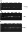
Alterations in gene expression in the midgut of female Culex pipiens quinquefasciatus exposed to blood meals containing 6.8 logs plaque-forming units/mL of West Nile virus (WNV) were studied by fluorescent differential display. Twenty-six different cDNAs exhibited reproducible differences after feeding on infected blood. Of these, 21 cDNAs showed an increase in expression, and 5 showed a decrease in expression as a result of WNV presence in the blood meal. GenBank database searches showed that one clone with increased expression, CQ G12A2, shares 94% identity with a leucine-rich repeat-containing protein from Cx. p. quinquefasciatus and 32% identity to Toll-like receptors from Aedes aegypti. We present the first cDNA clone isolated from female Cx. p. quinquefasciatus midgut tissue whose expression changes on exposure to WNV. This cDNA represents a mosquito gene that is an excellent candidate for interacting with WNV in Cx. p. quinquefasciatus and may play a role in disease transmission.
Figures


Similar articles
-
The Effect of West Nile Virus Infection on the Midgut Gene Expression of Culex pipiens quinquefasciatus Say (Diptera: Culicidae).Insects. 2016 Dec 19;7(4):76. doi: 10.3390/insects7040076. Insects. 2016. PMID: 27999244 Free PMC article.
-
Impact of extrinsic incubation temperature and virus exposure on vector competence of Culex pipiens quinquefasciatus Say (Diptera: Culicidae) for West Nile virus.Vector Borne Zoonotic Dis. 2007 Winter;7(4):629-36. doi: 10.1089/vbz.2007.0101. Vector Borne Zoonotic Dis. 2007. PMID: 18021028 Free PMC article.
-
Relationships between infection, dissemination, and transmission of West Nile virus RNA in Culex pipiens quinquefasciatus (Diptera: Culicidae).J Med Entomol. 2012 Jan;49(1):132-42. doi: 10.1603/me10280. J Med Entomol. 2012. PMID: 22308781 Free PMC article.
-
The insect-specific Palm Creek virus modulates West Nile virus infection in and transmission by Australian mosquitoes.Parasit Vectors. 2016 Jul 25;9(1):414. doi: 10.1186/s13071-016-1683-2. Parasit Vectors. 2016. PMID: 27457250 Free PMC article.
-
The contribution of Culex pipiens complex mosquitoes to transmission and persistence of West Nile virus in North America.J Am Mosq Control Assoc. 2012 Dec;28(4 Suppl):137-51. doi: 10.2987/8756-971X-28.4s.137. J Am Mosq Control Assoc. 2012. PMID: 23401954 Review.
Cited by
-
Alterations in the Aedes aegypti transcriptome during infection with West Nile, dengue and yellow fever viruses.PLoS Pathog. 2011 Sep;7(9):e1002189. doi: 10.1371/journal.ppat.1002189. Epub 2011 Sep 1. PLoS Pathog. 2011. PMID: 21909258 Free PMC article.
-
Can Horton hear the whos? The importance of scale in mosquito-borne disease.J Med Entomol. 2014 Mar;51(2):297-313. doi: 10.1603/me11168. J Med Entomol. 2014. PMID: 24724278 Free PMC article.
-
Immune-related transcripts, microbiota and vector competence differ in dengue-2 virus-infected geographically distinct Aedes aegypti populations.Parasit Vectors. 2023 May 19;16(1):166. doi: 10.1186/s13071-023-05784-3. Parasit Vectors. 2023. PMID: 37208697 Free PMC article.
-
The Effect of West Nile Virus Infection on the Midgut Gene Expression of Culex pipiens quinquefasciatus Say (Diptera: Culicidae).Insects. 2016 Dec 19;7(4):76. doi: 10.3390/insects7040076. Insects. 2016. PMID: 27999244 Free PMC article.
-
Transcriptome Analysis of the Midgut of the Chinese Oak Silkworm Antheraea pernyi Infected with Antheraea pernyi Nucleopolyhedrovirus.PLoS One. 2016 Nov 7;11(11):e0165959. doi: 10.1371/journal.pone.0165959. eCollection 2016. PLoS One. 2016. PMID: 27820844 Free PMC article.
References
-
- Peyrefitte CN, Pastorino B, Grau GE, Lou J, Tolou H, Couissinier-Paris P. Dengue virus infection of human microvascular endothelial cells from different vascular beds promotes both common and specific functional changes. J Med Virol. 2006;78:229–242. - PubMed
-
- Kramer LD, Styer LM, Ebel GD. A global perspective on the epidemiology of West Nile virus. Annu Rev Entomol. 2008;53:61–81. - PubMed
Publication types
MeSH terms
Substances
Grants and funding
LinkOut - more resources
Full Text Sources
