. 2009 Sep 18;284(38):25560-8.
doi: 10.1074/jbc.M109.007690. Epub 2009 Jul 24.

The Fanconi anemia protein FANCM is controlled by FANCD2 and the ATR/ATM pathways

Affiliations

The Fanconi anemia protein FANCM is controlled by FANCD2 and the ATR/ATM pathways

Alexandra Sobeck et al. J Biol Chem. .

Abstract

Genomic stability requires a functional Fanconi anemia (FA) pathway composed of an upstream "core complex" (FA proteins A/B/C/E/F/G/L/M) that mediates monoubiquitination of the downstream targets FANCD2 and FANCI. Unique among FA core complex members, FANCM has processing activities toward replication-associated DNA structures, suggesting a vital role for FANCM during replication. Using Xenopus egg extracts, we analyzed the functions of FANCM in replication and the DNA damage response. xFANCM binds chromatin in a replication-dependent manner and is phosphorylated in response to DNA damage structures. Chromatin binding and DNA damage-induced phosphorylation of xFANCM are mediated in part by the downstream FA pathway protein FANCD2. Moreover, phosphorylation and chromatin recruitment of FANCM is regulated by two mayor players in the DNA damage response: the cell cycle checkpoint kinases ATR and ATM. Our results indicate that functions of FANCM are controlled by FA- and non-FA pathways in the DNA damage response.


Figures
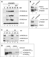
FIGURE 1.
FIGURE 1.
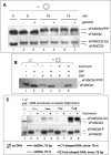
xFANCM binds chromatin in a replication-dependent, FA core complex-independent manner and is hyperphosphorylated during replication. A, chromatin binding of xFANCM is replication initiation-dependent, similar to xFANCA and xFANCD2 (14). Sperm chromatin was added to S phase extracts and reisolated at the indicated time points during replication. Extracts were either untreated (nt, lanes 1–4) or contained the replication initiation inhibitor geminin (gem, lanes 5 + 6). Chromatin fractions were analyzed for bound proteins (xFANCM-chr, xFANCD2-chr, and xFANCA-chr) by SDS-PAGE and immunoblotting. Chromatin-bound histone H3 (xH3-chr) was used as a loading control. B, immunodepletion of xFANCM from extracts partially co-depletes xFANCA and vice versa. S phase extracts were incubated with bead-coupled preimmune serum (mock, lane 1, upper and lower panels), with anti-xFANCA antibody (ΔxFANCA, upper panel, lane 2), or with anti-xFANCM antibody (ΔxFANCM, lower panel, lane 2). Extracts were then assayed for the presence of xFANCA and xFANCM as indicated. Nonspecific bands recognized by the anti-xFANCA and anti-xFANCM antibody, respectively, were used as a loading control. C, xFANCM binds chromatin in absence of the functional FA core complex. Sperm chromatin was added to egg extracts treated with preimmune serum (lanes 1 and 4), extracts depleted of xFANCM (lanes 2 and 5), or extracts depleted of xFANCA (lanes 3 and 6). Chromatin was reisolated at the indicated time points and assayed for the presence of xFANCM-chr and xFANCD2-chr by immunoblot. Chromatin-bound histone H3 (xH3-chr) was used as a loading control. D, chromatin-bound xFANCM is hyperphosphorylated during replication. Sperm chromatin (chrom.) was added to S phase extracts, and extracts were either left untreated (lanes 1 and 3) or supplemented with tautomycin (lanes 2 and 4) at 30 min following the addition of sperm chromatin. Nuclear fractions (lanes 1 and 2) or chromatin fractions (lanes 3 and 4) were reisolated at 60 min following the addition of sperm chromatin and analyzed for the presence of xFANCM by SDS-PAGE and immunoblot.
FIGURE 2.
FIGURE 2.
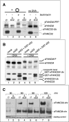
xFANCM is phosphorylated in response to ssDNA and dsDNA substrates. A, hyperphosphorylation of xFANCM (xFANCM-PPP) is induced in the presence of plasmid DNA, similarly to monoubiquitination of xFANCD2 (xFANCD2-Ub) (16). Egg extracts were incubated with circular plasmid DNA for the indicated time points. Extracts were either untreated (lanes 2, 4, and 6) or treated with geminin (lanes 3, 5, and 7). DNA-free extracts served as a negative control (lane 1). Following incubation, 1 μl of extract was analyzed for either xFANCM or xFANCD2 by immunoblotting. B, the DNA plasmid-induced mobility shift of xFANCM is due to phosphorylation. Extracts were incubated for 15 min in the absence (lanes 1 and 2) or presence (lanes 3–6) of plasmid DNA and supplemented with either H2O (lanes 1–4) or tautomycin (lanes 5 and 6). Following incubation, extracts were either untreated (lanes 1, 3, and 5) or treated with shrimp alkaline phosphatase (SAP, lanes 2, 4, and 6). Subsequently, 1 μl of extract was analyzed for xFANCM by SDS-PAGE and immunoblot. C, xFANCM is phosphorylated in response to linear and branched ssDNA and dsDNA structures. DNA structures ssDNA70 (lanes 2 and 3), dsDNA70 (lanes 4 and 5), Y-shaped DNA (lanes 6 and 7), and forked double-stranded DNA (lanes 8 and 9) were coupled to beads and incubated in egg extracts (extr.) for 30 min. Extracts were either untreated (lanes 1, 2, 4, 6, and 8) or supplemented with tautomycin (lanes 3, 5, 7, and 9). Following incubation, bead DNA substrates were separated from the extracts and analyzed for bound xFANCD2 and xFANCM by SDS-PAGE and immunoblot. 1 μl of DNA-free extract (lane 1) was used as a negative control.
FIGURE 3.
FIGURE 3.
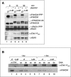
Chromatin binding and DNA-induced phosphorylation of xFANCM is partially dependent on xFANCD2. A, formation of xFANCM-PPP in response to plasmid DNA is partially controlled by xFANCD2. Egg extracts were either mock-depleted (lanes 1, 3, and 5) or depleted of xFANCD2 (lanes 2, 4, and 6) and incubated with plasmid DNA for 60 min. To further stabilize phosphorylated isoforms of xFANCM, extracts were supplemented with tautomycin (lanes 3 and 4). Following incubation of plasmid DNA, 1 μl of extract was subsequently analyzed for xFANCM and xFANCD2 by immunoblotting. B, recombinant xFANCD2 rescues xFANCM phosphorylation deficiency in xFANCD2-depleted extracts. Extracts were either mock-depleted (lanes 1 and 4) or depleted of xFANCD2 (lanes 2, 3, 5, and 6). xFANCD2-depleted extracts were either nonsupplemented (lanes 2 and 5) or supplemented with recombinant, glutathione S-transferase (GST)-tagged xFANCD2 (lanes 3 and 6). Extracts were either left DNA-free (lanes 1–3) or incubated with plasmid DNA (lanes 4–6) for 30 min, and 1 μl of extract was analyzed for xFANCD2 and xFANCM by SDS-PAGE and immunoblot. C, chromatin binding of xFANCM is partially controlled by xFANCD2. Sperm chromatin was added to S phase extracts that were either mock-depleted (lanes 1, 3, 5, and 7) or depleted of xFANCD2 (lanes 2, 4, 6, and 8) and incubated for the indicated time points. Following incubation, chromatin fractions were reisolated and analyzed for xFANCD2 and xFANCM by SDS-PAGE and immunoblot. A nonspecific band was used as a loading control.
FIGURE 4.
FIGURE 4.
xFANCM is partially regulated by ATR and ATM. A, DNA-induced xFANCM-PPP formation is partially controlled by xATR. Extracts were either mock-depleted (lanes 1, 3, 5, and 7) or depleted of xATR (lanes 2, 4, 6, and 8) and incubated with plasmid DNA for the indicated time points. Following incubation, 1 μl of extract was analyzed for xFANCM and xFANCD2 by SDS-PAGE and immunoblot. B, xATR kinase activity is required for efficient xFANCM-PPP formation. Extracts were either mock-depleted (lanes 2 and 3) or depleted of xATRIP (lanes 1, 5, and 6). A neutralizing anti-xATR antibody was added to mock-depleted or xATRIP-depleted extracts where indicated (lanes 4 and 6, respectively). Extracts were incubated with plasmid DNA for 30 min, and 1 μl of extract was analyzed for the indicated proteins by SDS-PAGE and immunoblot. Phosphorylated isoforms of xChk1 were detected as protein bands with lower mobility (xChk1-P) compared with nonphosphorylated xChk1. DNA free mock-depleted or xATRIP-depleted extracts (lanes 1 and 2, respectively) were used as negative control and as control for protein size and quantitative xATRIP depletion. C, chromatin binding of xFANCM, xFANCA, and xFANCD2 depends on xATR. Sperm chromatin was added to S phase extracts that were either mock-depleted (lanes 1, 3, and 5) or depleted of xATR (lanes 2, 4, and 6) and incubated for the indicated time points. Following incubation, chromatin fractions were reisolated and analyzed for chromatin-bound proteins (xFANCA-chr, xFANCD2-chr, and xFANCM-chr) by SDS-PAGE and immunoblot. Chromatin-bound histone H3 (xH3-chr) was used as a loading control. D, xATM is partially required for DNA-induced FANCM-PPP formation. Extracts were either untreated (lanes 1, 2, 4, 6, 7, and 9) or treated with the ATM kinase inhibitor, KU-55933 (lanes 3, 5, 8, and 10). To stabilize phosphorylated xFANCM isoforms, tautomycin was added to egg extracts where indicated (lanes 4, 5, 9, and 10). Extracts were incubated with plasmid DNA for the indicated time points, and 1 μl of extract was analyzed for xFANCM and xFANCD2 by SDS-PAGE and immunoblot. DNA free extracts (lanes 1 and 6) were used as a negative control.
FIGURE 5.
FIGURE 5.
xFANCM is not required for the dsDNA-induced, xATR-dependent checkpoint response. A, xFANCM depletion does not inhibit plasmid DNA-induced phosphorylation of xChk1, xRad1, or x-γH2AX. Extracts were either mock-depleted (lanes 1, 3, and 5) or depleted of xFANCM (lanes 2, 4, and 6) and incubated with plasmid DNA for the indicated time points. Following incubation, 1 μl of extract was analyzed for the indicated proteins by SDS-PAGE and immunoblot. DNA-free extracts (lanes 1 and 2) were used as a negative control and as a control for protein size and for quantitative depletion of xFANCM. B, xFANCM depletion does not inhibit dsDNA70-induced phosphorylation of Chk1. Extracts were either mock-depleted (lanes 1, 3, 4, 7, and 8) or depleted of xFANCM (lanes 2, 5, 6, 9, and 10) and incubated with either ssDNA70 (lanes 3, 5, 7, and 9) or dsDNA70 (lanes 4, 6, 8, and 10) for 30 min. Following incubation, 1 μl of extract was analyzed for the indicated proteins by SDS-PAGE and immunoblot. DNA-free extracts (lanes 1 and 2) were used as a negative control and as a control for protein size and for quantitative depletion of xFANCM. Phosphorylated xChk1 was detected using an anti-xChk1-PSer344 antibody. (Please note that the low DNA concentration (40 μg/ml) used in this assay triggers a robust xChk1-P response but is not sufficient to induce a robust xFANCM-PPP induction.)

Similar articles

Cited by

References

    1. Wang W. (2007) Nat. Rev. Genet. 8, 735–748 - PubMed
    1. Pichierri P., Rosselli F. (2004) EMBO J. 23, 1178–1187 - PMC - PubMed
    1. Andreassen P. R., D'Andrea A. D., Taniguchi T. (2004) Genes Dev. 18, 1958–1963 - PMC - PubMed
    1. Taniguchi T., Garcia-Higuera I., Xu B., Andreassen P. R., Gregory R. C., Kim S. T., Lane W. S., Kastan M. B., D'Andrea A. D. (2002) Cell 109, 459–472 - PubMed
    1. Smogorzewska A., Matsuoka S., Vinciguerra P., McDonald E. R., 3rd, Hurov K. E., Luo J., Ballif B. A., Gygi S. P., Hofmann K., D'Andrea A. D., Elledge S. J. (2007) Cell 129, 289–301 - PMC - PubMed

Publication types

MeSH terms