. 2009 Oct;83(19):9682-93.
doi: 10.1128/JVI.00715-09. Epub 2009 Jul 22.

Respiratory syncytial virus nonstructural proteins decrease levels of multiple members of the cellular interferon pathways

Affiliations

Respiratory syncytial virus nonstructural proteins decrease levels of multiple members of the cellular interferon pathways

Samer Swedan et al. J Virol. 2009 Oct.

Abstract

Viruses of the Paramyxoviridae family, such as the respiratory syncytial virus (RSV), suppress cellular innate immunity represented by type I interferon (IFN) for optimal growth in their hosts. The two unique nonstructural (NS) proteins, NS1 and NS2, of RSV suppress IFN synthesis, as well as IFN function, but their exact targets are still uncharacterized. Here, we investigate if either or both of the NS proteins affect the steady-state levels of key members of the IFN pathway. We found that both NS1 and NS2 decreased the levels of TRAF3, a strategic integrator of multiple IFN-inducing signals, although NS1 was more efficient. Only NS1 reduced IKKepsilon, a key protein kinase that specifically phosphorylates and activates IFN regulatory factor 3. Loss of the TRAF3 and IKKepsilon proteins appeared to involve a nonproteasomal mechanism. Interestingly, NS2 modestly increased IKKepsilon levels. In the IFN response pathway, NS2 decreased the levels of STAT2, the essential transcription factor for IFN-inducible antiviral genes. Preliminary mapping revealed that the C-terminal 10 residues of NS1 were essential for reducing IKKepsilon levels and the C-terminal 10 residues of NS2 were essential for increasing and reducing IKKepsilon and STAT2, respectively. In contrast, deletion of up to 20 residues of the C termini of NS1 and NS2 did not diminish their TRAF3-reducing activity. Coimmunoprecipitation studies revealed that NS1 and NS2 form a heterodimer. Clearly, the NS proteins of RSV, working individually and together, regulate key signaling molecules of both the IFN activation and response pathways.


Figures

FIG. 1.
FIG. 1.
Cytoplasmic location of recombinantly expressed NS proteins. A549 cells were transiently transfected with pCAGGS-FLAG-NS1 and -NS2 plasmids or with the vector alone (control) as indicated at the top. The proteins were immunostained with FLAG antibody (green) and the nuclei were stained with 4′,6-diamidino-2-phenylindole (DAPI; blue) as described in Materials and Methods.
FIG. 2.
FIG. 2.
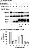
Reduction of TRAF3 levels by NS1. A549 cells in 10-cm2 wells were transfected with 4 μg FLAG-TRAF3 plasmid. In addition, they were transfected with the indicated amounts (in micrograms) of pCAGGS-FLAG-NS plasmids (left) or infected with wild-type (wt) or NS deletion-containing RSV (right) as indicated. In transfection experiments (left), the empty pCAGGS vector was used wherever needed, to keep the amount of DNA constant at 7 μg in every well. Cells were processed 24 h later for IB to detect the proteins as shown in panel A, with specific antibodies described in Materials and Methods. The TRAF3 band intensities were normalized against actin and plotted (B) as a percentage of the respective control lanes (lane 1 for the left side of the panel, lane 8 for the right side of the panel). Average values of three experiments and standard error bars are presented.
FIG. 3.
FIG. 3.
Reduction of TRAF3 levels by recombinant NS1 in the RSV-infected cell environment. (A) A549 cells in 10-cm2 wells were first infected with dual NS deletion-containing RSV. The medium was removed 3 h later, and the cell monolayers were rinsed with PBS. Fresh medium was then added, and the cells were transfected with 4 μg FLAG-TRAF3 plasmid and the indicated pCAGGS-FLAG-NS plasmids (3 μg for the single-NS plasmid, 1.5 μg each for dual transfection). Cells were processed 24 h later for IB to detect the proteins as shown, with the specific antibodies described in Materials and Methods. (B) The TRAF3 band intensities were normalized against actin and plotted as percentages of the no-NS control (lane 1). The mean ± the standard error of the mean of three experiments are presented.
FIG. 4.
FIG. 4.
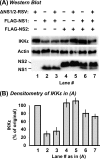
Reduction of IKKɛ levels by NS1. Experiments were performed essentially as described in the legend to Fig. 2, by substituting 2 μg of the myc-IKKɛ plasmid for the FLAG-TRAF3 plasmid. The total amount of transfected DNA was kept constant at 5 μg by using the empty pCAGGS vector wherever needed. wt, wild type.
FIG. 5.
FIG. 5.
Reduction of IKKɛ levels by recombinant NS1 in the RSV-infected cell environment. These experiments were performed essentially as described in the legend to Fig. 3, by substituting 2 μg of the myc-IKKɛ plasmid for the FLAG-TRAF3 plasmid. The total amount of transfected DNA was kept constant at 5 μg by using the empty pCAGGS vector wherever needed.
FIG. 6.
FIG. 6.
Reduction of endogenous STAT2 levels by recombinant NS2. A549 cells in 10-cm2 wells were transfected with the indicated amounts of pCAGGS-FLAG-NS plasmids. The empty pCAGGS vector was also transfected wherever needed to keep the amount of DNA constant at 7 μg in every well. Cells were processed 24 h later for IB to detect the proteins (top of each panel) with the specific antibodies described in Materials and Methods. The STAT2 band intensities were normalized against that of actin and plotted (bottom of each panel) as percentages of that of the control cells transfected with the empty pCAGGS vector (lanes 1 and 11 for the respective sets). The mean ± the standard error of the mean of three experiments are presented.
FIG. 7.
FIG. 7.
Expression of recombinant NS deletion mutant proteins. A549 cells in 10-cm2 wells were transfected with 3 μg of wild-type or C-terminal deletion-containing pCAGGS-FLAG-NS plasmid. Cells were processed 24 h later for IB with FLAG antibody. Note that the NS1 mutant protein with 30 amino acids deleted from the C terminus could not be detected.
FIG. 8.
FIG. 8.
Lack of a role for the C terminus of NS in TRAF3 reduction. (A, B) A549 cells in 10-cm2 wells were transfected with 4 μg of FLAG-TRAF3 plasmid. In addition, they were transfected with the indicated amounts of pCAGGS-FLAG-NS plasmids (or 3 μg of the empty vector in lane 1) or infected with RSV (lanes 2 and 12). Where indicated (B), MG132 was added 18 h later and the cells were incubated for another 6 h. (C) Degradation of IκBα by TNF-α. Where indicated, A549 cells were treated with TNF-α in the presence or absence of 10 μM MG132 for 20 min. All of the cells were then processed for IB with appropriate antibodies to detect actin, IκBα, or FLAG (for TRAF3 and NS). Note that ΔC10NS2 (lane 6) and ΔC20NS2 (lane 7) migrated either to the same spot as NS1 or too close to NS1 to be resolved clearly. (D) The TRAF3 band intensities were normalized against actin and plotted as percentages of the respective control lanes (lanes 1 and 11). The mean ± the standard error of the mean of three experiments are presented. wt, wild type.
FIG. 9.
FIG. 9.
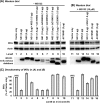
Restoration of IKKɛ levels upon deletion of the C termini of the NS proteins. These experiments were designed essentially as described for TRAF3 in the legend to Fig. 8. (A, B) A549 cells in 10-cm2 wells were transfected with 2 μg of myc-IKKɛ plasmid. In addition, they were transfected with the indicated amounts of pCAGGS-FLAG-NS plasmids (or 3 μg of the empty vector only in lanes 1 and 11). Where indicated (B), MG132 was added 18 h later and the cells were incubated for another 6 h. All of the cells were then processed for IB with appropriate antibodies to detect the myc tag (IKKɛ), actin, IκBα (not shown), or the FLAG tag (NS1/2). Note that the mobility of ΔC10NS2 was identical to that of NS1, and therefore, the two bands could not be resolved (lane 5). Lane 10 was taken from a different experiment, as indicated by the dotted line. (C) The band intensities were normalized against that of actin and plotted as percentages of the respective control lanes (lane 1 and 11). The mean ± the standard error of the mean of three experiments are presented. wt, wild type.
FIG. 10.
FIG. 10.
Destruction of STAT2 requires the C terminus of NS2. A549 cells in 10-cm2 wells were transfected with 3 μg of single plasmids (pCAGGS-FLAG-NS plasmids or the empty vector) or 1.5 μg of each plasmid when transfected as a mixture (lane 4). All cells were processed 24 h posttransfection for IB with STAT2 or actin antibody. The STAT2 band intensities were normalized against actin and plotted as percentages of the control (lane 1). Average values of three experiments and standard error bars are presented.
FIG. 11.
FIG. 11.
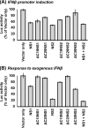
IFN suppression phenotypes of NS1 and NS2 deletion mutants. Infection and transfection were carried out as detailed in the legend to Fig. 3. In brief, A549 cells were first infected with dual NS deletion-containing RSV and 4 h later transfected with the indicated pCAGGS-FLAG-NS plasmids and either (A) an IFN promoter reporter or (B) an IFN response reporter firefly Luc plasmid. All of the cells were also cotransfected with the pCMV-Renilla Luc plasmid for a transfection control. Cells were processed 24 h later for a dual-luciferase assay (Promega), and firefly Luc activity was normalized against that of Renilla Luc. In each panel, the normalized activity with pCAGGS (vector only, no NS gene) was taken as 100 and all other activities are expressed as percentages thereof. The mean ± the standard error of the mean of three experiments are presented.
FIG. 12.
FIG. 12.
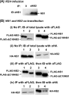
Coprecipitation of NS1 and NS2. (A) A549 cells were infected with wild-type RSV, and cell lysates made at 18 h p.i. were subjected to IP with our polyclonal rat antibody against NS2 or a no-antibody control (protein A-Sepharose beads only). Precipitated samples were resolved by sodium dodecyl sulfate-polyacrylamide gel electrophoresis, and IB was performed with the rat NS1 antibody. Note that our rat anti-NS2 antibody works only in IP, not in IB, and vice versa for the anti-NS1 antibody. (B) A549 cells were cotransfected with equal amounts of a mixture of pCAGGS-FLAG-NS and pCAGGS-HA-NS plasmids as follows: lane 1, FLAG-NS2 and HA-NS1; lane 2, FLAG-ΔC10NS2 and HA-NS1; lane 3, FLAG-NS1 and HA-NS2; lane 4, FLAG-ΔC10NS1 and HA-NS2. Cell lysates were made 24 h later, and portions were directly subjected to IB with (i) FLAG antibody or (ii) HA antibody. The remaining portions of the lysates were processed for IP with FLAG antibody, and the precipitate was subjected to IB with either (iii) FLAG antibody or (iv) HA antibody.
FIG. 13.
FIG. 13.
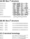
Sequence features of NS1 and NS2. (A) The BC Box homology domain, modeled after that reported by Elliot et al. (15), showing conservation among known members of this family (SOCS1 to -3, VHL). Conserved amino acids, present in at least one NS sequence and in another protein, are highlighted. The consensus 12-residue motif is shown at the bottom, with the invariant Val (or other aliphatic/hydrophobic residues), Leu/Ile, and Cys residues underlined and the intervening variable residues denoted as X. The nonconsensus Lys (K) residue, found in the NS proteins only, is in bold. 10, primary. (B) Local secondary (20) structures of the same 14-residue segment as in panel A are shown for the VHL and NS proteins: P, predicted by using the Porter algorithm (49); E, experimental (32, 63); c, coil (loop region); h, alpha helix; e, extended sheet (β sheet). (C) The homology at the C termini of NS1 and NS2 includes the underlined conserved terminal (F/Y)DLNP sequence. Residue numbers, corresponding to the full-length protein, are noted at the ends of selected sequences.

Similar articles

Cited by

References

    1. Andrejeva, J., K. S. Childs, D. F. Young, T. S. Carlos, N. Stock, S. Goodbourn, and R. E. Randall. 2004. The V proteins of paramyxoviruses bind the IFN-inducible RNA helicase, mda-5, and inhibit its activation of the IFN-β promoter. Proc. Natl. Acad. Sci. USA 101:17264-17269. - PMC - PubMed
    1. Atreya, P. L., M. E. Peeples, and P. L. Collins. 1998. The NS1 protein of human respiratory syncytial virus is a potent inhibitor of minigenome transcription and RNA replication. J. Virol. 72:1452-1461. - PMC - PubMed
    1. Bitko, V., A. Musiyenko, M. A. Bayfield, R. J. Maraia, and S. Barik. 2008. Cellular La protein shields nonsegmented negative-strand RNA viral leader RNA from RIG-I and enhances virus growth by diverse mechanisms. J. Virol. 82:7977-7987. - PMC - PubMed
    1. Bitko, V., and S. Barik. 2001. Phenotypic silencing of cytoplasmic genes using sequence-specific double-stranded short interfering RNA and its application in the reverse genetics of wild type negative-strand RNA viruses. BMC Microbiol. 1:34. - PMC - PubMed
    1. Bitko, V., O. Shulyayeva, B. Mazumder, A. Musiyenko, M. Ramaswamy, D. C. Look, and S. Barik. 2007. Nonstructural proteins of respiratory syncytial virus suppress premature apoptosis by an NF-κB-dependent, interferon-independent mechanism and facilitate virus growth. J. Virol. 81:1786-1795. - PMC - PubMed

Publication types