TARDBP 3'-UTR variant in autopsy-confirmed frontotemporal lobar degeneration with TDP-43 proteinopathy
- PMID: 19618195
- PMCID: PMC2783457
- DOI: 10.1007/s00401-009-0571-7
TARDBP 3'-UTR variant in autopsy-confirmed frontotemporal lobar degeneration with TDP-43 proteinopathy
Abstract
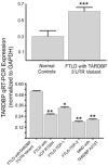
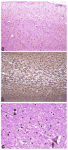
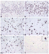
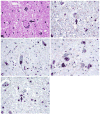
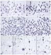
Pathogenic mutations in the gene encoding TDP-43, TARDBP, have been reported in familial amyotrophic lateral sclerosis (FALS) and, more recently, in families with a heterogeneous clinical phenotype including both ALS and frontotemporal lobar degeneration (FTLD). In our previous study, sequencing analyses identified one variant in the 3'-untranslated region (3'-UTR) of the TARDBP gene in two affected members of one family with bvFTD and ALS and in one unrelated clinically assessed case of FALS. Since that study, brain tissue has become available and provides autopsy confirmation of FTLD-TDP in the proband and ALS in the brother of the bvFTD-ALS family and the neuropathology of those two cases is reported here. The 3'-UTR variant was not found in 982 control subjects (1,964 alleles). To determine the functional significance of this variant, we undertook quantitative gene expression analysis. Allele-specific amplification showed a significant increase of 22% (P < 0.05) in disease-specific allele expression with a twofold increase in total TARDBP mRNA. The segregation of this variant in a family with clinical bvFTD and ALS adds to the spectrum of clinical phenotypes previously associated with TARDBP variants. In summary, TARDBP variants may result in clinically and neuropathologically heterogeneous phenotypes linked by a common molecular pathology called TDP-43 proteinopathy.
Conflict of interest statement
Figures

 , bvFTD
 , MND
, MND
 ; arrow proband. No DNA was available from either parent


Similar articles
-
TARDBP mutations in amyotrophic lateral sclerosis with TDP-43 neuropathology: a genetic and histopathological analysis.Lancet Neurol. 2008 May;7(5):409-16. doi: 10.1016/S1474-4422(08)70071-1. Epub 2008 Apr 7. Lancet Neurol. 2008. PMID: 18396105 Free PMC article.
-
Amyotrophic lateral sclerosis-frontotemporal lobar dementia in 3 families with p.Ala382Thr TARDBP mutations.Arch Neurol. 2010 Aug;67(8):1002-9. doi: 10.1001/archneurol.2010.173. Arch Neurol. 2010. PMID: 20697052 Free PMC article.
-
Molecular basis of amyotrophic lateral sclerosis.Prog Neuropsychopharmacol Biol Psychiatry. 2011 Mar 30;35(2):370-2. doi: 10.1016/j.pnpbp.2010.07.017. Epub 2010 Jul 23. Prog Neuropsychopharmacol Biol Psychiatry. 2011. PMID: 20655970 Review.
-
Frontotemporal lobar degeneration: Pathogenesis, pathology and pathways to phenotype.Brain Pathol. 2017 Nov;27(6):723-736. doi: 10.1111/bpa.12486. Epub 2017 Mar 2. Brain Pathol. 2017. PMID: 28100023 Free PMC article. Review.
-
TDP-43 is consistently co-localized with ubiquitinated inclusions in sporadic and Guam amyotrophic lateral sclerosis but not in familial amyotrophic lateral sclerosis with and without SOD1 mutations.Neuropathology. 2009 Dec;29(6):672-83. doi: 10.1111/j.1440-1789.2009.01029.x. Epub 2009 Jun 3. Neuropathology. 2009. PMID: 19496940
Cited by
-
Progranulin and TDP-43: mechanistic links and future directions.J Mol Neurosci. 2011 Nov;45(3):561-73. doi: 10.1007/s12031-011-9625-0. Epub 2011 Aug 24. J Mol Neurosci. 2011. PMID: 21863317 Free PMC article. Review.
-
TDP-43 regulates its mRNA levels through a negative feedback loop.EMBO J. 2011 Jan 19;30(2):277-88. doi: 10.1038/emboj.2010.310. Epub 2010 Dec 3. EMBO J. 2011. PMID: 21131904 Free PMC article.
-
Partial loss of TDP-43 function causes phenotypes of amyotrophic lateral sclerosis.Proc Natl Acad Sci U S A. 2014 Mar 25;111(12):E1121-9. doi: 10.1073/pnas.1322641111. Epub 2014 Mar 10. Proc Natl Acad Sci U S A. 2014. PMID: 24616503 Free PMC article.
-
A Clinical Guide to Frontotemporal Dementias.Focus (Am Psychiatr Publ). 2016 Oct;14(4):448-464. doi: 10.1176/appi.focus.20160018. Epub 2016 Oct 7. Focus (Am Psychiatr Publ). 2016. PMID: 31975825 Free PMC article.
-
FUS immunogold labeling TEM analysis of the neuronal cytoplasmic inclusions of neuronal intermediate filament inclusion disease: a frontotemporal lobar degeneration with FUS proteinopathy.J Mol Neurosci. 2011 Nov;45(3):409-21. doi: 10.1007/s12031-011-9549-8. Epub 2011 May 21. J Mol Neurosci. 2011. PMID: 21603978 Free PMC article.
References
-
- Consensus recommendations for the postmortem diagnosis of Alzheimer's disease. The National Institute on Aging and Reagan Institute Working Group on Diagnostic Criteria for the Neuropathological Assessment of Alzheimer's Disease. Neurobiol Aging. 1997;18(Suppl):S1–S2. - PubMed
-
- Arai T, Hasegawa M, Akiyama H, Ikeda K, Nonaka T, Mori H, Mann D, Tsuchiya K, Yoshida M, Hashizume Y, Oda T. TDP-43 is a component of ubiquitin-positive tau-negative inclusions in frontotemporal lobar degeneration and amyotrophic lateral sclerosis. Biochem Biophys Res Commun. 2006;351:602–611. - PubMed
-
- Ayala YM, Zago P, D'Ambrogio A, Xu YF, Petrucelli L, Buratti E, Baralle FE. Structural determinants of the cellular localization and shuttling of TDP-43. J Cell Sci. 2008;121:3778–3785. - PubMed
Publication types
MeSH terms
Substances
Grants and funding
- P30 AG013854-09/AG/NIA NIH HHS/United States
- P50 AG016574/AG/NIA NIH HHS/United States
- UL1 RR025741/RR/NCRR NIH HHS/United States
- P50 AG016574-11/AG/NIA NIH HHS/United States
- P30 AG013854-149003/AG/NIA NIH HHS/United States
- U01 AG016976-03/AG/NIA NIH HHS/United States
- P30-AG13854/AG/NIA NIH HHS/United States
- P50-AG05681/AG/NIA NIH HHS/United States
- P30 AG013854/AG/NIA NIH HHS/United States
- P50 AG16574/AG/NIA NIH HHS/United States
- P01-AG03991/AG/NIA NIH HHS/United States
- P30 NS057105/NS/NINDS NIH HHS/United States
- U01 AG016976/AG/NIA NIH HHS/United States
- P01 AG003991/AG/NIA NIH HHS/United States
- P50 AG005681/AG/NIA NIH HHS/United States
- U01-AG16976/AG/NIA NIH HHS/United States
- P01 AG003991-25/AG/NIA NIH HHS/United States
- P50 AG005681-26/AG/NIA NIH HHS/United States
- P30-NS057105/NS/NINDS NIH HHS/United States
LinkOut - more resources
Full Text Sources
Other Literature Sources
Miscellaneous

