. 2009 Oct;27(10):2527-38.
doi: 10.1002/stem.178.

3D timelapse analysis of muscle satellite cell motility

Affiliations
Free PMC article

3D timelapse analysis of muscle satellite cell motility

Ashley L Siegel et al. Stem Cells. 2009 Oct.
Free PMC article

Abstract

Skeletal muscle repair and regeneration requires the activity of satellite cells, a population of myogenic stem cells scattered throughout the tissue and activated to proliferate and differentiate in response to myotrauma or disease. While it seems likely that satellite cells would need to navigate local muscle tissue to reach damaged areas, relatively little data on such motility exist, and most studies have been with immortalized cell lines. We find that primary satellite cells are significantly more motile than myoblast cell lines, and that adhesion to laminin promotes primary cell motility more than fourfold over other substrates. Using timelapse videomicroscopy to assess satellite cell motility on single living myofibers, we have identified a requirement for the laminin-binding integrin alpha 7 beta 1 in satellite cell motility, as well as a role for hepatocyte growth factor in promoting directional persistence. The extensive migratory behavior of satellite cells resident on muscle fibers suggests caution when determining, based on fixed specimens, whether adjacent cells are daughters from the same mother cell. We also observed more persistent long-term contact between individual satellite cells than has been previously supposed, potential cell-cell attractive and repulsive interactions, and migration between host myofibers. Based on such activity, we assayed for expression of "pathfinding" cues, and found that satellite cells express multiple guidance ligands and receptors. Together, these data suggest that satellite cell migration in vivo may be more extensive than currently thought, and could be regulated by combinations of signals, including adhesive haptotaxis, soluble factors, and guidance cues.


Figures

Figure 1
Figure 1
Primary satellite cells and cell lines display differential adhesion and migration on purified extracellular matrix (ECM) substrates; neutralization of α7β1 integrin blocks motility on laminin. (A): Fluorescence micrographs of f-actin (visualized with Alexa 488-phalloidin) and nuclei (visualized with DAPI) of each myogenic cell type plated on different ECM substrates. (B): Final images from representative videos of each cell type on collagen, fibronectin, and laminin; individual cell traces used to calculate average cellular velocity are highlighted in unique colors. Scale bar = 100 μm. Inclusive time on all videos is 14 hours. Movies 1b1, 1b2, and 1b3 correspond to the displayed laminin-adhered cell movies. Number of cells tracked per condition = 12. (C): Primary satellite cells are significantly more motile than cell lines on all substrates; MM14s and primary satellite cells are significantly more motile on laminin than other substrates. Number of cells tracked per condition = 12. (D): Inhibition of integrin α7, integrin β1, or both by neutralizing antibody significantly decreases motility of MM14 and primary satellite cells on laminin. * = p < .05; ** = p < .01; *** = p < .001. Number of cells tracked per condition = 12. Abbreviation: PSC, primary satellite cells.
Figure 2
Figure 2
Primary satellite cells express a larger number of integrin mRNAs than cell lines: C2C12 cells express some integrin proteins differently compared to primary cells and MM14 cells. (A): Expression of integrin mRNAs in C2C12, MM14, and primary satellite cell-derived myoblasts; identical samples were processed simultaneously. Unlike immortalized cell lines, primary cells are positive for most or all known integrin chains (some samples of primary cells were positive for expression of integrins αE and αL, but not all were, including the one shown here). Intron-spanning primer sequences may be found in Supporting Table 1. (B): Expression of integrin protein in primary satellite cells (PSC), MM14, and C2C12 cells: all blots were stripped and reprobed with IP90 (calnexin) as a loading control; shown is anti-αV after stripping and reprobing.
Figure 3
Figure 3
Fiber-associated satellite cells are sublaminar immediately after harvest, but emerge from the sublaminar niche and adhere to the exterior of the myofiber after activation. (A): Reference still images extracted from movie 3a. A satellite cell 12 hours after fiber harvest exits from a depression in the myofiber (its presumptive quiescent location, indicated by arrowhead). Movie 3a: inclusive time 12 to 21 hours after activation; stills taken at 12:00, 12:30; 13:00; 13:10; 13:20; and 14:00 hours. (B): Reference still images extracted from movie 3b containing Z-scans and/or volume renderings of satellite cells at 4, 24, and 48 hours after fiber harvest, stained with anti-laminin (red) and anti-syndecan-4 (green). Although cells are sublaminar at 4 hours, they appear to have left the sublaminar niche by 24 hours, and are clearly outside the basal lamina by 48 hours.
Figure 4
Figure 4
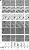
Fiber-associated satellite cells exhibit multiple novel “behaviors.” Figures are reference stills taken from the associated movies; magnified regions are shown below full-scale images. (A): Two movies showing recently-divided sister cells remaining closely associated for extended periods while moving along the fiber. Movie 4a1: Inclusive time 24 to 48 hours after activation, stills taken at 28:30; 28:40; 30:10; 32:10; 33:20; 45:40 hours. Movie 4a2: inclusive time 24 to 51 hours after activation, stills taken at 30:00; 30:30; 32:20; 34:30; 35:20; 48:50 hours. (B): Beginning at 12 hours (prior to any cell division), two presumably unrelated satellite cells migrate toward each other, associate, and remain closely associated for an extended period. Movie 4b: inclusive time 12 to 36 hours after activation, stills taken at 21:40; 25:20; 25:50; 26:30; 29:20; 36:00 hours. (C): Multiple satellite cells remain very closely associated for an extended time, interacting extensively but not fusing. Movie 4c: Inclusive time 48 to 72 hours after activation, stills taken at 48:00; 54:20; 59:20; 61:30; 69:10; 72:00 hours. Note that while some apparent “planar” cell divisions can be (as in still 2) other cell associations that appear to be planar cell divisions are almost certainly not (as in still 3). (D): A cell repeatedly extends protrusions into the collagen matrix, then moves off the fiber; another cell moves out along the first cell, divides, and the daughter cells continue in opposite directions. Movie 4d: inclusive time 48 to 72 hours after activation; stills taken at 52:20; 63:20; 65:20; 66:40; 67:10; 68:00 hours. (E): A cell in the collagen matrix moves parallel to a fiber, turns, and migrates onto the fiber. Movie 4e: Inclusive time 62 to 70 hours after activation, stills taken at 64:00; 65:10; 66:50; 67:20; 68:00; 68:50 hours. Note: area of magnified image is moved from frame to frame. (F): Relative frequencies of described activities in FGF-2-treated cultures from 24 to 48 hours after harvest. See Supporting Table 2, supporting movie Information, and additional Quicktime movies for more detailed information.
Figure 5
Figure 5
Exogenous cytokines can influence satellite cell motility: hepatocyte growth factor (HGF) increases directional persistence and decreases tortuosity. (A): Motility of 255 individual FGF-2 treated, fiber-associated satellite cells from 24 to 48 hours after isolation, arranged by increasing average velocity. All cells tested in all conditions showed displacement of at least 50 μm over 24 hours. (B): Average velocity in the presence of FGF-2 (control), HGF, lysophosphatidic acid (LPA), S1P, and pairwise combinations of factors. Number of cells tracked per condition ranged from 11 to 35. No treatment produced a statistically significant change in cell velocity from FGF-2 treatment. (C): Percent of FGF-2 or HGF-treated cells binned by total motility in 300 μm increments. A larger fraction of HGF-treated cells populate the high-motility bins. (D): Stills showing the final frames of movies 5b1 and 5b2. Tracings of fiber-associated satellite cells from the same mouse, with similar total velocity, over an identical time period, at an identical scale, treated with either FGF-2 or HGF illustrate that cells treated with HGF tend to have increased directional persistence. Inclusive time of both movies was 24 to 48 hours. (E): Comparison of tortuosity between tracks of FGF2-treated cells and HGF-treated cells: HGF treatment significantly decreases cell turning (p < .001). Number of cells tracked is 270 for FGF, 70 for HGF.
Figure 6
Figure 6
Blocking α7β1 integrin with neutralizing antibodies specifically inhibits satellite cell motility on myofibers. (A, B, C): figures are still images extracted from movies 6a, b, c.; inclusive time for all movies is 32 to 48 hours after activation. (A): Cells in 3D myofiber culture treated with neutralizing antibody to α7 integrin, stills taken at 32:00; 33:40; 36:00; 40:40; 43:00, and 48:00 hours. (B): Cells in 3D myofiber culture treated with neutralizing antibody to β1 integrin, stills taken at 32:00; 34:50; 36:00; 41:40; 43:00, and 48:00 hours. (C): Cells in 3D myofiber culture treated with neutralizing antibodies to α7 and β1 integrins, stills taken at 32:00; 36:00, 41:20; and 48:00 hours. Note that cell proliferation and cell-cell interactions are not blocked by integrin neutralization. (D): Quantitative representation of satellite cell motility in 3D myofiber culture after treatment with neutralizing antibody to α6 integrin, α7 integrin, αV integrin, β1 integrin, cyclic RGD, and pairwise combinations. Neutralization of α7 and β1 integrin, but neither neutralization of α6 or αV nor cyclic RGD, decreases satellite cell motility. * = p < .05; ** = p < .01; *** = p < .001. Number of cells tracked per condition ranged from 6 to 35.
Figure 7
Figure 7
Satellite cell-derived myoblasts express mRNAs for multiple guidance receptors. (A): Reverse transcriptase polymerase chain reaction for gene products associated with migration guidance receptors; multiple classes of guidance receptor are represented. Intron-spanning primer sequences can be found in Supporting Table 1. (B): Immunohistochemistry for Robo1, a transmembrane guidance molecule receptor (Abcam); expression is exclusive to the satellite cells and appears localized to the satellite cell membrane contacting the myofiber surface. Scale bar = 100 μm. Abbreviation: HGF, hepatocyte growth factor.

Similar articles

Cited by

References

    1. Hawke TJ, Garry DJ. Myogenic satellite cells: Physiology to molecular biology. J Appl Physiol. 2001;91:534–551. - PubMed
    1. Shi X, Garry DJ. Muscle stem cells in development, regeneration, and disease. Genes Dev. 2006;20:1692–1708. - PubMed
    1. Partridge TA, Morgan JE, Coulton GR, et al. Conversion of mdx myofibres from dystrophin-negative to -positive by injection of normal myoblasts. Nature. 1989;337:176–179. - PubMed
    1. Péault B, Rudnicki M, Torrente Y, et al. Stem and progenitor cells in skeletal muscle development, maintenance, and therapy. Mol Ther. 2007;15:867–877. - PubMed
    1. Skuk D, Goulet M, Roy B, et al. Dystrophin expression in muscles of duchenne muscular dystrophy patients after high-density injections of normal myogenic cells. J Neuropathol Exp Neurol. 2006;65:371–386. - PubMed

Publication types

MeSH terms