. 2009 Jul 15;23(14):1689-98.
doi: 10.1101/gad.1776209.

Removal of maternal retinoic acid by embryonic CYP26 is required for correct Nodal expression during early embryonic patterning

Affiliations

Removal of maternal retinoic acid by embryonic CYP26 is required for correct Nodal expression during early embryonic patterning

Masayuki Uehara et al. Genes Dev. .

Abstract

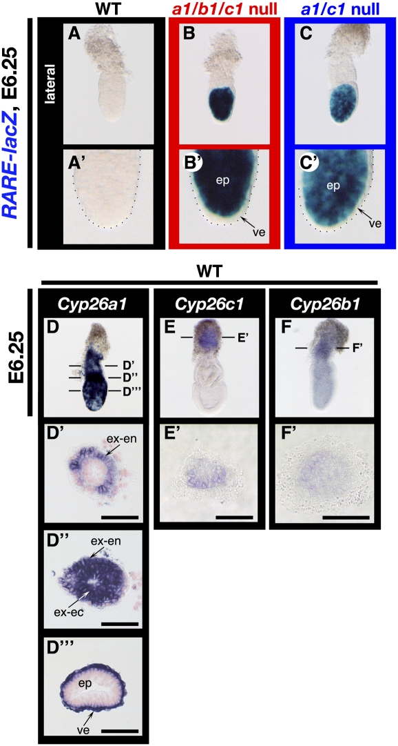
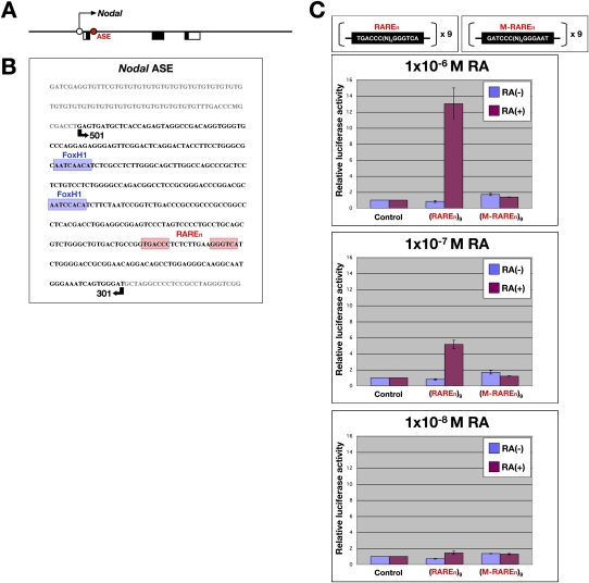
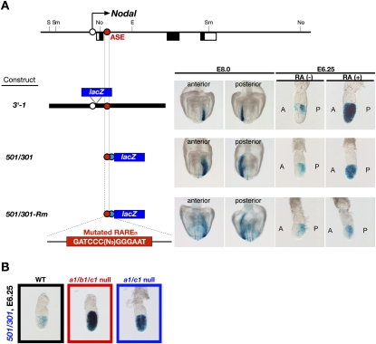
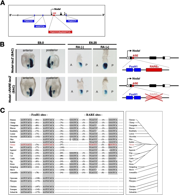
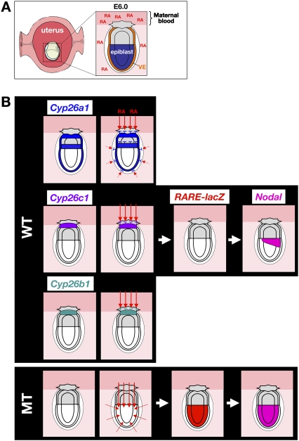
The abundance of retinoic acid (RA) is determined by the balance between its synthesis by retinaldehyde dehydrogenase (RALDH) and its degradation by CYP26. In particular, the dynamic expression of three CYP26 genes controls the regional level of RA within the body. Pregastrulation mouse embryos express CYP26 but not RALDH. We now show that mice lacking all three CYP26 genes manifest duplication of the body axis as a result of expansion of the Nodal expression domain throughout the epiblast. Mouse Nodal was found to contain an RA-responsive element in intron 1 that is highly conserved among mammals. In the absence of CYP26, maternally derived RA activates Nodal expression in the entire epiblast of pregastrulation embryos via this element. These observations suggest that maternal RA must be removed by embryonic CYP26 for correct Nodal expression during embryonic patterning.


Figures

Figure 1.
Figure 1.
Duplication of the body axis in Cyp26a1b1c1−/− and Cyp26a1c1−/− embryos. (A–D) Lateral views of wild-type (A), Cyp26a1b1c1−/− (B,C), and Cyp26a1/c1−/− (D) embryos at E9.25. The anterior side is on the left. About two-fifths of Cyp26a1b1c1−/− (B,C) or one-fourth of Cyp26a1/c1−/− (D) embryos were severely deformed, showing complete or partial duplication of the body axis. (A′–D′) Hematoxylin-eosin (HE) staining of sections of the embryos shown in A through D, respectively. Two sets of neural tubes or grooves (red arrowheads) were apparent in the mutant embryos, compared with the single neural tube (black arrowhead) in the wild-type embryo. The planes of the sections are indicated by the horizontal lines through the embryos in A–D. The boxed region in C′ is shown at higher magnification in C″. Bars, 100 μm. (E–L) Lateral views of wild-type (E,I), Cyp26a1b1c1−/− (F,G,J,K), and Cyp26a1c1−/− (H,L) embryos at E9.0 (EH) or E8.25 (IL) that had been subjected to whole-mount in situ hybridization for analysis of the expression of Shh (EH) or T (Brachury) (IL). The anterior side is on the left. (E′–L′) Ventral (E′–H′) and posterior (I′–L′) views of the embryos in E–H and in I–L, respectively. The expression domain of Shh was partially or completely duplicated in the mutant embryos (red arrowheads in F′H′). Mutant embryos expressed T in two distinct primitive streaks (red arrowheads in J′ and L′) or ectopically at the embryonic–extraembryonic boundary (red arrowheads in K′).
Figure 2.
Figure 2.
Epiblast patterning defects in Cyp26a1b1c1−/− and Cyp26a1c1−/− embryos. (A–I,G′–I′) Expression of Nodal, Cripto, and Lefty1 in wild-type (A,D,G,G′), Cyp26a1b1c1−/− (B,E,H,H′), and Cyp26a1c1−/− (C,F,I,I′) embryos at E6.25 or E6.5, as indicated, was examined by whole-mount in situ hybridization. Lateral views with the anterior side on the left are shown, with the exception of the frontal views shown in G–I. Nodal was expressed in the posterior region of the wild-type embryo (A) but was expressed throughout the epiblast of Cyp26a1b1c1−/− (B) or Cyp26a1c1−/− (C) embryos. Whereas Cripto expression was most prominent in the posterior region of the epiblast of the wild-type embryo (D), it was up-regulated and extended to the entire epiblast in the mutant embryos (E,F). The expression domain of Lefty1, which marks the AVE in wild-type embryos (G,G′), was maintained in the mutant embryos (H,H′,I,I′), although the level of Lefty1 expression was increased in the mutants (H,I). (J–M,K′,M′) Expression of Nodal (J,K,K′) and the RARE-hsplacZ transgene (L,M,M′) in untreated (J,L) or RA-treated (K,K′,M,M′) wild-type embryos at E6.25 was examined by whole-mount in situ hybridization or staining with the β-galactosidase substrate X-gal. Nodal was expressed in the posterior region of the epiblast in the untreated embryo (J) but was expressed in the entire epiblast of the RA-treated embryo (K,K′). Expression of RARE-hsplacZ was not detected in the untreated embryo (L) but was pronounced throughout the epiblast of the RA-treated embryo (M,M′). The images in K and M are shown at higher magnification in K′ and M′, respectively. (ep) Epiblast; (ve) visceral endoderm.
Figure 3.
Figure 3.
Contribution of each CYP26 isozyme to RA metabolism in pregastrulation embryos. (A–C,A′–C′) Expression of the RARE-hsplacZ transgene in wild-type (A), Cyp26a1b1c1−/− (B), and Cyp26a1c1−/− (C) embryos at E6.25 as revealed by X-gal staining. Lateral views are shown with the anterior side on the left. Each image is shown at higher magnification in A′–C′, respectively. Expression of the transgene was not detected in the wild-type embryo (A,A′) but was pronounced in the entire epiblast (ep) of the mutants (B,B′,C,C′). (D–F,D′–F′,D″,D″′) Expression patterns of Cyp26a1 (D), Cyp26c1 (E), and Cyp26b1 (F) in wild-type embryos at E6.25 as revealed by whole-mount in situ hybridization. Lateral views are shown with the anterior side on the left. The planes of sections shown in D′, D″, D″′, E′, and F′ are indicated by the horizontal lines through the embryos in D–F. Cyp26a1 was highly expressed in the extraembryonic endoderm (ex-en) (D′,D″), the extraembryonic ectoderm (ex-ec) (D″), and the visceral endoderm (ve) (D′″). Cyp26c1 and Cyp26b1 were expressed in the ectoplacental cone, albeit at a lower level (E,E′,F,F′). Bars, 50 μm.
Figure 4.
Figure 4.
Rescue of embryonic patterning defects in Nodal+/lacZ, Cyp26a1c1−/− and Nodalneo/neo, Cyp26a1c1−/− embryos. (A–E,A′–E′) Expression of T (Brachury) in wild-type (A,A′), Cyp26a1/c1−/− (B,B′,C,,C′), Nodal+/lacZ, Cyp26a1c1−/− (D,D′), and Nodalneo/neo, Cyp26a1c1−/− (E,E′) embryos at E8.25 was examined by whole-mount in situ hybridization. Lateral views with the anterior side on the left are shown in A through E, and posterior views are shown in A′ through E′. Exon 1 of Nodal is replaced by lacZ in the NodallacZ allele. Whereas ∼35% of Cyp26a1c1−/− embryos expressed T in two distinct primitive streaks (arrowheads in B′) or ectopically at the embryonic–extraembryonic boundary (arrowheads in C′), only 6% of the Nodal+/lacZ, Cyp26a1c1−/− embryos (not shown) did so. (E′) Furthermore, none of the Nodalneo/neo, Cyp26a1c1−/− embryos showed the embryonic patterning defects.
Figure 5.
Figure 5.
Direct regulation of Nodal expression by RA through an RARE in intron 1. (A) Localization of the ASE in intron 1 of Nodal. The arrow indicates the direction of transcription; open and closed boxes denote noncoding and coding regions of exons, respectively; the ASE is depicted by the red circle. (B) Nucleotide sequence of the Nodal ASE, which contains a RARE-like sequence, TGACCCN9GGGTCA (RAREn, red shading), in addition to two FoxH1-binding sequences (FASTs, blue shading). The region included in the 501/301 transgene (see Fig. 6A) is indicated by the arrows. (C) Luciferase reporter assay performed with P19 cells transiently transfected with reporter constructs containing tandem repeats of TGACCCN9GGGTCA [(RAREn)9] or the mutated sequence GATCCCN9GGGAAT [(M-RAREn)9] and treated with the indicated concentrations of RA or with dimethyl sulfoxide as vehicle. Data are means of triplicates from a representative experiment.
Figure 6.
Figure 6.
Nodal as a target of RA signaling in pregastrulation embryos. (A) Structure of three lacZ constructs and their expression in mouse embryos. (E) EcoRI; (No) NotI; (S) SalI; (Sm) SmaI. The open black and closed blue circles indicate the proximal promoter of Nodal and the Hsp68 promoter, respectively. Staining with X-gal revealed that both 3′-1 and 501/301 transgenes were expressed in the posterior region of the epiblast (at E6.25) and in the left lateral plate (at E8.0) of untreated [RA(−)] wild-type embryos, whereas they were expressed in the entire epiblast of RA-treated wild-type embryos at E6.25. In contrast, the expression of 501/301-Rm in wild-type embryos was not affected by RA treatment. (B) Expression of the 501/301 transgene in wild-type, Cyp26a1b1c1−/−, and Cyp26a1c1−/− embryos at E6.25 as revealed by X-gal staining. Lateral views are shown for each embryo with the anterior side on the left. The transgene was expressed in the posterior region of the wild-type embryo but was expressed in the entire epiblast of the mutant embryos.
Figure 7.
Figure 7.
Role of RAREn in regulation of Nodal expression in the epiblast and its structural conservation among mammals. (A) Relative positions of RAREn (red) and three RARE-like half sites (blue) in mouse Nodal. (B) X-gal staining of E8.0 and E6.25 embryos harboring the indicated lacZ (BAC) constructs. The expression pattern of Nodal-lacZ (BAC) mimicked that of endogenous Nodal and responded to the administration of exogenous RA at E6.0. Expression of Nodal-ΔRARE-lacZ (BAC) failed to respond to exogenous RA. (C, left panel) Evolutionary conservation of RARE-like and FoxH1-binding sequences in intron 1 of vertebrate Nodal genes; numbers in parentheses indicate nucleotide spacing. The vertebrate evolutionary tree is shown in the right panel.
Figure 8.
Figure 8.
Model for the role of CYP26 genes in early embryonic patterning. (A) In the pregastrulation mouse embryo, RA appears to be of maternal origin. Raldh2 is expressed in the decidua and RA is present in serum. (VE) Visceral endoderm. (B) The expression domains of the three CYP26 genes in the wild-type embryo at E6.25 are shown in the top panels. (Bottom panels) In mutant (MT) embryos lacking all three CYP26 genes, maternally derived RA activates Nodal expression in the entire epiblast via RAREn. Red arrows indicate the route of maternally derived RA. The removal of maternal RA by embryonic CYP26 is thus required for correct Nodal expression during embryonic patterning. See the text for details.

Similar articles

Cited by

References

    1. Abu-Abed S, Dolle P, Metzger D, Beckett B, Chambon P, Petkovich M. The retinoic acid-metabolizing enzyme, CYP26A1, is essential for normal hindbrain patterning, vertebral identity, and development of posterior structures. Genes & Dev. 2001;15:226–240. - PMC - PubMed
    1. Abu-Abed S, MacLean G, Fraulob V, Chambon P, Petkovich M, Dolle P. Differential expression of the retinoic acid-metabolizing enzymes CYP26A1 and CYP26B1 during murine organogenesis. Mech Dev. 2002;110:173–177. - PubMed
    1. Adachi H, Saijoh Y, Mochida K, Ohishi S, Hashiguchi H, Hirao A, Hamada H. Determination of left/right asymmetric expression of nodal by a left side-specific enhancer with sequence similarity to a lefty-2 enhancer. Genes & Dev. 1999;13:1589–1600. - PMC - PubMed
    1. Brennan J, Lu CC, Norris DP, Rodriguez TA, Beddington RS, Robertson EJ. Nodal signalling in the epiblast patterns the early mouse embryo. Nature. 2001;411:965–969. - PubMed
    1. Collignon J, Varlet I, Robertson EJ. Relationship between asymmetric nodal expression and the direction of embryonic turning. Nature. 1996;381:155–158. - PubMed

Publication types

MeSH terms

LinkOut - more resources