. 2009 Aug;136(16):2725-33.
doi: 10.1242/dev.038596. Epub 2009 Jul 15.

UNC-83 is a nuclear-specific cargo adaptor for kinesin-1-mediated nuclear migration

Affiliations

UNC-83 is a nuclear-specific cargo adaptor for kinesin-1-mediated nuclear migration

Marina Meyerzon et al. Development. 2009 Aug.

Abstract

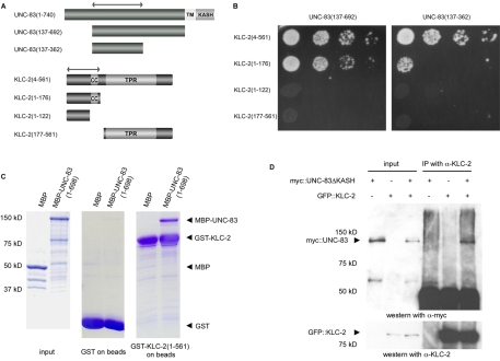
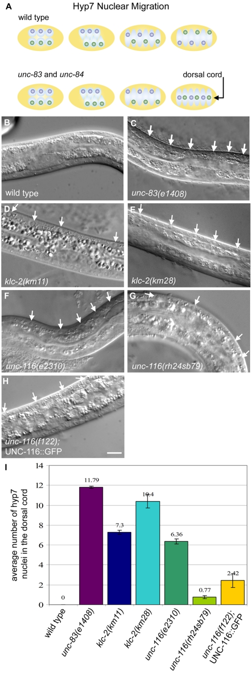
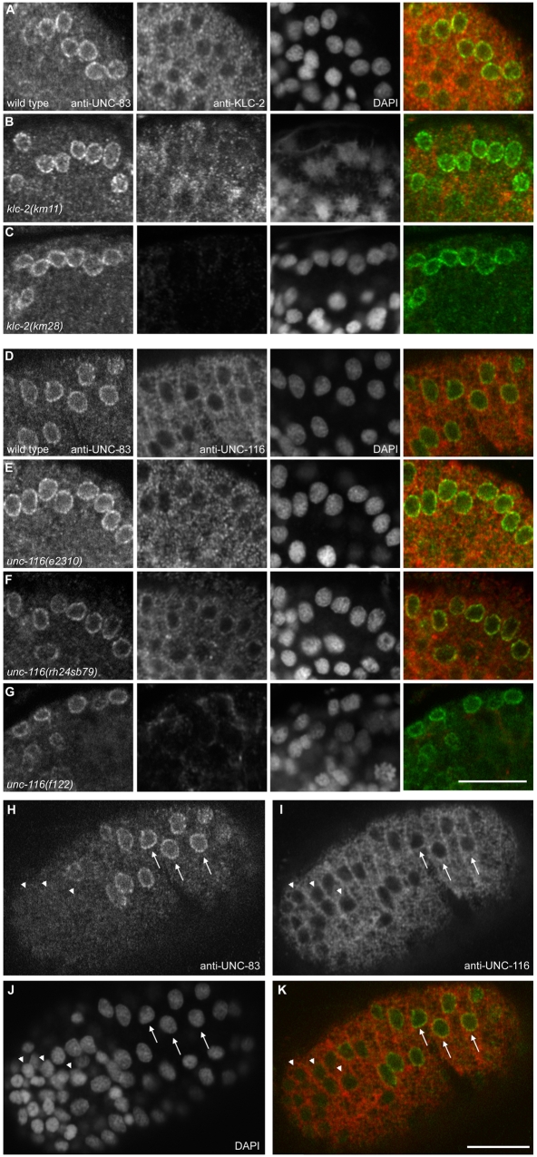
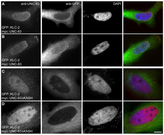
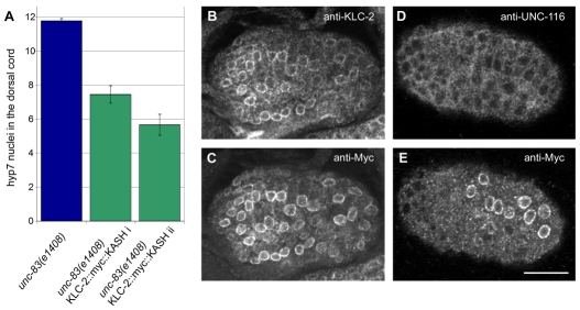
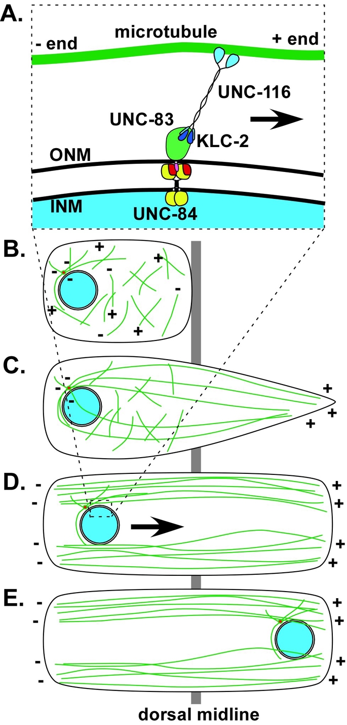
Intracellular nuclear migration is essential for many cellular events including fertilization, establishment of polarity, division and differentiation. How nuclei migrate is not understood at the molecular level. The C. elegans KASH protein UNC-83 is required for nuclear migration and localizes to the outer nuclear membrane. UNC-83 interacts with the inner nuclear membrane SUN protein UNC-84 and is proposed to connect the cytoskeleton to the nuclear lamina. Here, we show that UNC-83 also interacts with the kinesin-1 light chain KLC-2, as identified in a yeast two-hybrid screen and confirmed by in vitro assays. UNC-83 interacts with and recruits KLC-2 to the nuclear envelope in a heterologous tissue culture system. Additionally, analysis of mutant phenotypes demonstrated that both KLC-2 and the kinesin-1 heavy chain UNC-116 are required for nuclear migration. Finally, the requirement for UNC-83 in nuclear migration could be partially bypassed by expressing a synthetic outer nuclear membrane KLC-2::KASH fusion protein. Our data support a model in which UNC-83 plays a central role in nuclear migration by acting to bridge the nuclear envelope and as a kinesin-1 cargo-specific adaptor so that motor-generated forces specifically move the nucleus as a single unit.


Figures

Fig. 1.
Fig. 1.
UNC-83 interacts with KLC-2. (A) Constructs of C. elegans UNC-83 and KLC-2 used for yeast two-hybrid and GST in vitro interaction analysis. The cytoplasmic portion of UNC-83 is dark gray. The transmembrane (TM) and KASH domains of UNC-83 and the coiled-coil (CC) and tetratricopeptide repeat (TPR) domains of KLC-2 are indicated. Double-headed arrows indicate the KLC-2-binding domain of UNC-83 and the UNC-83-binding domain of KLC-2. (B) Yeast two-hybrid interactions between UNC-83c (137-362) or UNC-83c (137-692) as bait and the indicated regions of KLC-2 as prey. Serial dilutions of log-phase cultures are shown. (C) Coomassie Blue stained SDS-PAGE gels demonstrating the interactions between GST-KLC-2 (1-561) and MBP-UNC-83 (1-698). In the left-hand panel, 1% of the input proteins are shown. The middle panel shows the amount of proteins pulled down by beads with GST only, to show background. The right-hand panel shows MBP and MBP-UNC-83 fusion proteins pulled down by GST-KLC-2 beads. Arrowheads mark the expected positions of the indicated constructs. (D) myc::UNC-83ΔKASH was co-immunoprecipitated with anti-KLC-2 antibodies. A western blot probed with anti-myc antibodies to recognize myc::UNC-83ΔKASH is shown in the top panel. Lanes 1-3 show transfected HeLa cell extracts used as inputs. Lanes 4-6 show immunoprecipitation by anti-KLC-2 antibodies from the same extracts. Beneath is shown the same blot probed with anti-KLC-2 antibodies. Size standards and the UNC-83 and KLC-2 fusion proteins are indicated on the left.
Fig. 2.
Fig. 2.
Loss-of-function alleles of klc-2 or unc-116 cause defects in hyp7 nuclear migration. (A) Dorsal view of a pre-comma stage C. elegans embryo illustrating intercalation and nuclear migration of hyp7 precursors in wild-type and unc-83 or unc-84 embryos. The embryo is depicted in yellow. The cytoplasm of the hyp7 precursors is light blue, nuclei that migrate from right to left are dark blue, and nuclei that migrate from left to right are green. Anterior is on the left, right is up. (B-H) Lateral view of L1 hermaphrodites; dorsal is up. (B) Wild type. (C) unc-83(e1408). (D) klc-2(km11). (E) klc-2(km28). (F) unc-116(e2310). (G) unc-116(rh24sb79). (H) unc-116(f122); UNC-116::GFP. White arrows mark hyp7 nuclei in the dorsal cord. Scale bar: 10 μm. (I) The average number of hyp7 nuclei counted in the dorsal cord. Numbers above bars indicate averages; error bars indicate s.e.
Fig. 3.
Fig. 3.
Specificity of the anti-KLC-2 and anti-UNC-116 antibodies. (A) A western blot (developed on film) with anti-UNC-116 antibodies. A single band of ∼97 kDa is detected in immunoblots of mixed-stage wild-type worms (lane 1) as well as unc-116(rh24sb79) (lane 2) and unc-116(e2310) (lane 3) animals. A single band of ∼113 kDa in immunoblots of mixed-stage unc-116(f122); UNC-116::GFP animals corresponds to the expected size of UNC-116::GFP (lane 4). (B) A western blot (imaged by digital camera) with anti-KLC-2 antibodies. Bands were detected of ∼66 kDa in wild-type embryos (lane 1) and 97 kDa in klc-2(km28); KLC-2::GFP embryos, corresponding to KLC-2::GFP (lane 2). (C,D) Wide-field fluorescence images of wild-type embryos stained with the anti-KLC-2 or UNC-116 antibodies. (C) KLC-2 expression is low in early embryos, such as in the 4-cell stage embryo shown (arrowhead). Expression increases greatly in the pre-elongation stage (arrow). (D) UNC-116 expression in early embryos is relatively low and diffuse, such as in the 8-cell embryo shown (arrowhead). Expression is much higher and more organized in the pre-elongation stage (arrow). Scale bar: 10 μm.
Fig. 4.
Fig. 4.
Kinesin-1 is expressed in hyp7 cells during nuclear migration. (A-G) Single-slice confocal images of dorsal views of pre-elongation C. elegans embryos undergoing hyp7 nuclear migration. Immunolocalization of UNC-83 is shown in the left-most column, KLC-2 (A-C) or UNC-116 (D-G) in the second column, followed by DAPI staining of DNA in the third column, and a merge of UNC-83 (green) and KLC-2 or UNC-116 (red) localization in the right-most column. (A-C) KLC-2 localization in (A) wild-type, (B) klc-2(km11) and (C) klc-2(km28) embryos. (D-G) UNC-116 localization in (D) wild-type, (E) unc-116(e2310), (F) unc-116(rh24sb79) and (G) unc-116(f122) embryos. (H-K) A single-slice confocal image of a dorsal view of the pre-elongation wild-type embryo shown in D. (H) UNC-83 immunostaining (green in merge); (I) UNC-116 immunostaining (red in merge); (J) DAPI staining of DNA; (K) merge of H and I. Arrows indicate UNC-83-positive hyp7 nuclei and arrowheads mark other hypodermal nuclei. In all images, anterior is to the left. Scale bars: 10 μm.
Fig. 5.
Fig. 5.
UNC-83 recruits kinesin-1 to the nuclear envelope. HeLa cells transiently transfected with DNA encoding GFP::KLC-2 and (A,B) full-length myc::UNC-83 or (C,D) myc::UNC-83ΔKASH. Shown are immunolocalization of UNC-83 (column 1) or KLC-2 (column 2), DAPI staining of DNA (column 3) and a merge of UNC-83 (red), KLC-2 (green) and DAPI (blue) straining (column 4). UNC-83 can be seen enriched at the nuclear envelope with KLC-2. UNC-83ΔKASH is partially imported into the nucleus and, in this case, KLC-2 remains cytoplasmic. Scale bar: 10 μm.
Fig. 6.
Fig. 6.
The KLC-2::KASH construct can partially bypass the requirement of UNC-83 for hyp7 nuclear migration. (A) The average number of hyp7 nuclei counted in the dorsal cord of unc-83(e1408) null animals (n=71) and two lines of unc-83(e1408); ycEx117,118[pSL440(KLC-2::myc::KASH)] (KLC-2::myc::KASH i, ii) (n=20). Error bars indicate s.e. (B-E) Single-slice confocal images of pre-elongation embryos. (B) An unc-83(e1408); ycEx118[pSL440(KLC-2::myc::KASH)] (KLC-2::myc::KASH ii) embryo stained with the anti-KLC-2 antibody. (C) The same embryo and focal plane as in B, stained with an anti-myc antibody. (D) An unc-83(e1408); ycEx118[pSL440(KLC-2::myc::KASH)] (KLC-2::myc::KASH ii) embryo stained with the anti-UNC-116 antibody. (E) The same embryo and focal plane as in D, stained with an anti-myc antibody. Dorsal views with anterior on the left. Scale bar: 10 μm.
Fig. 7.
Fig. 7.
A model of nuclear migration in C. elegans hyp7 cells. (A) The nuclear envelope during nuclear migration. UNC-84 (red and yellow) localizes to the inner nuclear membrane (INM) and UNC-83 (green and pink) localizes to the outer nuclear membrane (ONM). The UNC-84 SUN domain (red) interacts with the UNC-83 KASH domain (pink) in the perinuclear space. The cytoplasmic domain of UNC-83 (green) interacts with KLC-2 (dark blue). The motor activity of UNC-116 (light blue) moves hyp7 nuclei toward the plus ends of polarized microtubules (green). (B) Microtubules (green) resemble a meshwork at the beginning of cell intercalation (plus and minus ends of microtubules marked). (C) As intercalation proceeds across the dorsal midline (gray), microtubules nucleated from the centrosome (red) at the stationary end of the cell (left) extend into the growing tip (right) of the cell. (D) At the completion of intercalation, most microtubules are arranged in polarized, centrosome-independent bundles parallel to the cell boundaries. (E) Nuclei (blue) move along these polarized microtubule bundles (in the direction of the arrow in D).

Similar articles

Cited by

References

    1. Bowman, A. B., Kamal, A., Ritchings, B. W., Philp, A. V., McGrail, M., Gindhart, J. G. and Goldstein, L. S. (2000). Kinesin-dependent axonal transport is mediated by the sunday driver (SYD) protein. Cell 103, 583-594. - PubMed
    1. Fan, J. and Beck, K. A. (2004). A role for the spectrin superfamily member Syne-1 and kinesin II in cytokinesis. J. Cell Sci. 117, 619-629. - PubMed
    1. Gindhart, J. G. (2006). Towards an understanding of kinesin-1 dependent transport pathways through the study of protein-protein interactions. Brief. Funct. Genomic. Proteomic. 5, 74-86. - PubMed
    1. Glater, E. E., Megeath, L. J., Stowers, R. S. and Schwarz, T. L. (2006). Axonal transport of mitochondria requires milton to recruit kinesin heavy chain and is light chain independent. J. Cell Biol. 173, 545-557. - PMC - PubMed
    1. Guo, Y., Jangi, S. and Welte, M. A. (2005). Organelle-specific control of intracellular transport: distinctly targeted isoforms of the regulator Klar. Mol. Biol. Cell 16, 1406-1416. - PMC - PubMed

Publication types

MeSH terms

LinkOut - more resources