. 2009 Jul 10;35(1):11-25.
doi: 10.1016/j.molcel.2009.06.013.

FAK phosphorylation by ERK primes ras-induced tyrosine dephosphorylation of FAK mediated by PIN1 and PTP-PEST

Affiliations

FAK phosphorylation by ERK primes ras-induced tyrosine dephosphorylation of FAK mediated by PIN1 and PTP-PEST

Yanhua Zheng et al. Mol Cell. .

Abstract

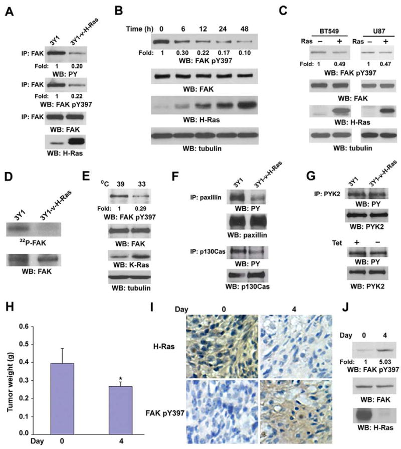
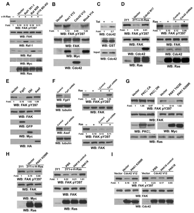
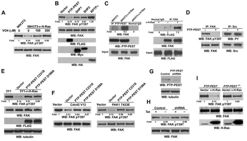
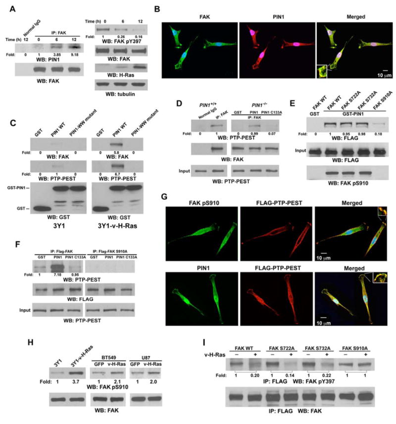
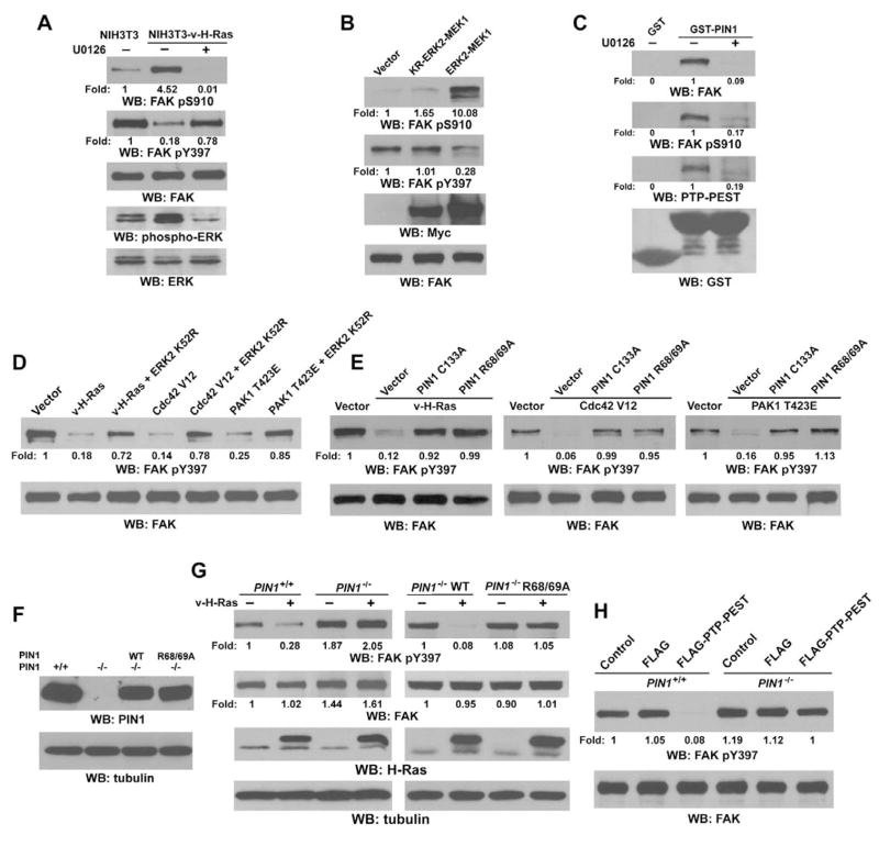
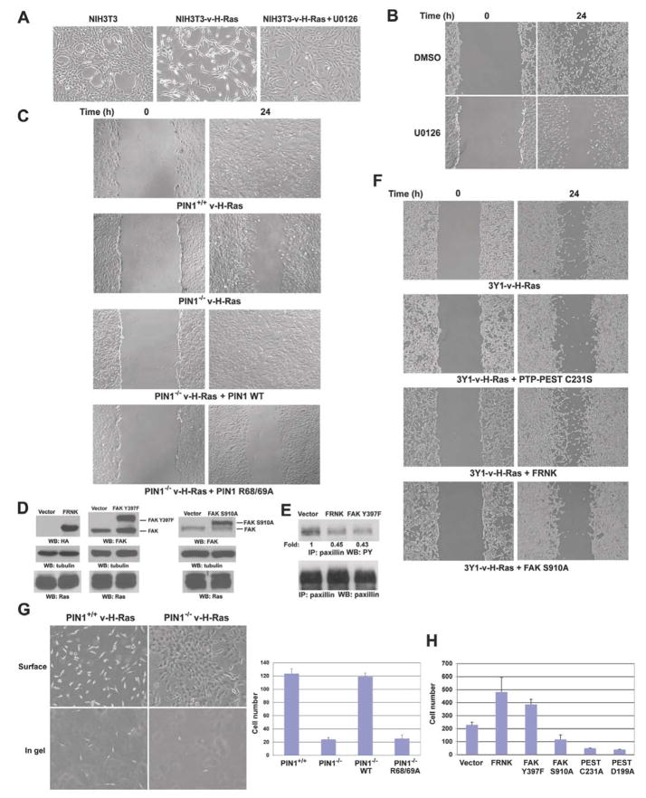
Activated Ras has been found in many types of cancer. However, the mechanism underlying Ras-promoted tumor metastasis remains unclear. We demonstrate here that activated Ras induces tyrosine dephosphorylation and inhibition of FAK mediated by the Ras downstream Fgd1-Cdc42-PAK1-MEK-ERK signaling cascade. ERK phosphorylates FAK S910 and recruits PIN1 and PTP-PEST, which colocalize with FAK at the lamellipodia of migrating cells. PIN1 binding and prolyl isomerization of FAK cause PTP-PEST to interact with and dephosphorylate FAK Y397. Inhibition of FAK mediated by this signal relay promotes Ras-induced cell migration, invasion, and metastasis. These findings uncover the importance of sequential modification of FAK-by serine phosphorylation, isomerization, and tyrosine dephosphorylation--in the regulation of FAK activity and, thereby, in Ras-related tumor metastasis.


Figures

Figure 1
Figure 1. FAK Dephosphorylation at Y397 and FAK Inhibition by Activated Ras Correlate with Tumor Progression
(A–G, J) Western blotting (WB) analyses with the indicated antibodies. Tubulin was used as a loading control. PY, phospho-tyrosine. (A) Cell lysates were prepared from 3Y1 or 3Y1-v-H-Ras cells. IP, immunoprecipitation. (B) NIH3T3-v-H-Ras (Tet-off) cells were cultured with or without withdrawal of doxycycline (1 μg/ml) for the indicated time. (C) BT549 cells (left) and U87 cells (right) were infected with adeno-green fluorescent protein (GFP) or adeno-v-H-Ras virus for 6 h. (D) FAK was immunoprecipitated from the indicated cells and analyzed by in vitro kinase assay. (E) Rat thyroid epithelial cells transformed by a temperature-sensitive mutant of the Kirsten murine sarcoma virus were shifted from 39°C to 33°C for 24 h. (F) Paxillin (upper panel) and p130Cas (lower panel) were immunoprecipitated from the indicated cells. (G) PYK2 was immunoprecipitated from 3Y1 and 3Y1-v-H-Ras cells (upper panel) or from NIH3T3-v-H-Ras (Tet-off) cells cultured with or without withdrawal of doxycycline (1 μg/ml) for 48 h (lower panel). (H) MK14 cells were injected subcutaneously into the right flanks of athymic nude mice. The mice were fed with water containing 2 mg/ml doxycycline. When the tumor size reached 0.8–1.2 cm in diameter, doxycycline was withdrawn from the water for 4 days. The tumor masses were removed and weighed. Six mice in each group were used, and the standard errors represent the variation in tumor weight. *P<0.05, indicating significant differences on post hoc comparisons to the group of mice without withdrawal of doxycycline. (I, J) The removed tumor masses were immunostained (I) or immunoblotted (J) with the indicated antibodies.
Figure 2
Figure 2. Ras-induced FAK Dephosphorylation is Mediated by Fgd1, Cdc42, and PAK1
Western blotting analyses with the indicated antibodies. Immunoprecipitation of FAK preceded immunoblotting with anti-FAK or anti-FAK pY397 antibodies. (A, B) 293T cells were transiently transfected with plasmids expressing v-H-Ras, dominant-negative Raf1 301-1, Ral N28, or Myc-p110 BD-KD mutants (A), or constitutively active Myc-Rac1 V12, Myc-RhoA V14, or Cdc42 V12 mutants (B). (C) NIH3T3-v-H-Ras (Tet-off) cells were cultured with or without withdrawal of doxycycline (1 μg/ml) for 48 h. GST pull-down assay was performed with incubating GST-PBD with cell lysates. (D) Cell lysates were prepared from 3Y1, 3Y1-v-H-Ras stably expressing a control vector or Cdc42N17 (left panel), or 293T cells transiently expressing control shRNA or Cdc42 shRNA (right panel). (E) 293T cells were transiently transfected with plasmids expressing constitutively active pEGFP-C1-Fgd1, pRK5-Myc-Dbl, or pcDNA-HA-Asef-ΔAPC. (F) 293T cells stably expressing control shRNA, Asef shRNA, or Fgd1 shRNA were transiently transfected with plasmids expressing v-H-Ras. (G) 293T cells were transiently transfected with the indicated plasmids. CA, constitutively active; DN, dominant-negative. (H) Cell lysates were prepared from the indicated cell lines. 3Y1-v-H-Ras cells were treated with PBS, PAK18 (10 μM), or PAK18 control (10 μM) for 48 h. (I) 293T cells were transiently transfected with the indicated plasmids. Cells were treated with PBS, PAK18 (10 μM), or PAK18 control (10 μM) for 48 h.
Figure 3
Figure 3. Ras-induced FAK Dephosphorylation is Mediated by PTP-PEST
Western blotting analyses with the indicated antibodies. Immunoprecipitation of FAK preceded immunoblotting with anti-FAK or anti-FAK pY397 antibodies. (A) NIH3T3 or NIH3T3-v-H-Ras cells were treated with or without pervanadate at the indicated doses for 1 h. (B) 293T cells were transiently transfected with plasmids expressing FLAG-PTP-PEST, Myc-SHP1, Myc-SHP2, or HA-RPTPα. (C) Immunoprecipitation of PTP-PEST from NIH3T3 or NIH3T3-v-H-Ras cells (left panel) or immunoprecipitation of FAK from 293T cells transiently transfected with FLAG-PTP-PEST and control vector or with FLAG-PTP-PEST and v-H-Ras (right panel) was followed with immunoblotting with the indicated antibodies. (D) An in vitro phosphatase assay was performed with incubation of immunoprecipitated FAK or Src from 3Y1-v-H-Ras cells with eluted FLAG peptide or FLAG-PTP-PEST immunoprecipitated from 293T cells transiently expressing pcDNA3.1 FLAG vector or pcDNA3.1 FLAG PTP-PEST. (E) Cell lysates were prepared from 3Y1, 3Y1-v-H-Ras, and 3Y1-v-H-Ras cells stably expressing PTP-PEST mutants. (F) 293T cells were transiently transfected with the indicated plasmids. (G, H) NIH3T3-v-H-Ras (Tet-off) cells were stably transfected with pGIPZ control shRNA or pGIPZ PTP-PEST shRNA (G). These cells were cultured with or without withdrawal of doxycycline (1 μg/ml) for 48 h (H). (I) Cell lysates were prepared from PTP-PEST−/− and PTP-PEST−/− cells re-expressing WT PTP-PEST without or with stable expression of v-H-Ras.
Figure 4
Figure 4. FAK Phosphorylation at S910 Results in Binding of PIN1 and PTP-PEST to FAK at Lamellipodia and FAK Dephosphorylation at Y397
(A, C–F, H, I) Western blotting analyses with the indicated antibodies. (A) FAK was immunoprecipitated from NIH3T3-v-H-Ras (Tet-off) cells cultured with or without withdrawal of doxycycline (1 μg/ml) for the indicated time. (B) Immunofluorescence analysis of 3Y1-v-H-Ras cells with the indicated antibodies. (C) GST pull-down assays were performed by incubating purified GST, GST-PIN1, or GST-PIN1 WW mutant proteins with lysates of 3Y1 or 3Y1-v-H-Ras cells. (D) Immunoprecipitates with control (rabbit IgG) or a FAK antibody from PIN1+/+ and PIN1−/− fibroblasts infected with adeno-v-H-Ras virus were incubated with or without purified PIN1 or PIN1 C133A followed by incubation with purified PTP-PEST. After incubation, immunoprecipitates were washed three times with cell lysate buffer. (E) GST pull-down assays were performed by incubating GST or GST-PIN1 agarose beads with lysate of fak−/− cells transiently expressing FLAG-FAK WT or FLAG-FAK mutants, as indicated. (F) FLAG-FAK and FLAG-FAK S910A immunoprecipitated with anti-FLAG antibody from 3Y1-v-H-Ras cells stably expressing FLAG-FAK and FLAG-FAK S910A was incubated with purified PTP-PEST in the presence of either soluble GST, GST-PIN1, or GST-PIN1 C133A protein, and then immunoprecipitates were washed three times with cell lysate buffer. (G) Immunofluorescence analysis of 3Y1-v-H-Ras cells transiently expressing FLAG-PTP-PEST with the indicated antibodies. (H) Cell lysates were prepared from 3Y1 cells, 3Y1-v-H-Ras cells (left panel), BT549 cells, and U87 cells infected with adeno-GFP or adeno-v-H-Ras virus (middle and right panel). (I) pDCR or pDCR-v-H-Ras was transiently cotransfected with pCDNA3.1 FLAG-FAK WT or FLAG-FAK mutants, as indicated, into 293T cells.
Figure 5
Figure 5. ERK-Dependent FAK Phosphorylation at S910 Regulates PIN1-mediated FAK Dephosphorylation at Y397
Western blotting analyses with the indicated antibodies. Immunoprecipitation of FAK preceded immunoblotting with anti-FAK or anti-FAK pY397 antibodies. (A) NIH3T3 or NIH3T3–v-H-Ras cells were treated with or without U0126 (20 μM) for 24 h. (B) 293T cells were transiently transfected with pCMV5 (Vector), pCMV5-Myc-KR-ERK2–MEK1, or pCMV5-Myc-ERK2–MEK1. (C) GST pull-down assays were performed by incubating GST or GST-PIN1 agarose beads with lysate of 3Y1-v-H-Ras cells treated with or without U0126 (20 μM) for 24 h. (D, E) 293T cells were transiently transfected with the indicated plasmids. (F) The cell lysates were prepared from PIN1+/+, PIN1−/−, and PIN1−/− stably expressing PIN1 WT or PIN1 R68/69A fibroblasts. (G) PIN1+/+, PIN1−/−, and PIN1−/− fibroblasts stably expressing PIN1 WT or PIN1 R68/69A were infected with adeno-GFP or adeno-v-H-Ras virus for 24 h. (H) An in vitro phosphatase assay was performed with incubation of immunoprecipitated FAK from adeno-v-H-Ras-infected PIN1+/+ and PIN1−/− fibroblasts without or with eluted FLAG peptide or FLAG-PTP-PEST immunoprecipitated from 293T cells transiently expressing pcDNA3.1 FLAG or pcDNA3.1 FLAG PTP-PEST.
Figure 6
Figure 6. Y397 Dephosphorylation and Inhibition of FAK Mediated by ERK, PIN1, and PTP-PEST Promotes Ras-induced Cell Migration, Invasion, and Metastasis
(A–C, F, G) Pictures were taken with a digital camera mounted on a microscope with 100× (A, G) or 50× (B, C, F) magnification. (A) NIH3T3 or NIH3T3-v-H-Ras cells were treated with or without U0126 (20 μM) for 24 h. (B) NIH3T3-v-H-Ras cells wounded by scraping with a micropipette tip were treated with dimethyl sulfoxide (DMSO) or U0126 (20 μM) for 24 h. (C) PIN1+/+, PIN1−/−, and PIN1−/− fibroblasts stably expressing PIN1 WT or PIN1 R68/69A were infected with adeno-v-H-Ras for 24 h before the migration assay. (D) Immunoblotting analyses of 3Y1-v-H-Ras cells stably expressing HA-FRNK, CD2-FAK Y397F, or FLAG-FAK S910A with the indicated antibodies. (E) Immunoblotting of immunoprecipitated paxillin from 3Y1-v-H-Ras cells with or without stable expression of HA-FRNK or CD2-FAK Y397F with the indicated antibodies. (F) 3Y1-v-H-Ras cells with or without stably expressed PTP-PEST C231S, HA-FRNK, or FLAG-FAK S910A were analyzed with the migration assay. (G) PIN1+/+, PIN1−/−, and PIN1−/− fibroblasts stably re-expressing PIN1 WT or PIN1 R68/69A were infected with adeno-v-H-Ras for 24 h. These cells were plated on the surface of collagen gel. Five days after plating, cells were photographed either at the top surface of the gel (left top panel) or at a focal plane beneath the surface to visualize cells that had penetrated the gel (left bottom panel) for PIN1+/+ and PIN1−/− cells. The numbers of invading cells in 10 photographic fields from three separate experiments were counted (right panel). Data represent the mean ± standard deviation of three independent experiments. (H) 3Y1-v-H-Ras cells with or without stably expressed FRNK, FAK Y397F, FAK S910A, PTP-PEST C231S, or PTP-PEST D199A were analyzed with the collagen-gel invasion assay.
Figure 7
Figure 7. Inhibition of FAK Promotes Ras-Induced Cell Metastasis
(A) 3Y1-v-H-Ras cells or 3Y1-v-H-Ras cells expressing FRNK, FAK Y397F, FAK S910A were injected into the lateral tail veins of six athymic nude mice. After 3 weeks, mice were sacrificed and lung metastasis nodules were counted under a low-power dissecting microscope. Data represent the mean ± standard deviation of two independent experiments. *P<0.05, indicating significant differences on post hoc comparisons to the group of mice injected with 3Y1-v-H-Ras cells. An arrow points to a metastasis nodule. (B) A mechanism of Ras-induced cancer cell metastasis. Activated Ras promotes tumor cell migration, invasion, and metastasis by inhibition of FAK, which is mediated by ERK-dependent FAK phosphorylation at S910. FAK phosphorylation at S910 results in PIN1-dependent prolyl isomerization of FAK and subsequent recruitment of PTP-PEST for FAK dephosphorylation at Y397.

Comment in

  • Ras and the FAK paradox.
    Antonyak MA, Cerione RA. Antonyak MA, et al. Mol Cell. 2009 Jul 31;35(2):141-2. doi: 10.1016/j.molcel.2009.07.005. Mol Cell. 2009. PMID: 19647511 Free PMC article.

Similar articles

Cited by

References

    1. Angers-Loustau A, Cote JF, Charest A, Dowbenko D, Spencer S, Lasky LA, Tremblay ML. Protein tyrosine phosphatase-PEST regulates focal adhesion disassembly, migration, and cytokinesis in fibroblasts. The Journal of cell biology. 1999;144:1019–1031. - PMC - PubMed
    1. Ayaki M, Komatsu K, Mukai M, Murata K, Kameyama M, Ishiguro S, Miyoshi J, Tatsuta M, Nakamura H. Reduced expression of focal adhesion kinase in liver metastases compared with matched primary human colorectal adenocarcinomas. Clin Cancer Res. 2001;7:3106–3112. - PubMed
    1. Ayala I, Giacchetti G, Caldieri G, Attanasio F, Mariggio S, Tete S, Polishchuk R, Castronovo V, Buccione R. Faciogenital dysplasia protein Fgd1 regulates invadopodia biogenesis and extracellular matrix degradation and is up-regulated in prostate and breast cancer. Cancer research. 2009;69:747–752. - PubMed
    1. Basson MD, Sanders MA, Gomez R, Hatfield J, Vanderheide R, Thamilselvan V, Zhang J, Walsh MF. Focal adhesion kinase protein levels in gut epithelial motility. American journal of physiology. 2006;291:G491–499. - PubMed
    1. Beeser A, Jaffer ZM, Hofmann C, Chernoff J. Role of group A p21-activated kinases in activation of extracellular-regulated kinase by growth factors. The Journal of biological chemistry. 2005;280:36609–36615. - PubMed

Publication types

MeSH terms