. 2009 Sep;83(18):9045-56.
doi: 10.1128/JVI.00728-09. Epub 2009 Jul 8.

A 49-kilodalton isoform of the adenovirus type 5 early region 1B 55-kilodalton protein is sufficient to support virus replication

Affiliations

A 49-kilodalton isoform of the adenovirus type 5 early region 1B 55-kilodalton protein is sufficient to support virus replication

Kathrin Kindsmüller et al. J Virol. 2009 Sep.

Abstract

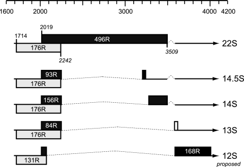
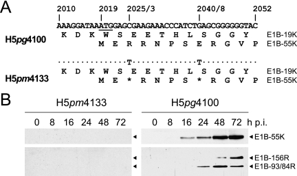
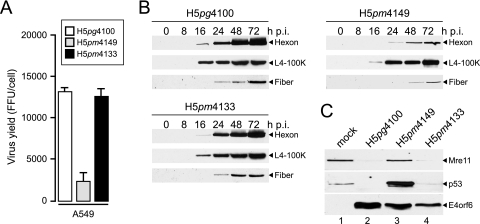
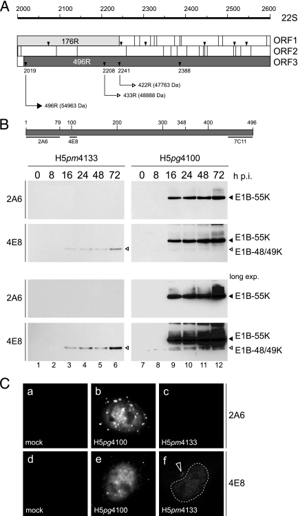
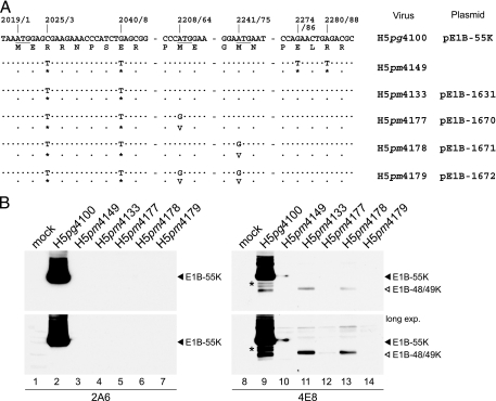
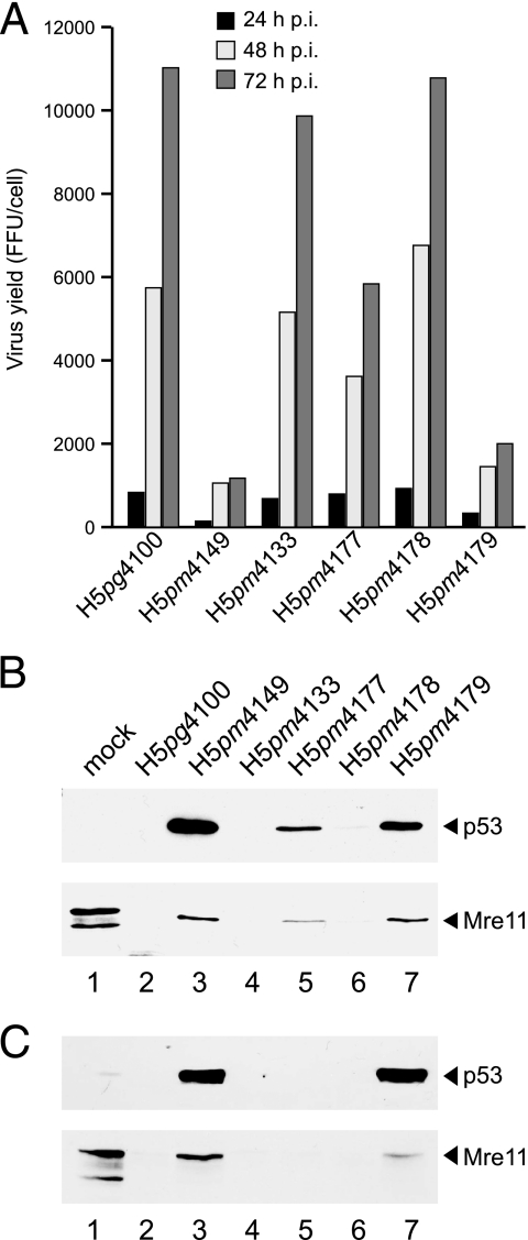
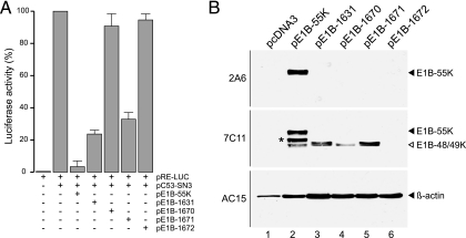
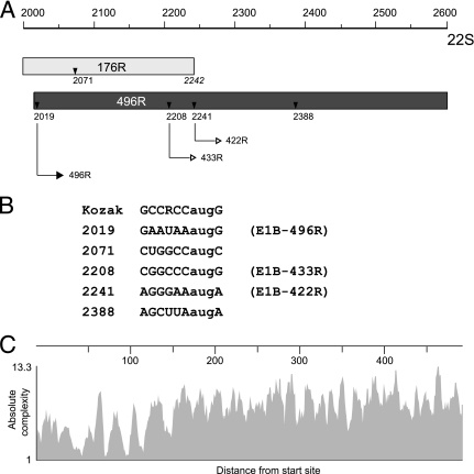
The adenovirus type 5 (Ad5) early region 1B 55-kDa (E1B-55K) protein is a multifunctional regulator of cell-cycle-independent virus replication that participates in many processes required for maximal virus production. As part of a study of E1B-55K function, we generated the Ad5 mutant H5pm4133, carrying stop codons after the second and seventh codons of the E1B reading frame, thereby eliminating synthesis of the full-length 55K product and its smaller derivatives. Unexpectedly, phenotypic studies revealed that H5pm4133 fully exhibits the characteristics of wild-type (wt) Ad5 in all assays tested. Immunoblot analyses demonstrated that H5pm4133 and wt Ad5 produce very low levels of two distinct polypeptides in the 48- to 49-kDa range, which lack the amino-terminal region but contain segments from the central and carboxy-terminal part of the 55K protein. Genetic and biochemical studies with different Ad5 mutants show that at least one of these isoforms consists of two closely migrating polypeptides of 433 amino acid residues (433R) and 422R, which are produced by translation initiation at two downstream AUG codons of the 55K reading frame. Significantly, a virus mutant producing low levels of the 433R isoform alone replicated to levels comparable to those of wt Ad5, demonstrating that this polypeptide provides essentially all functions of E1B-55K required to promote maximal virus growth in human tumor cells. Altogether, these results extend previous findings that the wt Ad5 E1B region encodes a series of smaller isoforms of E1B-55K and demonstrate that very low levels of at least one of these novel proteins (E1B-433R) are sufficient for a productive infection.


Figures

FIG. 1.
FIG. 1.
Ad5 E1B transcription unit. The positions (nucleotide numbers) on the Ad5 DNA sequence from 1600 to 4200 are indicated on the top. The Ad5 E1B mRNAs are indicated by thin lines and arrows; introns are indicated by dotted lines. The reading frames predicted from DNA sequences and mapping studies are identified by boxes. Included also are the relevant nucleotides for translation start sites. Positions of translation stop sites are shown in italics. The figure was assembled based on material in several publications cited in the text.
FIG. 2.
FIG. 2.
Effect of mutations on expression of E1B-55K and its related products. (A) Mutations in virus H5pm4133. Numbers refer to positions of nucleotides in the wt H5pg4100 DNA sequence, and numbers after the diagonal slash denote the positions of the amino acid residues. The first initiation codon of the 55K reading frame is underlined. Segments of the amino acid sequences of E1B-19K (176R) and E1B-55K (496R) are shown below. The positions of the point mutations in H5pm4133 are indicated, and dots identify identical nucleotides. Asterisks denote the introduced opal and amber codons in the 55K reading frame from H5pm4133. (B) Steady-state expression levels of E1B-55K proteins in mutant and wt virus-infected A549 cells. A549 cells were infected at a multiplicity of 20 focus-forming units per cell. Cells were harvested at the indicated times postinfection (p.i.), and total cell extracts were prepared. Proteins (50-μg samples) from each time point were separated by SDS-12% PAGE and subjected to immunoblotting using anti-E1B-55K mouse MAb 2A6.
FIG. 3.
FIG. 3.
Virus growth and viral late protein synthesis. (A) A549 cells were infected with wt H5pg4100 or E1B mutant viruses at a multiplicity of 50 focus-forming units (FFU) per cell. Viral particles were harvested 72 h postinfection, and virus yield was determined by quantitative E2A-72K immunofluorescence staining on 293 cells. The results represent the averages from three independent experiments. Error bars indicate the standard errors of the means. (B) Viral late protein synthesis. A549 cells were infected with the wt or mutant viruses at a multiplicity of 50 FFU per cell. Total cell extracts were prepared at the indicated times postinfection (p.i.). Proteins (10-μg samples) were separated by SDS-12% PAGE, transferred to nitrocellulose membranes, and probed with the anti-Ad5 rabbit polyclonal serum L133. Bands corresponding to the hexon, L4-100K, and fiber proteins are indicated on the right. (C) Steady-state concentrations of Mre11, p53, and E4orf6 in infected cells. A549 cells were infected with wt H5pg4100 or E1B mutant viruses at a multiplicity of 50 FFU per cell. Whole-cell extracts were prepared at 48 h after infection, and proteins (50-μg samples) were separated on SDS-12% polyacrylamide gels and subjected to immunoblotting using anti-Mre11 rabbit polyclonal antibody pNB-100-142, anti-p53 mouse MAb DO-1, or anti-E4orf6 mouse MAb RSA3.
FIG. 4.
FIG. 4.
wt H5pg4100 and H5pm4133 express a novel 49-kDa isoform of the E1B-55K product. (A) Schematic drawing of the E1B-55K coding region surrounding the starting methionine at nucleotide position 2019. Numbers refer to positions of nucleotides in the wt E1B 22S mRNA. Open reading frame (ORF) maps spanning the region from nt 2000 to 2600 of the E1B 22S mRNA are shown. The three reading frames (ORF1 to ORF3) of the E1B sense strand are shown below. Gray-shaded regions indicate parts of the 19K and 55K protein coding sequences. Vertical lines bisecting each reading frame indicate stop codons, whereas small triangles denote methionine codons. Numbers below refer to nucleotide positions of the methionine codons, and thin-lined arrows indicate translation of the corresponding E1B 496R, 433R, and 422R polypeptides. Numbers in parentheses designate the theoretical molecular mass calculated from the protein sequences. (B) Top: schematic drawing of the E1B-55K 496R protein. Numbers refer to positions of amino acid residues in the viral polypeptide, and regions of 55K recognized by MAbs 2A6, 4E8, and 7C11 are indicated below by thin lines. Bottom: Immunoblot analysis of total-cell lysates from wt and mutant virus-infected cells. A549 cells were infected at a multiplicity of 50 FFU per cell. Cells were harvested at the indicated times postinfection, and total cell extracts were prepared. Proteins (50-μg samples) from each time point were separated by SDS-12% PAGE and subjected to immunoblotting using anti-E1B-55K mouse MAb 2A6 or anti-E1B-55K rat MAb 4E8. The positions of the E1B proteins are indicated on the right, and their molecular masses were estimated from their electrophoretic mobilities. Long exp., longer exposure. (C) Steady-state localization of E1B-55K and its isoforms in wt and mutant virus-infected cells. A549 cells were mock infected or infected with wt or E1B mutant virus H5pm4133 at a multiplicity of 50 FFU per cell. Cells were fixed at 24 h after infection and labeled with anti-E1B-55K mouse MAb 2A6 (panels a, b, and c) or anti-E1B-55K rat MAb 4E8 (panels d, e, and f) and FITC- and Texas Red-conjugated secondary antibodies. In panel d, the nucleus is indicated by a dotted line. Magnification, ×7,600.
FIG. 5.
FIG. 5.
The AUGs at positions 64 and 75 initiate translation of two E1B isoforms. (A) Mutations in the 5′ coding region of the E1B-55K mutant viruses (Virus) and plasmid constructs (Plasmid). In wt virus H5pg4100 and plasmid pE1B-55K (indicated on the right), numbers refer to positions of nucleotides in the Ad5 wt DNA sequence and numbers after the diagonal slash denote the positions of the amino acid residues. The first three initiation codons of the 55K reading frame are underlined. The amino acid sequence is shown below. The positions of the point mutations in the virus mutants and plasmid constructs are indicated, and dots identify identical nucleotides and amino acid residues, respectively. Asterisks denote the introduced opal, amber, and ochre codons in the 55K reading frame. (B) Steady-state expression levels of E1B-55K proteins in wt and mutant virus-infected cells. A549 cells were infected with a multiplicity of 50 FFU per cell. Whole-cell extracts were prepared, and samples containing 50 μg of protein were examined by SDS-12% PAGE followed by immunoblotting with antibody 2A6 (left) or 4E8 (right). Lower panels represent longer exposures (long exp.) of the same immunoblots to visualize expression of the 422R E1B isoform in H5pm4177-infected cells. The designations of the viruses and plasmids are indicated on the right. The asterisk in lane 9 indicates two bands in the 52-to-53-kDa range.
FIG. 6.
FIG. 6.
Effect of amino acid substitutions in E1B-55K on virus growth and proteasomal degradation of p53 and Mre11. (A) A549 cells were infected with wt H5pg4100 or E1B mutant viruses at a multiplicity of 50 FFU per cell. Viral particles were harvested at the indicated time points after infection, and virus yield was determined by quantitative E2A-72K immunofluorescence staining on 293 cells. The results represent the averages from two independent experiments. FFU, focus-forming units. (B) A549 cells were infected as described above, and whole-cell extracts were prepared. Proteins (50-μg samples) from the 48-h-p.i. time point were separated on SDS-12% polyacrylamide gels and subjected to immunoblotting using anti-p53 mouse MAb DO-1 or anti-Mre11 rabbit polyclonal antibody pNB-100-142. (C) A549 cells were infected at a multiplicity of 50 FFU per cell (H5pg4100, H5pm4149, H5pm4133, H5pm4178, and H5pm4179) or 500 FFU per cell (H5pm4177), and steady-state concentrations of p53 and Mre11 were determined by immunoblotting using MAbs DO-1 and pNB-100-142, respectively.
FIG. 7.
FIG. 7.
Effect of mutations on inhibition of p53-stimulated transcription. (A) Subconfluent H1299 cells were transfected with reporter and effector plasmids as described in Materials and Methods. The mean and standard deviation are presented for three independent experiments, each performed in duplicate. (B) Expression of E1B proteins in transfected H1299 cells. Luciferase samples were immunoblotted by normalizing the amount of extract used according to Renilla luciferase activity and probing the immunoblots with MAbs 2A6, 7C11, and AC-15. The asterisk in lane 2 indicates a slower-migrating band in the 52-to-53-kDa range.
FIG. 8.
FIG. 8.
Ad5 E1B transcription unit may encode two additional isoforms of the E1B-55K protein. (A) Schematic drawing of the E1B-55K coding region from nt 2000 to 2600. Numbers refer to positions of nucleotides in the wt E1B 22S mRNA. Gray-shaded regions indicate parts of the 19K (176R) and 55K (496R) coding sequences. Small triangles denote methionine codons. Numbers below refer to nucleotide positions of the methionine codons, and thin-lined arrows indicate translation of the corresponding E1B 496R, 433R, and 422R polypeptides. The position of the translation stop site of E1B-176R is shown in italics below the coding sequence. (B) Comparison of sequences flanking the AUG codons at positions 2019, 2071, 2208, 2241, and 2238 with the Kozak consensus sequence. R, adenine or guanine. (C) Absolute complexity (y axis) of aligned E1B sequences from subgroup A to G adenoviruses was calculated using the Vector NTI software program (Invitrogen) in terms of average pairwise alignment scores. Higher values in the y axis show higher sequence conservation. The x axis indicates the distance from the start site. Amino acid sequences of large E1B proteins were derived from Ad subtypes 1, 2, 3, 4, 5, 7, 8, 9, 11, 12, 14, 16, 17, 19, 21, 26, 34, 35, 37, 40, 41, 46, 48, 49, 50, 52, and 53.

Similar articles

Cited by

References

    1. Alemany, R. 2007. Cancer selective adenoviruses. Mol. Aspects Med. 28:42-58. - PubMed
    1. Anderson, C. W., R. C. Schmitt, J. E. Smart, and J. B. Lewis. 1984. Early region 1B of adenovirus 2 encodes two coterminal proteins of 495 and 155 amino acid residues. J. Virol. 50:387-396. - PMC - PubMed
    1. Araujo, F. D., T. H. Stracker, C. T. Carson, D. V. Lee, and M. D. Weitzman. 2005. Adenovirus type 5 E4orf3 protein targets the Mre11 complex to cytoplasmic aggresomes. J. Virol. 79:11382-11391. - PMC - PubMed
    1. Baker, A., K. J. Rohleder, L. A. Hanakahi, and G. Ketner. 2007. The adenovirus E4 34k and E1b 55k oncoproteins target host DNA ligase IV for proteasomal degradation. J. Virol. 13:7034-7040. - PMC - PubMed
    1. Barker, D. D., and A. J. Berk. 1987. Adenovirus proteins from both E1B reading frames are required for transformation of rodent cells by viral infection and DNA transfection. Virology 156:107-121. - PubMed

Publication types