

Crystal structures of the reduced, sulfenic acid, and mixed disulfide forms of SarZ, a redox active global regulator in Staphylococcus aureus
- PMID: 19586910
- PMCID: PMC2749125
- DOI: 10.1074/jbc.M109.015826
Crystal structures of the reduced, sulfenic acid, and mixed disulfide forms of SarZ, a redox active global regulator in Staphylococcus aureus
Abstract
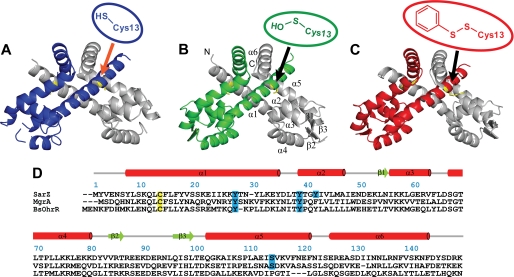
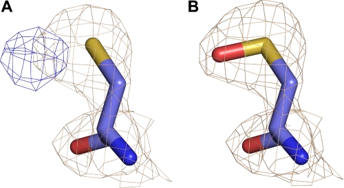
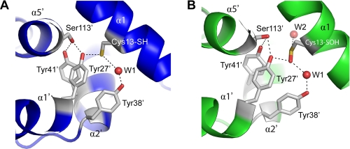
SarZ is a global transcriptional regulator that uses a single cysteine residue, Cys(13), to sense peroxide stress and control metabolic switching and virulence in Staphylococcus aureus. SarZ belongs to the single-cysteine class of OhrR-MgrA proteins that play key roles in oxidative resistance and virulence regulation in various bacteria. We present the crystal structures of the reduced form, sulfenic acid form, and mixed disulfide form of SarZ. Both the sulfenic acid and mixed disulfide forms are structurally characterized for the first time for this class of proteins. The Cys(13) sulfenic acid modification is stabilized through two hydrogen bonds with surrounding residues, and the overall DNA-binding conformation is retained. A further reaction of the Cys(13) sulfenic acid with an external thiol leads to formation of a mixed disulfide bond, which results in an allosteric change in the DNA-binding domains, disrupting DNA binding. Thus, the crystal structures of SarZ in three different states provide molecular level pictures delineating the mechanism by which this class of redox active regulators undergoes activation. These structures help to understand redox-mediated virulence regulation in S. aureus and activation of the MarR family proteins in general.
Figures


Similar articles
-
A new oxidative sensing and regulation pathway mediated by the MgrA homologue SarZ in Staphylococcus aureus.Mol Microbiol. 2009 Jan;71(1):198-211. doi: 10.1111/j.1365-2958.2008.06518.x. Epub 2008 Nov 5. Mol Microbiol. 2009. PMID: 19007410 Free PMC article.
-
The oxidation-sensing regulator (MosR) is a new redox-dependent transcription factor in Mycobacterium tuberculosis.J Biol Chem. 2012 Nov 2;287(45):37703-12. doi: 10.1074/jbc.M112.388611. Epub 2012 Sep 18. J Biol Chem. 2012. PMID: 22992749 Free PMC article.
-
sarZ, a sarA family gene, is transcriptionally activated by MgrA and is involved in the regulation of genes encoding exoproteins in Staphylococcus aureus.J Bacteriol. 2009 Mar;191(5):1656-65. doi: 10.1128/JB.01555-08. Epub 2008 Dec 19. J Bacteriol. 2009. PMID: 19103928 Free PMC article.
-
Protein cysteine oxidation in redox signaling: Caveats on sulfenic acid detection and quantification.Arch Biochem Biophys. 2017 Mar 1;617:26-37. doi: 10.1016/j.abb.2016.09.013. Epub 2016 Sep 28. Arch Biochem Biophys. 2017. PMID: 27693037 Free PMC article. Review.
-
Protein sulfenic acids in redox signaling.Annu Rev Pharmacol Toxicol. 2004;44:325-47. doi: 10.1146/annurev.pharmtox.44.101802.121735. Annu Rev Pharmacol Toxicol. 2004. PMID: 14744249 Review.
Cited by
-
Regulation of transcription by eukaryotic-like serine-threonine kinases and phosphatases in Gram-positive bacterial pathogens.Virulence. 2014;5(8):863-85. doi: 10.4161/21505594.2014.983404. Virulence. 2014. PMID: 25603430 Free PMC article. Review.
-
Staphylococcal response to oxidative stress.Front Cell Infect Microbiol. 2012 Mar 16;2:33. doi: 10.3389/fcimb.2012.00033. eCollection 2012. Front Cell Infect Microbiol. 2012. PMID: 22919625 Free PMC article. Review.
-
The AGXX® Antimicrobial Coating Causes a Thiol-Specific Oxidative Stress Response and Protein S-bacillithiolation in Staphylococcus aureus.Front Microbiol. 2018 Dec 11;9:3037. doi: 10.3389/fmicb.2018.03037. eCollection 2018. Front Microbiol. 2018. PMID: 30619128 Free PMC article.
-
Chemical approaches to detect and analyze protein sulfenic acids.Mass Spectrom Rev. 2014 Mar-Apr;33(2):126-46. doi: 10.1002/mas.21384. Epub 2013 Sep 17. Mass Spectrom Rev. 2014. PMID: 24105931 Free PMC article. Review.
-
Redox-Sensing Under Hypochlorite Stress and Infection Conditions by the Rrf2-Family Repressor HypR in Staphylococcus aureus.Antioxid Redox Signal. 2018 Sep 1;29(7):615-636. doi: 10.1089/ars.2017.7354. Epub 2018 Jan 30. Antioxid Redox Signal. 2018. PMID: 29237286 Free PMC article.
References
Publication types
MeSH terms
Substances
Associated data
- Actions
- Actions
- Actions
Grants and funding
LinkOut - more resources
Full Text Sources
