. 2009 Jul 1;23(13):1559-70.
doi: 10.1101/gad.524209.

The SESA network links duplication of the yeast centrosome with the protein translation machinery

Affiliations

The SESA network links duplication of the yeast centrosome with the protein translation machinery

Bengü Sezen et al. Genes Dev. .

Abstract

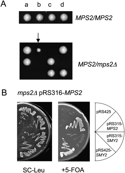
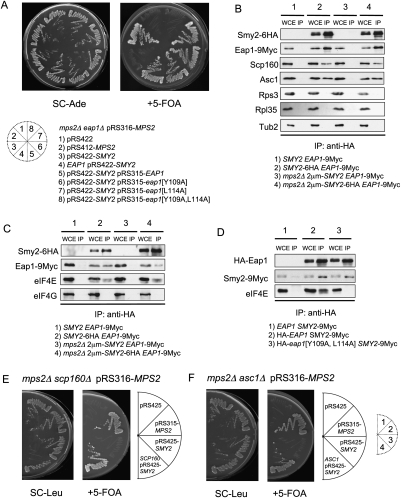
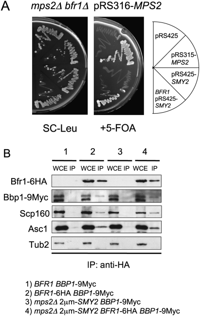
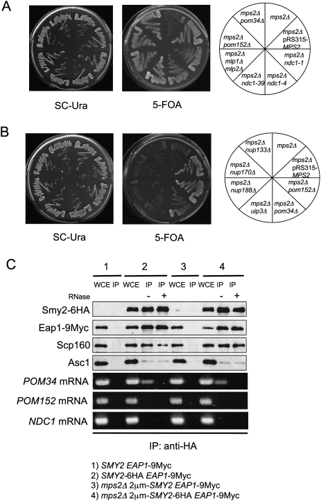
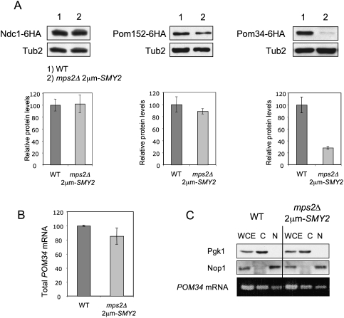
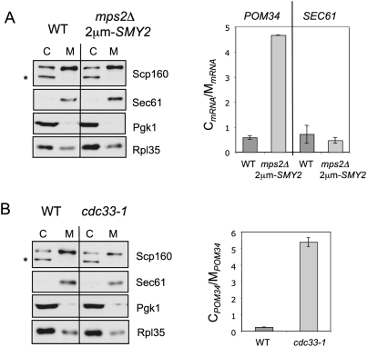
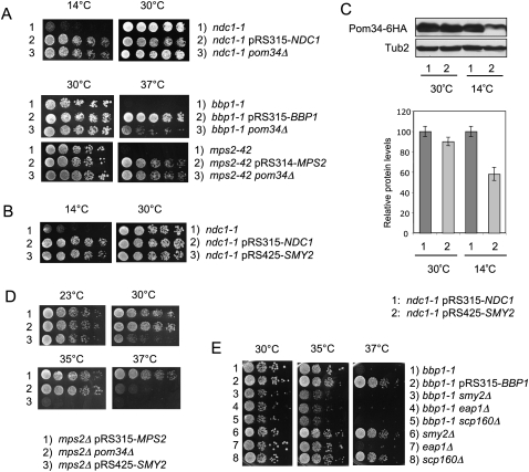
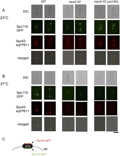
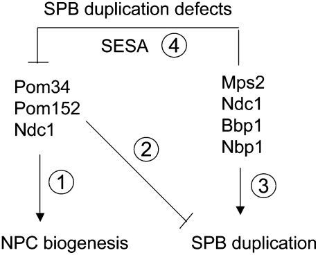
The yeast spindle pole body (SPB), the functional equivalent of mammalian centrosome, duplicates in G1/S phase of the cell cycle and then becomes inserted into the nuclear envelope. Here we describe a link between SPB duplication and targeted translation control. When insertion of the newly formed SPB into the nuclear envelope fails, the SESA network comprising the GYF domain protein Smy2, the translation inhibitor Eap1, the mRNA-binding protein Scp160 and the Asc1 protein, specifically inhibits initiation of translation of POM34 mRNA that encodes an integral membrane protein of the nuclear pore complex, while having no impact on other mRNAs. In response to SESA, POM34 mRNA accumulates in the cytoplasm and is not targeted to the ER for cotranslational translocation of the protein. Reduced level of Pom34 is sufficient to restore viability of mutants with defects in SPB duplication. We suggest that the SESA network provides a mechanism by which cells can regulate the translation of specific mRNAs. This regulation is used to coordinate competing events in the nuclear envelope.


Figures

Figure 1.
Figure 1.
Suppressor screen for the lethal phenotype of MPS2 deletion. (A) Spore analysis of MPS2/MPS2 and mps2Δ/MPS2 cells. a, b, c, and d indicate cells that developed from spores of one complete tetrad. The cells marked by an arrow did not contain the MPS2 gene. (B) SMY2 is a multicopy suppressor of the lethality of mps2Δ cells. mps2Δ pRS316-MPS2 cells were transformed with the indicated plasmids. Transformants were grown on SC-Leu or 5-FOA plates for 3 d at 23°C.
Figure 2.
Figure 2.
EAP1, SCP160, and ASC1 are essential components of the SMY2 pathway. (A) Dependency of suppression on EAP1. mps2Δ eap1Δ pRS316-MPS2 cells were transformed with the listed plasmids and tested for growth on SC-Ade and 5-FOA plates. eap1[Y109A], eap1[L114A], and eap1[Y109A,L114A] code for Eap1 proteins with mutations that partially (eap1[Y109A], eap1[L114A]) or totally (eap1[Y109A,L114A]) disrupt binding to eIF4E (Fig. 2D; Ibrahimo et al. 2006). (B) Coimmunoprecipitation of Eap1, Scp160, and Asc1 by Smy2. Extracts from cells expressing SMY2 EAP1-9Myc or SMY2-6HA EAP1-9Myc were immunoprecipitated by anti-HA-coated magnetic beads and analyzed by immunoblotting with the indicated antibodies. (WCE) Whole-cell extract, 10% of the immunoprecipitation input; (IP) immunoprecipitation. (C) Coimmunoprecipitation of Eap1, eIF4E, and eIF4G by Smy2. Extracts from the yeast cells expressing SMY2 EAP1-9Myc or SMY2-6HA EAP1-9Myc were immunoprecipitated by anti-HA-coated magnetic beads and analyzed by immunoblotting with the indicated antibodies. Abbreviations as in B. (D) Coimmunoprecipitation of Smy2 and Eap1 and mutated Eap1 protein that fails to bind to eIF4E. Extracts from yeast cells with the indicated genotypes were immunoprecipitated by anti-HA-coated magnetic beads and analyzed by immunoblotting with anti-HA, anti-Myc, and anti-eIF4E antibodies. Abbreviations are as in B. (E,F) Dependency of suppression on SCP160 and ASC1. mps2Δ scp160Δ pRS316-MPS2 (E) or mps2Δ asc1Δ pRS316-MPS2 cells (F) were transformed with the indicated plasmids and tested for growth on SC-Leu and 5-FOA at 23°C.
Figure 3.
Figure 3.
BFR1 is essential for viability of cells lacking MPS2. (A) Dependency of suppression on BFR1. mps2Δ bfr1Δ pRS316-MPS2 cells were transformed with the indicated plasmids and tested for growth on SC-Leu and 5-FOA plates at 23°C. (B) Coimmunoprecipitation of Bbp1 by Bfr1. Extracts from the yeast cells with the indicated genotypes were subjected to immunoprecipitation with anti-HA-coated magnetic beads and analyzed by immunoblotting with the indicated antibodies. Abbreviations as in Figure 2B.
Figure 4.
Figure 4.
POM34 mRNA binds to SESA components. (A,B) The indicated yeast cells were tested for growth on SC-Ura and 5-FOA plates at 23°C. All strains harbored initially the pRS316-MPS2 plasmid. (C) Coimmunoprecipitation of POM34 mRNA together with Eap1, Scp160, and Asc1 by Smy2. Extracts from yeast cells were incubated with anti-HA-coated magnetic beads, with or without RNaseA treatment, and analyzed by immunoblotting with the indicated antibodies. RNA was isolated from the immunoprecipitates and analyzed by RT–PCR using primers specific to POM34, POM152, and NDC1. In this immunoprecipitation experiment the efficiency of Asc1 coimmunoprecipitation was reduced probably because of the longer incubation time due to RNaseA treatment (cf. Figs. 2 and 4C). Abbreviations are as in Figure 2B.
Figure 5.
Figure 5.
Strongly reduced amount of Pom34 in mps2Δ 2μm-SMY2 cells. (A) Pom34 protein level is reduced in mps2Δ 2μm-SMY2 cells. Total cell extracts from yeast strains expressing NDC1-6HA, POM152-6HA, or POM34-6HA were analyzed by immunoblotting using anti-HA antibodies. Anti-Tub2 antibodies were used as loading control. The graphs underneath the immunoblots show the quantification of three independent experiments, normalized for the wild-type protein levels. Bars are standard deviations around the mean value. (B) POM34 mRNA levels are similar in wild-type and mps2Δ 2μm-SMY2 cells. Total RNA extracts from wild-type (WT) and mps2Δ 2μm-SMY2 cells were analyzed by quantitative RT–PCR using primers specific to POM34 mRNA. (C) mps2Δ 2μm-SMY2 cells do not show mRNA export defect. Wild-type and mps2Δ 2μm-SMY2 cells were fractionated into nuclear and cytoplasmic fractions and analyzed by immunoblotting with anti-Pgk1 and anti-Nop1 antibodies. RNA was isolated from the nuclear and cytoplasmic fractions and analyzed by RT–PCR using primers specific to POM34.
Figure 6.
Figure 6.
POM34 mRNA accumulates in the cytoplasm in mps2Δ 2μm-SMY2 cells. (A) Total cell extracts from wild-type and mps2Δ 2μm-SMY2 cells were fractionated into cytosolic and membrane-bound fractions. Fractions were analyzed by immunoblotting using the indicated antibodies. The lower molecular weight band (asterisk) is a cytoplasmic degradation product of Scp160 (Frey et al. 2001). POM34 and SEC61 mRNA levels were determined by quantitative RT–PCR from three independent experiments. The graph shows the ratio of cytosolic POM34 mRNA levels to the membrane-bound POM34 mRNA levels. The bars indicate standard deviation of the results from the mean value. (C) Cytosolic fraction; (M) membrane-bound fraction. (B) Subcellular localization of POM34 mRNA in the translation initiation mutant cdc33-1. Cells were grown in YPAD at 23°C and were shifted for 2 h to 37°C. The cellular fractionation and analysis was performed as in A.
Figure 7.
Figure 7.
Genetic interactions between SESA and POM34 with genes involved in SPB duplication. (A) Deletion of POM34 suppresses ndc1-1, bbp1-1, and mps2-42. The listed yeast cells were tested for growth on YPAD plates at indicated temperatures. (B) SMY2 is a multicopy suppressor of the cold-sensitive phenotype of ndc1-1. ndc1-1 cells transformed with pRS315-NDC1 or pRS425-SMY2 were tested for growth on YPAD plates at 14°C and 30°C. (C) Pom34 protein level is reduced in ndc1-1 2μm-SMY2 cells. Total cell extracts from yeast strains expressing POM34-6HA were analyzed by immunoblotting using anti-HA antibodies. Anti-Tub2 antibodies were used as loading control. The graph shows the quantification of three independent experiments, normalized for the wild-type protein levels. Bars are standard deviations around the mean value. (D) mps2Δ 2μm-SMY2 and mps2Δ pom34Δ cells are temperature sensitive for growth. The listed yeast cells were tested for growth on YPAD plates at indicated temperatures. (E) Deletion of SESA components enhances bbp1-1 growth defects. The listed yeast cells were tested for growth on YPAD plates at the indicated temperatures.
Figure 8.
Figure 8.
SPBs are inserted into the nuclear envelope in mps2-42 pom34Δ cells. (A,B) Analysis of wild-type, mps2-42, and mps2-42 pom34Δ cells with SPC110-GFP SPC42-eqFP611 by fluorescence and phase contrast (DIC) microscopy at 23°C (A) and 37°C (B) (2 h). Note that in B Spc110-GFP is only associated with one of the two Spc42-eqFP611-marked SPBs of mps2-42 cells. This is the typical phenotype of cells with a defect in duplication plaque insertion (Schramm et al. 2000; Jaspersen and Winey 2004). Bars, 5 μm. (C) Shown is a cartoon of the SPB with the localization of Spc42 and Spc110 relative to the nuclear envelope (NE) (Adams and Kilmartin 1999).
Figure 9.
Figure 9.
Model for the function of SESA network. (Step 1) Pom34, Pom152, and Ndc1 form a complex that functions in NPC biogenesis (Chial et al. 1998; Madrid et al. 2006; Alber et al. 2007a,b; Onischenko et al. 2009). (Step 3) Ndc1 has a dual role and together with Mps2, Bbp1, and Nbp1 it also functions in SPB duplication (Winey et al. 1993; Araki et al. 2006). (Step 2) Deletion of POM34 or POM152 or mutations in ndc1 rescue SPB duplication defects (Fig. 4; Chial et al. 1998), suggesting an inhibitory role of the Pom34–Pom152–Ndc1 complex in SPB duplication. In response to SPB duplication defects, SESA down-regulates translation of POM34 (Figs. 4–7). (Step 4) This in turn rescues the defect and allows for SPB duplication (Fig. 8; Supplemental Fig. S12).

Similar articles

Cited by

References

    1. Adams IR, Kilmartin JV. Localization of core spindle pole body (SPB) components during SPB duplication in Saccharomyces cerevisiae . J Cell Biol. 1999;145:809–823. - PMC - PubMed
    1. Alber F, Dokudovskaya S, Veenhoff LM, Zhang W, Kipper J, Devos D, Suprapto A, Karni-Schmidt O, Williams R, Chait BT, et al. Determining the architectures of macromolecular assemblies. Nature. 2007a;450:683–694. - PubMed
    1. Alber F, Dokudovskaya S, Veenhoff LM, Zhang W, Kipper J, Devos D, Suprapto A, Karni-Schmidt O, Williams R, Chait BT, et al. The molecular architecture of the nuclear pore complex. Nature. 2007b;450:695–701. - PubMed
    1. Araki Y, Lau CK, Maekawa H, Jaspersen SL, Giddings JTH, Schiebel E, Winey M. The Saccharomyces cerevisiae spindle pole body (SPB) component Nbp1p is required for SPB membrane insertion and interacts with the integral membrane proteins Ndc1p and Mps2p. Mol Biol Cell. 2006;17:1959–1970. - PMC - PubMed
    1. Baum S, Bittins M, Frey S, Seedorf M. Asc1p, a WD40-domain containing adaptor protein, is required for the interaction of the RNA-binding protein Scp160p with polysomes. Biochem J. 2004;380:823–830. - PMC - PubMed

Publication types

MeSH terms