

Mice with altered myelin proteolipid protein gene expression display cognitive deficits accompanied by abnormal neuron-glia interactions and decreased conduction velocities
- PMID: 19571127
- PMCID: PMC6665647
- DOI: 10.1523/JNEUROSCI.3216-08.2009
Mice with altered myelin proteolipid protein gene expression display cognitive deficits accompanied by abnormal neuron-glia interactions and decreased conduction velocities
Abstract
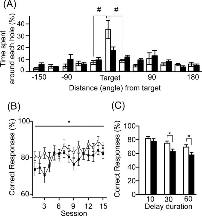
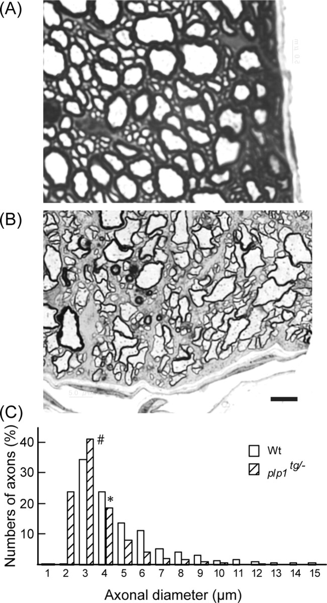
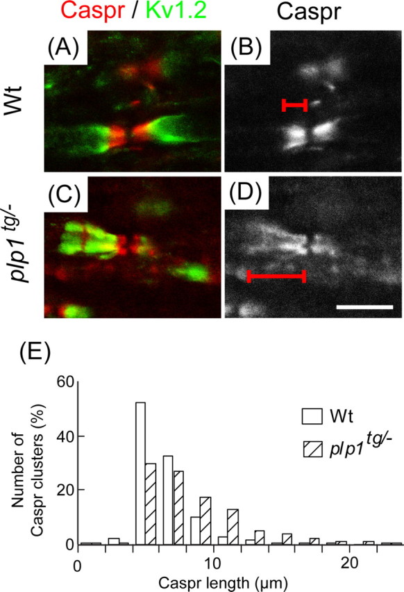
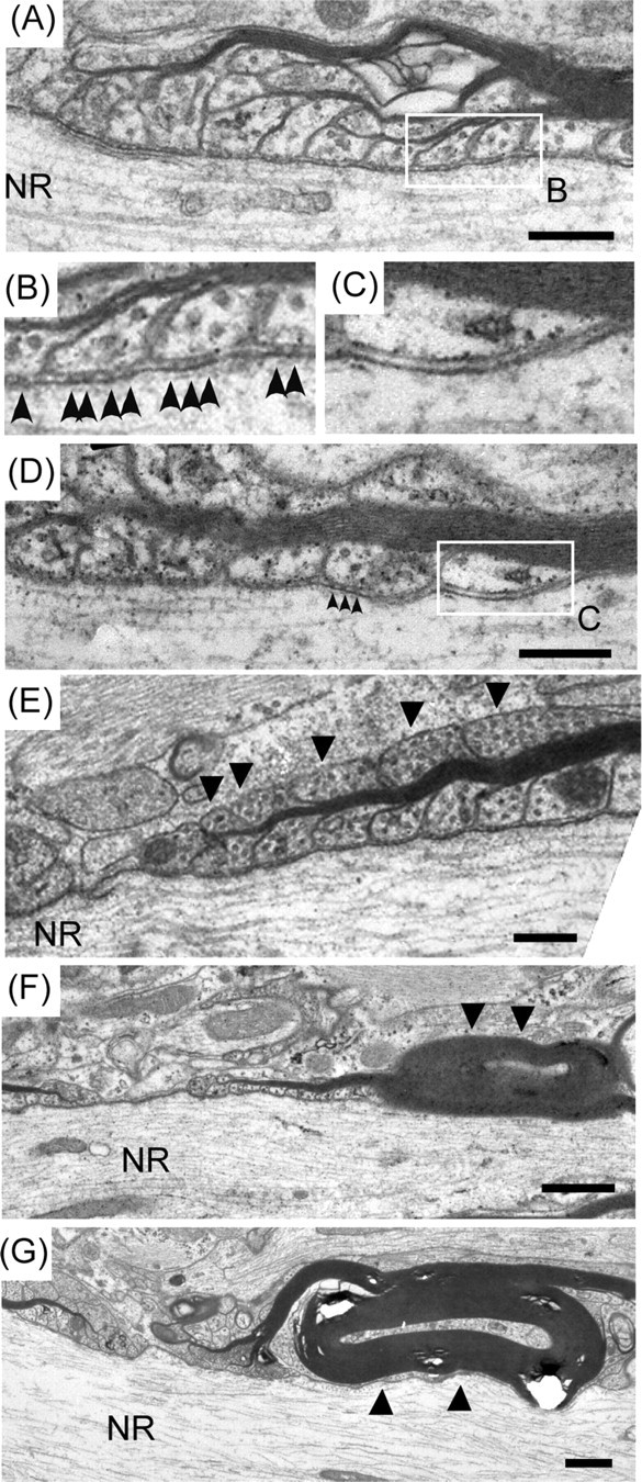
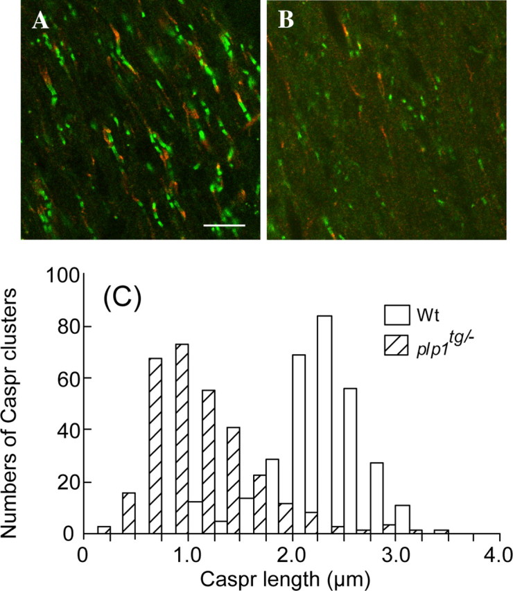
Conduction velocity (CV) of myelinated axons has been shown to be regulated by oligodendrocytes even after myelination has been completed. However, how myelinating oligodendrocytes regulate CV, and what the significance of this regulation is for normal brain function remain unknown. To address these questions, we analyzed a transgenic mouse line harboring extra copies of the myelin proteolipid protein 1 (plp1) gene (plp1(tg/-) mice) at 2 months of age. At this stage, the plp1(tg/-) mice have an unaffected myelin structure with a normally appearing ion channel distribution, but the CV in all axonal tracts tested in the CNS is greatly reduced. We also found decreased axonal diameters and slightly abnormal paranodal structures, both of which can be a cause for the reduced CV. Interestingly the plp1(tg/-) mice showed altered anxiety-like behaviors, reduced prepulse inhibitions, spatial learning deficits and working memory deficit, all of which are schizophrenia-related behaviors. Our results implicate that abnormalities in the neuron-glia interactions at the paranodal junctions can result in reduced CV in the CNS, which then induces behavioral abnormalities related to schizophrenia.
Figures
Similar articles
-
Spatiotemporal ablation of myelinating glia-specific neurofascin (Nfasc NF155) in mice reveals gradual loss of paranodal axoglial junctions and concomitant disorganization of axonal domains.J Neurosci Res. 2009 Jun;87(8):1773-93. doi: 10.1002/jnr.22015. J Neurosci Res. 2009. PMID: 19185024 Free PMC article.
-
Progressive disorganization of paranodal junctions and compact myelin due to loss of DCC expression by oligodendrocytes.J Neurosci. 2014 Jul 16;34(29):9768-78. doi: 10.1523/JNEUROSCI.0448-14.2014. J Neurosci. 2014. PMID: 25031414 Free PMC article.
-
The raft-associated protein MAL is required for maintenance of proper axon--glia interactions in the central nervous system.J Cell Biol. 2004 Aug 30;166(5):731-42. doi: 10.1083/jcb.200406092. J Cell Biol. 2004. PMID: 15337780 Free PMC article.
-
Glial regulation of the axonal membrane at nodes of Ranvier.Curr Opin Neurobiol. 2006 Oct;16(5):508-14. doi: 10.1016/j.conb.2006.08.003. Epub 2006 Sep 1. Curr Opin Neurobiol. 2006. PMID: 16945520 Review.
-
Neuron-glia interactions at the node of Ranvier.Results Probl Cell Differ. 2006;43:129-49. doi: 10.1007/400_014. Results Probl Cell Differ. 2006. PMID: 17068970 Review.
Cited by
-
The effects of psychosis risk variants on brain connectivity: a review.Front Psychiatry. 2012 Mar 9;3:18. doi: 10.3389/fpsyt.2012.00018. eCollection 2012. Front Psychiatry. 2012. PMID: 22416237 Free PMC article.
-
RIT1 regulation of CNS lipids RIT1 deficiency Alters cerebral lipid metabolism and reduces white matter tract oligodendrocytes and conduction velocities.Heliyon. 2023 Sep 23;9(10):e20384. doi: 10.1016/j.heliyon.2023.e20384. eCollection 2023 Oct. Heliyon. 2023. PMID: 37780758 Free PMC article.
-
Function and mechanism of axonal targeting of voltage-sensitive potassium channels.Prog Neurobiol. 2011 Jul;94(2):115-32. doi: 10.1016/j.pneurobio.2011.04.009. Epub 2011 Apr 22. Prog Neurobiol. 2011. PMID: 21530607 Free PMC article. Review.
-
Association between chronic stress-induced structural abnormalities in Ranvier nodes and reduced oligodendrocyte activity in major depression.Sci Rep. 2016 Mar 15;6:23084. doi: 10.1038/srep23084. Sci Rep. 2016. PMID: 26976207 Free PMC article.
-
White matter abnormalities and animal models examining a putative role of altered white matter in schizophrenia.Schizophr Res Treatment. 2011;2011:826976. doi: 10.1155/2011/826976. Epub 2011 Aug 11. Schizophr Res Treatment. 2011. PMID: 22937274 Free PMC article.
References
-
- Akbarian S, Kim JJ, Potkin SG, Hetrick WP, Bunney WE, Jr, Jones EG. Maldistribution of interstitial neurons in prefrontal white matter of the brains of schizophrenic patients. Arch Gen Psychiatry. 1996;53:425–436. - PubMed
-
- American Psychiatric Association. Diagnostic and statistical manual of mental disorders: DSM-IV-TR. 4th ed. Washington DC: American Psychiatric Association; 2000.
-
- Braff DL, Geyer MA. Sensorimotor gating and schizophrenia. Human and animal model studies. Arch Gen Psychiatry. 1990;47:181–188. - PubMed
-
- Crawley JN. New York: Wiley; 2000. What's wrong with my mouse? Behavioral phenotyping of transgenic and knockout mice.
-
- Davis KL, Stewart DG, Friedman JI, Buchsbaum M, Harvey PD, Hof PR, Buxbaum J, Haroutunian V. White matter changes in schizophrenia: evidence for myelin-related dysfunction. Arch Gen Psychiatry. 2003;60:443–456. - PubMed
Publication types
MeSH terms
Substances
LinkOut - more resources
Full Text Sources
Molecular Biology Databases
Research Materials
Miscellaneous
