. 2009 Sep 25;284(39):26427-38.
doi: 10.1074/jbc.M109.014845. Epub 2009 Jun 30.

Numb regulates post-endocytic trafficking and degradation of Notch1

Affiliations

Numb regulates post-endocytic trafficking and degradation of Notch1

Melanie A McGill et al. J Biol Chem. .

Abstract

Notch is a transmembrane receptor that controls cell fate decisions during development and tissue homeostasis. Both activation and attenuation of the Notch signal are tightly regulated by endocytosis. The adaptor protein Numb acts as an inhibitor of Notch and is known to function within the intracellular trafficking pathways. However, a role for Numb in regulating Notch trafficking has not been defined. Here we show that mammalian Notch1 is constitutively internalized and trafficked to both recycling and late endosomal compartments, and we demonstrate that changes in Numb expression alter the dynamics of Notch1 trafficking. Overexpression of Numb promotes sorting of Notch1 through late endosomes for degradation, whereas depletion of Numb facilitates Notch1 recycling. Numb mutants that do not interact with the ubiquitin-protein isopeptide ligase, Itch, or that lack motifs important for interaction with endocytic proteins fail to promote Notch1 degradation. Our data suggest that Numb inhibits Notch1 activity by regulating post-endocytic sorting events that lead to Notch1 degradation.


Figures
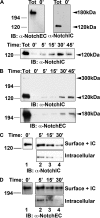
FIGURE 1.
FIGURE 1.
Notch1 is constitutively trafficked independent of ligand. A, subconfluent C2C12 cells expressing endogenous Notch1 receptor were labeled with biotin at 4 °C. After 1 h, one plate of cells was lysed to examine total surface biotin-labeled Notch1 (Tot), and a second was treated with MeSNa (0′) to strip the biotin label. Cells were then incubated at 37 °C and at the indicated times stripped of biotin remaining at the cell surface. Intracellular pools of biotinylated Notch1 were recovered from whole cell lysates using streptavidin beads and analyzed by Western blot using antisera specific for the carboxyl terminus of Notch1, anti-NotchIC, or for the extracellular epidermal growth factor-like repeat region of Notch1, anti-NotchEC, as indicated. IB, immunoblot. B, HEK293T cells transfected with FL-Notch1 were labeled with biotin, and intracellular pools of biotin-labeled FL-Notch1 were examined as described above. C and D, FL-Notch1 is recycled. HEK293T cells expressing FL-Notch1 were biotinylated, incubated at 18 °C for 2 h to internalize biotin-labeled FL-Notch1, and then treated with MeSNa to remove surface biotin. Cells were then returned to 37 °C to resume trafficking. One set of cells was treated a second time with MeSNa followed by lysis (intracellular, lower panel), although the second set was lysed without MeSNa treatment (surface + IC, intracellular; upper panel). Pools of biotinylated FL-Notch1 were recovered with streptavidin beads and analyzed by Western blot using antisera specific for either the intracellular (C) or extracellular domain (D) of Notch1.
FIGURE 2.
FIGURE 2.
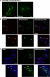
Surface-labeled Notch1 is constitutively endocytosed into vesicles that colocalize with FITC-dextran and HRS. A, C2C12 cells that stably express HA-tagged Notch1 were surface-labeled with Al488anti-HA at 4 °C (left panel), washed, and incubated at 37 °C for 60 min to allow for endocytosis (right panel). B, HEK293T cells transiently expressing HA-Notch1 were incubated for 18 h with 1 mg/ml FITC-dextran (70,000 molecular weight; Molecular Probes) in normal growth media at 37 °C. The cells were washed and chased with media containing anti-HA for 2 h, fixed, permeabilized, and then stained with Cy3-α-mouse to identify α-HA-labeled Notch1 and with rabbit α-NbC (Cy5-α-rabbit secondary) to mark the plasma membrane. C, HEK293T cells transiently expressing HA-Notch1 were incubated with Alexa488-α-HA (green) at 37 °C for 1 h. Following fixation with 2% paraformaldehyde, cells were permeabilized and immunostained with rabbit αHRS (Cy5-labeled donkey α-rabbit) and guinea pig αNbC (Cy3 labeled donkey anti-guinea pig). Images were captured using a Zeiss Axiovert 200 microscope equipped with a Hamamatsu Orca AG CCD camera and spinning disk confocal scan head. Images were processed using Volocity software, and figures were prepared using Photoshop. Scale bars represent 10 μm. Images are representative of three independent experiments.
FIGURE 3.
FIGURE 3.
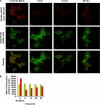
Surface-labeled Notch1 is constitutively recycled to the plasma membrane. A, C2C12 HA-N1 cells were incubated with Al488anti-HA (green) in growth media at 37 °C for 30 min to accumulate surface-labeled HA-Notch1 in intracellular vesicles (0 min No Block). Cy3-labeled anti-mouse-binding sites on surface Al488anti-HA were blocked with excess unlabeled anti-mouse IgG at 4 °C (0 min). Cells were then re-warmed to 37 °C to allow for continued trafficking of internalized HA-Notch1 (10 min and 20 min). Prior to fixation, all cells were incubated with Cy3-labeled anti-mouse (red) to visualize unblocked, Al488-anti-HA tagged, and HA-Notch1 at the cell surface. Cells were imaged using a ×25 water objective and spinning disk confocal microscope as described under “Materials and Methods.” Scale bars represent 20 μm. Images are representative of three independent experiments. B, average total cellular fluorescence intensity in each channel was measured in 45–55 cells for each condition shown in A. The mean intensities for each condition are shown, allowing comparison of the changes in the total labeled HA-Notch1 (green bars, left) and surface-labeled HA-Notch1 (red bars, right).
FIGURE 4.
FIGURE 4.
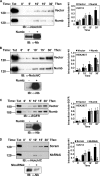
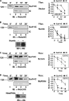
Numb regulates the dynamics of Notch1 receptor trafficking. A, C2C12 cells expressing endogenous Notch1 receptor transfected with empty vector or Numb were labeled with biotin at 4 °C for 1 h, allowed to traffic at 37 °C, and the intracellular pools of biotin-labeled Notch1 (biotin-N1) examined using streptavidin beads and Western blot analysis as described previously. IB, immunoblot; Tfxn, transfection. B, HEK293T cells were transfected with FL-Notch1 alone or FL-Notch1 and Numb and the trafficking of the FL-Notch1 receptor examined. C, Numb does not alter trafficking of EGFR. HEK293T were cotransfected with EGFR alone or with Numb, and accumulation of intracellular biotin-labeled EGFR examined as described above. D, loss of Numb protein disrupts intracellular trafficking of Notch1. C2C12 cells were transfected with Numb-specific RNA duplexes or control scrambled RNA duplexes, and the trafficking of endogenous Notch1 receptor was examined 48 h post-transfection as described above. Whole cell lysates were examined for Numb protein expression using antisera specific for Numb. Scram, Scrambled siRNA. Representative Western blots are shown. Films from three independent experiments were scanned and quantitated using an Alpha Innotech Fluorochem 8000 imaging system (see “Material and Methods”), and results are shown in graphs (right). Data are expressed relative to total (Tot) surface-biotinylated Notch1 (mean ± S.D.; n = 3). Significance was determined by Student's t test; **, p < 0.01; *, p < 0.05 compared with control.
FIGURE 5.
FIGURE 5.
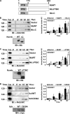
PTB domain and tripeptide endocytic motifs of Numb are required to regulate Notch1 trafficking. A, schematic representation of Numb and Numb mutants. NbΔPTBC is missing 88 amino acids in the carboxyl-terminal half of the PTB domain. NbΔC lacks the carboxyl-terminal 41 amino acids, including NPF and DPF motifs. Tfxn, transfection; IB, immunoblot. B, NbΔC does not affect Notch1 receptor trafficking. HEK293T cells were cotransfected with FL-Notch1 and wild type Numb or NbΔC and labeled with biotin at 4 °C. The total amount of biotin-labeled FL-Notch1 (biotin-N1) at the cell surface (Tot) and the efficiency of MeSNa treatment (0′) were monitored as described previously. Cells were incubated at 37 °C, and surface biotin stripped at indicated time points. Equivalent amounts of protein lysates were incubated with streptavidin beads and the intracellular pool of biotin-labeled FL-Notch1 analyzed by Western blot. Whole cell lysates were examined for equal expression of NbWT and NbΔC. Tfxn, transfection. C, accumulation of intracellular FL-Notch1 upon overexpression of NbΔPTBC. HEK293T cells were cotransfected with FL-Notch1 and NbWT or NbΔPTBC, and the intracellular pools of biotin-labeled FL-Notch1 were examined as described above. Whole cell lysates were examined for equal expression of NbWT and NbΔPTBC. D, Itch disrupts trafficking of FL-Notch1. HEK293T cells expressing FL-Notch1 and wild type Itch or a ligase-dead mutant were biotinylated, and trafficking of FL-Notch1 was examined. Whole cell lysates were examined for equal expression of ItchWT and ItchC830A. Representative Western blots are shown. Films from three independent experiments were scanned and quantitated using an Alpha Innotech Fluorochem 8000 imaging system (see “Materials and Methods”) and results are shown in the graphs (right). Data are expressed relative to total (Tot) surface-biotinylated Notch1 (mean ± S.D.; n = 3). Significance was determined by Student's t test; **, p < 0.01; *, p < 0.05 compared with control.
FIGURE 6.
FIGURE 6.
Numb regulates intracellular sorting of Notch1. A, overexpression of Numb does not affect internalization of Notch1 from the cell surface. HEK293T cells expressing FL-Notch1 with or without overexpressed Numb were biotinylated at 4 °C and then incubated at 18 °C. Total biotin-labeled FL-Notch1 (biotin-N1) at the cell surface (Tot) and the efficiency of MeSNa treatment (0′) was monitored as described previously. Biotin remaining at the cell surface was stripped at the indicated time points, and intracellular pools of biotin-labeled FL-Notch1 were recovered using streptavidin beads and analyzed by Western blot. Tfxn, transfection; IB, immunoblot. B, loss of Numb protein using Numb-specific siRNA had no effect on Notch1 internalization compared with C2C12 cells expressing scrambled siRNA duplexes. Representative Western blots are shown. Numb overexpression or depletion was confirmed by immunoblotting of whole cell lysates with anti-Numb. Films from three independent experiments were scanned and quantitated using an Alpha Innotech Fluorochem 8000 imaging system (see “Materials and Methods”) and results are shown in graphs (right). Data are expressed relative to total (Tot) surface-biotinylated Notch1 (mean ± S.D.; n = 3). Significance was determined by Student's t test.
FIGURE 7.
FIGURE 7.
Numb promotes Notch1 degradation. A and B, overexpression of Numb promotes Notch1 degradation not recycling. HEK293T cells expressing FL-Notch1 alone (A) or FL-Notch1 and Numb (B) were biotinylated, incubated at 18 °C for 2 h to accumulate biotin-labeled FL-Notch1 intracellularly, and then stripped of remaining surface biotin with MeSNa. Cells were then returned to 37 °C to resume trafficking. One set of cells was treated a second time with MeSNa followed by lysis to examine intracellular pools of biotinylated FL-Notch1 (IC, lower panels), whereas the second set was lysed without MeSNa treatment to examine total pools of biotinylated FL-Notch1 remaining (Surf + IC, upper panels). Pools of biotinylated FL-Notch1 were recovered with streptavidin beads and analyzed by Western blot using antisera specific for Notch1. Tfxn, transfection; IB, immunoblot. C and D, knockdown of Numb protein using Numb-specific RNA interference increases FL-Notch1 recycling back to the plasma membrane. C2C12 cells expressing a scrambled control siRNA duplex (C) or Numb-specific siRNA duplex (D) were analyzed as described above for intracellular and total pools of biotinylated FL-Notch1. Whole cell lysates were quantified and analyzed for Numb expression. Scram, scrambled siRNA; IC, intracellular; Surf + IC, surface and intracellular. Representative Western blots are shown. Films from three independent experiments were scanned and quantitated using an Alpha Innotech Fluorochem 8000 imaging system (see “Materials and Methods”), and results are shown in graphs (right). Data are expressed relative to total (Tot) surface-biotinylated Notch1 (mean ± S.D.; n = 3). Significance was determined by Student's t test; **, p < 0.01;*, p < 0.05 compared with respective controls shown in A and C.

Similar articles

Cited by

References

    1. Artavanis-Tsakonas S., Rand M. D., Lake R. J. (1999) Science 284, 770–776 - PubMed
    1. Schweisguth F. (2004) Curr. Biol. 14, R129–R138 - PubMed
    1. Lai E. C. (2004) Development 131, 965–973 - PubMed
    1. Kidd S., Lieber T., Young M. W. (1998) Genes Dev. 12, 3728–3740 - PMC - PubMed
    1. Schroeter E. H., Kisslinger J. A., Kopan R. (1998) Nature 393, 382–386 - PubMed

Publication types

MeSH terms