. 2009 Jun 29;185(7):1209-25.
doi: 10.1083/jcb.200811005.

Cholesterol sensor ORP1L contacts the ER protein VAP to control Rab7-RILP-p150 Glued and late endosome positioning

Affiliations

Cholesterol sensor ORP1L contacts the ER protein VAP to control Rab7-RILP-p150 Glued and late endosome positioning

Nuno Rocha et al. J Cell Biol. .

Abstract

Late endosomes (LEs) have characteristic intracellular distributions determined by their interactions with various motor proteins. Motor proteins associated to the dynactin subunit p150(Glued) bind to LEs via the Rab7 effector Rab7-interacting lysosomal protein (RILP) in association with the oxysterol-binding protein ORP1L. We found that cholesterol levels in LEs are sensed by ORP1L and are lower in peripheral vesicles. Under low cholesterol conditions, ORP1L conformation induces the formation of endoplasmic reticulum (ER)-LE membrane contact sites. At these sites, the ER protein VAP (VAMP [vesicle-associated membrane protein]-associated ER protein) can interact in trans with the Rab7-RILP complex to remove p150(Glued) and associated motors. LEs then move to the microtubule plus end. Under high cholesterol conditions, as in Niemann-Pick type C disease, this process is prevented, and LEs accumulate at the microtubule minus end as the result of dynein motor activity. These data explain how the ER and cholesterol control the association of LEs with motor proteins and their positioning in cells.


Figures
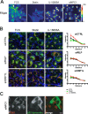
Figure 1.
Figure 1.
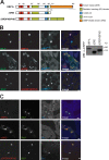
Chemical and genetic cholesterol manipulations and LEs. (A) Modulation of intracellular cholesterol levels. Mel JuSo cells were cultured in normal medium (FCS), cholesterol-depleted medium supplemented with lovastatin (statin), or normal medium supplemented with U-18666A, as indicated. Fixed cells were stained with filipin to detect cholesterol. A color look up table (LUT) shows fluorescence intensities. n > 100 for each condition. (B) Effects of intracellular cholesterol manipulation (control [CTRL], U-18666A, or statin treatment) on LE positioning in RILP- or ORP1L-silenced Mel JuSo cells. Cells were stained for the LE marker CD63 (red), endogenous ORP1L (green), and actin (blue). The position of the CD63-positive vesicles relative to the nucleus (radial distribution) was determined for the various conditions (>50 cells per condition), binned, and plotted (right). The distributions were statistically different according to the Jonckheere-Terpstra test for control siRNA (P = 1.52 × 10⁻⁵ and 7.76 × 10⁻⁶ for CD63 and ORP1L, respectively) only. No difference was detected for siRILP (P = 0.20 and 0.54 for CD63 and ORP1L, respectively) and for siORP1L (P = 0.29 for CD63). (C) Dynein motor activity and clustering of LEs in NPC1-silenced cells. Mel JuSo cells were transfected with siRNA for NPC1 for 72 h and, after 48 h, transfected with an expression construct for p50dynamitin to disrupt the dynein–dynactin motor. Fixed cells were stained for CD63 and p50dynamitin to mark the overexpressing cells. n > 100. Bars: (A and C)10 µm; (B) 20 µm.
Figure 2.
Figure 2.
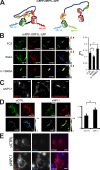
ORP1L controls recruitment of p150Glued to the Rab7–RILP receptor. (A) ORP1L domain structure and constructs. Five domains are predicted in ORP1L. Numbers indicate amino acid residue positions. Constructs were N-terminally tagged with mRFP. The ΔORDPHDPHD chimera had ORD exchanged for a tandem PH domain derived from ORP1L. (B) Effect of ORP1L deletion or chimeric constructs on RILP-mediated p150Glued recruitment. (left) Mel JuSo cells transfected with GFP-RILP and mRFP-ORP1L constructs and stained with anti-p150Glued antibodies before CLSM. For pixel analyses of the images, see Fig. S2 E. n > 200 for each condition. (right) Mel JuSo cells transfected with the indicated mRFP-ORP1L constructs and whole cell lysates analyzed by immunoblotting with anti-mRFP antibodies (WB: α-mRFP). Molecular standards are indicated. WB, Western blot. (C) Effect of ORP1L constructs on RILP recruitment of the p150Glued-associated dynein motor adapter DIC. Mel JuSo cells were transfected with GFP-RILP and the mRFP-ORP1L constructs indicated and stained with anti-DIC antibodies. n > 100 for each condition. Bars, 10 µm.
Figure 3.
Figure 3.
ORP1L controls cholesterol-dependent LE positioning. (A) Cholesterol-dependent ORP1L vesicle positioning. Mel JuSo cells expressing mRFP-ORP1L were cultured under control conditions (FCS) or conditions causing decreased (statin) or increased (U-18666A) cholesterol levels. Actin was stained with falloidin (green) to mark the cell boundaries before analyses by CLSM. n > 100. (B) ORP1L is dominant in vesicle repositioning as the result of cholesterol manipulations. Mel JuSo cells expressing mRFP-ΔORDPHDPHD or mRFP-ΔORD were treated and imaged as described in A. n > 100. Bars, 10 µm.
Figure 4.
Figure 4.
LE cholesterol alters the conformation of ORP1L. (A) Intramolecular FRET for mRFP-ORP1L-GFP. GFP and mRFP are attached to the same molecule, allowing FRET from donor GFP to acceptor mRFP. FRET depends on distance and orientation and thus indicates conformational changes. FRET can be detected by sensitized emission. GFP is excited by 488-nm light, and then, after energy transfer, 582–675-nm light emission by mRFP is detected. (B) Cholesterol effects on ORP1L conformation detected by sensitized emission. Mel JuSo cells expressing mRFP-ORP1L-GFP were cultured under control (FCS) or cholesterol-depleting (statin) or -enhancing (U-18666A) conditions before imaging by CLSM for FRET determination. Panels show the GFP signal, the mRFP signal, calculated FRET, and FRET related to donor fluorophore input: the donor FRET efficiency (ED). The color LUT visualizes the differences in the ED panels. (right) quantification of the donor FRET efficiency detected for mRFP-ORP1L-GFP under the different conditions of cholesterol manipulation. The mean and SD from two experiments (>10 cells analyzed) are shown (*, P = 0.05; **, P = 0.03). (C) ORP1L controls LE positioning in NPC1-silenced cells. Mel JuSo cells were transfected with mRFP-ORP1L, -ΔORD, or -ΔORDPHDPHD and siRNA for NPC1 and analyzed by CLSM. n > 100. (D) Sensitized emission and ORP1L conformation in NPC1-deficient cells. mRFP-ORP1L-GFP–expressing MelJuSo cells were transfected with control (siCTRL) or NPC1 (siNPC1) siRNAs before imaging by confocal FRET. (right) Donor FRET efficiencies determined in >10 control siRNA– or NPC1 siRNA–transfected cells. The mean ± SD is shown (**, P = 5.1 × 10⁻⁶). (E) NPC1, cholesterol, and LE clustering. Mel JuSo cells were transfected with control or NPC1 siRNA and then mRFP-ΔORD before staining with filipin for cholesterol. Pixel analyses are shown in Fig. S2 E. n > 50 for each condition. Bars, 10 µm.
Figure 5.
Figure 5.
ORP1L conformation and LE positioning. (A and B, top) Mel JuSo cells expressing mRFP-ORP1L-GFP (A) or mRFP-ΔORD-GFP (B) were imaged for FRET by CLSM. The GFP image, mRFP image, calculated FRET, and donor FRET efficiency ED images are indicated. ED values are shown in false colors (the LUT shows the corresponding values). The position of the nucleus (N) and cell boundaries are drawn in the GFP channel. The cytosol is divided by five concentric rings, as indicated in the ED panel. (bottom) ED values for the constructs were collected and binned for the different rings. Values are shown for the different rings under the corresponding image. Mean ± SEM of ED values collected for >10 cells analyzed per construct are shown. The asterisks indicate a statistically significant difference in ED of that bin compared to bin 1 (the most internal ring; *, P = 0.024; **, P = 0.026; ***, P = 0.0063). Bars, 10 µm.
Figure 6.
Figure 6.
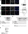
ORP1L requires VAP-A to remove p150Glued from Rab7–RILP. (A) Effect of the FFAT motif in ΔORD on RILP-mediated p150Glued recruitment. (left) Mel JuSo cells were transfected with GFP-RILP and mRFP-ΔORD containing a point mutation in its FFAT motif, ΔORDFFAT(D478A). Fixed cells were stained with anti-p150Glued antibodies. n > 200. (right) Mel JuSo cells were transfected with the mRFP-ΔORD or mRFP-ΔORDFFAT(D478A) constructs, and whole cell lysates were analyzed by immunoblotting with anti-mRFP antibodies (WB: α-mRFP). (B) p150Glued exclusion by ΔORD and VAP-A. VAP-A was silenced by siRNA (siVAP-A) in cells expressing GFP-RILP and mRFP-ΔORD. (left) Cells stained for p150Glued. Pixel analyses are shown in Fig. S2 E. n > 100 for each condition. (right) Western blot analysis of cells transfected with transfection reagent (mock), control (siCTRL), or VAP-A siRNA (siVAP-A) and probed for α-tubulin (as loading control) and anti–VAP-A antibodies. (C) VAP-A removes p150Glued from Rab7–RILP. GTP-locked His-Rab7(Q67L) was GTP loaded and coupled to Talon beads before adding purified RILP, ORP1L, and p150Glued(C25) fragments in equimolar amounts to form a preassembled ORP1L–Rab7–RILP–p150Glued(C25) complex. Subsequently, the complex was incubated in the presence or absence of purified VAP-A. After washing, the bead-bound proteins were analyzed by SDS-PAGE and Western blotting. (D) ORP1L requirements for p150Glued removal by VAP-A. GTP-loaded His-Rab7(Q67L) was coupled to Talon Co2+ beads before adding the isolated proteins indicated in equimolar amounts to form a preassembled complex. After washing, these complexes were exposed to isolated VAP-A or the p150Glued(C25) fragment, as indicated, and the effects on the preformed complex were assessed by SDS-PAGE and Western blot analyses with antibodies, as indicated. (E) VAP-A interacts with p150Glued. GST or GST–VAP-A was coupled to beads before exposure to the p150Glued(C25) fragment. After washing, the complexes were analyzed by SDS-PAGE and Western blotting. (A–E) Molecular masses are indicated. WB, Western blot. Bars, 10 µm.
Figure 7.
Figure 7.
Cholesterol-dependent ORP1L-mediated contacts between LEs and the ER. (A) ΔORD recruits endogenous VAP-A. Mel JuSo cells expressing mRFP-ΔORD, -ORP1L, or -ΔORDPHDPHD were stained for VAP-A. The merge panels show an overlay of the two channels, and the boxes indicate the zoomed-in areas. Pixel analyses of the zoomed-in areas for fluorescence distribution of the mRFP-ORP1L variants and VAP-A are shown. n > 100. (B) Mel JuSo cells expressing TAP1-GFP (red) were exposed to different cholesterol manipulating conditions, as indicated. Cells were stained with antibodies for CD63 (green). Colocalization of the markers was determined by computational pixel analysis and visualized in blue. (right) Quantification of the colocalizing surface of the CD63-positive area. The mean ± SEM for >20 cells analyzed per condition is shown (*, P = 0.068; **, P = 1.55 × 10⁻⁶; ***, P = 0.0003). Bars: (A) 10 µm; (B) 5 µm.
Figure 8.
Figure 8.
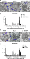
Cholesterol-dependent MCSs between LEs and the ER allowing in trans VAP interactions with the Rab7–RILP complex. (A) Electron micrographs of multivesicular bodies in Mel JuSo cells expressing HA-VAP and different variants of GFP-ORP1L, as indicated. Sections were stained with anti-HA antibodies detected with 15-nm gold particles. The gold particles are highlighted by red dots, and the ER membrane (containing HA-VAP) is indicated by blue lines. The original images are shown in Fig. S5 C. (B) The percentage of multivesicular bodies contacting ER structures and the contact area between the ER and LE membranes was determined in Mel JuSo cells expressing the ORP1L variants as shown in A. Over 50 multivesicular bodies were considered per condition. The data on contact area from two independent quantifications were binned as indicated, and error bars show SEM. (C) Electron micrographs of multivesicular bodies in HA-VAP– and GFP-ORP1L–expressing MelJuSo cells exposed to different cholesterol-manipulating conditions, as indicated. Sections were stained with anti-HA antibodies marked by 15-nm gold particles. The gold particles are highlighted by red dots, and the ER membrane (containing HA-VAP) is indicated by blue lines. The original images are shown in Fig. S5 F. (D) The percentage of multivesicular bodies contacting ER structures and the contact area between the ER and LE membranes from MelJuSo cells expressing ORP1L under various conditions of cholesterol manipulation (as shown in C). Over 50 multivesicular bodies were considered per condition. The data on contact area from two independent quantifications were binned as indicated, and error bars show SEM. Bars, 200 nm.
Figure 9.
Figure 9.
Model showing control of Rab7–RILP–p150Glued motor protein complexes by the ER protein VAP, cholesterol, and the sensor ORP1L. Rab7 recruits the homodimeric effector protein RILP to LEs. RILP binds the p150Glued subunit of the dynein–dynactin motor. ORP1L also binds to the Rab7–RILP complex. ORP1L has a C-terminal cholesterol-sensing ORD that exists in different conformational states determined by cholesterol in LEs. At low cholesterol levels, ORD adopts a conformation in which the adjacent FFAT motif is exposed and which can be detected by the ER protein VAP in ER–LE MCSs. VAP binds in trans to the Rab7–RILP–p150Glued complex and removes p150Glued, thus preventing minus end–directed transport. High cholesterol conditions initiate a different ORP1L conformation, preventing formation of ER–LE MCSs, and VAP fails to interact with the Rab7–RILP–p150Glued complex. Thus, cholesterol levels in LEs determine the conformation of ORP1L and thereby VAP recruitment in ER–LE contact sites. The ER protein VAP then controls p150Glued binding to Rab7–RILP, resulting in the scattering of cholesterol-poor LEs and clustering of cholesterol-laden LEs, as in Niemann-Pick type C disease.

Similar articles

Cited by

References

    1. Bastiaens P.I., Squire A. 1999. Fluorescence lifetime imaging microscopy: spatial resolution of biochemical processes in the cell.Trends Cell Biol. 9:48–52 - PubMed
    1. Brown C.L., Maier K.C., Stauber T., Ginkel L.M., Wordeman L., Vernos I., Schroer T.A. 2005. Kinesin-2 is a motor for late endosomes and lysosomes.Traffic. 6:1114–1124 - PubMed
    1. Burkhardt J.K., Echeverri C.J., Nilsson T., Vallee R.B. 1997. Overexpression of the dynamitin (p50) subunit of the dynactin complex disrupts dynein-dependent maintenance of membrane organelle distribution.J. Cell Biol. 139:469–484 - PMC - PubMed
    1. Calafat J., Janssen H., Stahle-Backdahl M., Zuurbier A.E., Knol E.F., Egesten A. 1997. Human monocytes and neutrophils store transforming growth factor-alpha in a subpopulation of cytoplasmic granules.Blood. 90:1255–1266 - PubMed
    1. Calleja V., Ameer-Beg S.M., Vojnovic B., Woscholski R., Downward J., Larijani B. 2003. Monitoring conformational changes of proteins in cells by fluorescence lifetime imaging microscopy.Biochem. J. 372:33–40 - PMC - PubMed

Publication types

MeSH terms