. 2009 Aug;297(2):H874-86.
doi: 10.1152/ajpheart.00311.2009. Epub 2009 Jun 26.

Resveratrol blocks interleukin-18-EMMPRIN cross-regulation and smooth muscle cell migration

Affiliations

Resveratrol blocks interleukin-18-EMMPRIN cross-regulation and smooth muscle cell migration

Balachandar Venkatesan et al. Am J Physiol Heart Circ Physiol. 2009 Aug.

Abstract

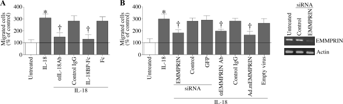
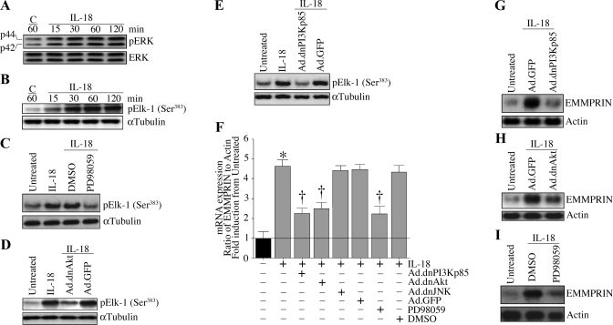
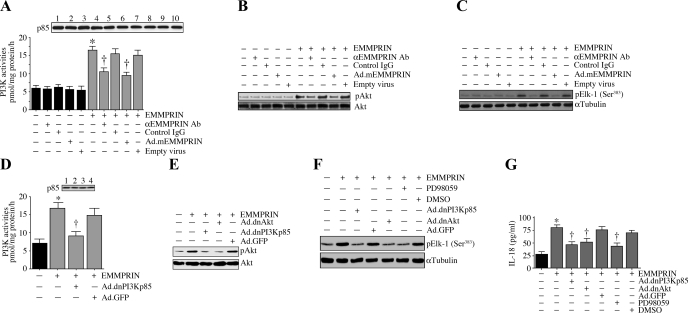
Vascular smooth muscle cell (SMC) migration is an important mechanism in atherogenesis and postangioplasty arterial remodeling. Previously, we demonstrated that the proinflammatory cytokine interleukin (IL)-18 is a potent inducer of SMC migration. Since extracellular matrix metalloproteinase inducer (EMMPRIN) stimulates ECM degradation and facilitates cell migration, we investigated whether IL-18 and EMMPRIN regulate each other's expression, whether their cross talk induces SMC migration, and whether the phytoalexin resveratrol inhibits IL-18-EMMPRIN signaling and SMC migration. Our studies demonstrate that 1) IL-18 induces EMMPRIN mRNA and protein expressions and stimulates EMMPRIN secretion from human aortic SMCs; 2) IL-18 stimulates EMMPRIN expression via oxidative stress and phosphatidylinositol 3-kinase (PI3K)-Akt-ERK signaling; 3) IL-18-stimulated SMC migration is significantly blunted by EMMPRIN knockdown, EMMPRIN function-blocking antibodies, or adenoviral transduction of mutant EMMPRIN; 4) conversely, EMMPRIN stimulates IL-18 expression and secretion via PI3K, Akt, and ERK; and 5) resveratrol attenuates IL-18- and EMMPRIN-mediated PI3K, Akt, and ERK activations; blunts IL-18-mediated oxidative stress; blocks IL-18-EMMPRIN cross-regulation; and inhibits SMC migration. Collectively, our results demonstrate that the coexpression and regulation of IL-18 and EMMPRIN in the vessel wall may amplify the inflammatory cascade and promote atherosclerosis and remodeling. Resveratrol, via its antioxidant and anti-inflammatory properties, has the potential to inhibit the progression of atherosclerosis by blocking IL-18 and EMMPRIN cross-regulation and SMC migration.


Figures

Fig. 1.
Fig. 1.
IL-18 stimulates smooth muscle cell (SMC) migration via extracellular matrix metalloproteinase inducer (EMMPRIN). A: IL-18 stimulates SMC migration. Cultured SMCs were trypsinized, suspended in Ham's F12 medium and 0.5% bovine serum albumin, and layered on Matrigel basement membrane matrix-coated filters. Cells were stimulated with IL-18 (10 ng/ml) in both upper and lower chambers. Plates were incubated at 37°C for 12 h to allow cell migration. Cells migrating to the other side of the membrane were quantified using 3-(4,5-dimethylthiazol-2-yl)-2,5-diphenoltetrazolium bromide (MTT) assay. Specificity of IL-18 was verified by incubating the cells with IL-18-neutralizing antibodies or IL-18-binding protein (BP)-fragment crystallizable region (Fc) (10 μg/ml) for 1 h before IL-18 addition. Normal mouse IgG and Fc served as respective controls. *P < 0.001 vs. untreated; †P < 0.01 vs. IL-18 (n = 12 experiments). B: IL-18 stimulates SMC migration via EMMPRIN. SMCs were treated with anti-EMMPRIN function-blocking antibody (10 μg/ml for 1 h), EMMPRIN-specific small interfering (si)RNA (100 nM for 48 h), or transduced with adenoviral vector expressing an inhibitory mutant of EMMPRIN [Ad.mEMMPRIN; Δ208–269; 100 multiplicity of infection (MOI) for 48 h] before IL-18 addition (10 ng/ml for 24 h). Normal IgG, an irrelevant siRNA and green fluorescent protein (GFP)-specific siRNA and adenoviral transduction of empty vector served as respective controls. Knockdown of EMMPRIN was confirmed by RT-PCR (B, right). Actin served as a loading control. *P < 0.001 vs. untreated; †P < at least 0.05 vs. IL-18 + respective controls (n = 12 experiments).
Fig. 2.
Fig. 2.
IL-18 induces EMMPRIN expression. A: IL-18 induces EMMPRIN mRNA expression in a time-dependent manner. Quiescent SMCs were treated with recombinant human IL-18 (10 ng/ml for up to 72 h). At the indicated time periods, DNA-free total RNA was extracted and analyzed for EMMPRIN mRNA expression by RT-qPCR (n = 6 experiments). *P < 0.05 and **P < 0.001 vs. untreated control at 24 h. C, control. B: IL-18 induces EMMPRIN expression. Specificity of IL-18 was verified by preincubating cells with IL-18-neutralizing antibodies (10 μg/ml for 1 h) or IL-18BP:Fc chimera (10 μg/ml for 1 h) before IL-18 addition (10 ng/ml for 24 h). Normal mouse IgG and Fc served as respective controls. EMMPRIN mRNA expression was analyzed as in A (n = 6 experiments). *P < 0.001 vs. untreated; †P < 0.01 vs. IL-18 + control IgG or Fc. C: IL-18-mediated EMMPRIN mRNA expression was confirmed by Northern blot analysis. Total RNA obtained in B was analyzed for EMMPRIN mRNA expression by Northern blot analysis. Actin served as a loading control. A representative of 3 independent experiments is shown. D: IL-18 induces EMMPRIN protein expression. Quiescent SMCs treated as in A were analyzed for EMMPRIN protein levels in cleared cell lysates. α-Tubulin served as a loading control (n = 3 experiments). E: IL-18 stimulates EMMPRIN secretion. Culture supernatants from B were analyzed for soluble EMMPRIN by an ELISA (n = 6 experiments). *P < 0.01 vs. untreated; †P < 0.01 vs. IL-18 + control IgG or Fc.
Fig. 3.
Fig. 3.
IL-18 stimulates phosphatidylinositol 3-kinase (PI3K)-dependent Akt activation. A: IL-18 induces PI3K activation. Quiescent SMCs were incubated with IL-18 (10 ng/ml) for 30 min. p85α-associated PI3K activities were analyzed by ELISA as described in materials and methods. *P < 0.01 vs. untreated (n = 6 experiments). PI3K ELISA was performed as described under materials and methods. A, top: immunoblot analysis of the same samples with anti-p85 antibody. B: IL-18 activates Akt. Quiescent SMCs were incubated with IL-18 for up to 2 h. Cleared cell lysates were immunoblotted with phospho-(p)Akt or Akt antibodies. A representative of 3 independent experiments is shown. C: IL-18 stimulates Akt kinase activity. Quiescent SMCs treated as in B, but for up to 1 h, were analyzed for Akt kinase activity using immunecomplex kinase assays. GSK3 served as a substrate (n = 3 experiments). D and E: adenoviral transduction of dominant negative (dn)Akt (D) or treatment with Akt inhibitor d-3-deoxy-2-O-methyl-myo-inositol 1-[(R)-2-methoxy-3-(octadecyloxy)propyl hydrogen phosphate] (SH-5; E) blocks IL-18 induced Akt activation. SMCs were transduced with Ad.dnAkt (100 MOI for 24 h; D) or treated with SH-5 (1 μM in DMSO for 1 h; E) before IL-18 addition (10 ng/ml for 1 h) and then processed for Akt activation as in B (n = 3 experiments). F: inhibition of PI3K blocks IL-18-mediated Akt activation. SMCs were transduced with Ad.dnPI3Kp85 (100 MOI for 24 h) before IL-18 addition (10 ng/ml for 1 h) and then processed for Akt activation as in B (n = 3 experiments).
Fig. 4.
Fig. 4.
IL-18 stimulates EMMPRIN expression via PI3K-Akt-ERK signaling. A: IL-18 induces ERK activation. Quiescent SMCs were incubated with IL-18 (10 ng/ml) for up to 2 h. Cleared cell lysates were immunoblotted with pERK or ERK antibodies (n = 3 experiments). B: IL-18 stimulates ERK activity. Quiescent SMC treated as in A were analyzed for ERK activity using immunecomplex kinase assays. E-26-like protein (Elk) served as a substrate (n = 3 experiments). α-Tubulin served as a control. C: PD-98059 blocks IL-18-mediated ERK activity. Quiescent SMCs treated with PD-98059 (10 μM in DMSO for 1 h) before IL-18 addition were analyzed for ERK activity as described in B (n = 3 experiments). D: adenoviral transduction of dnAkt inhibits IL-18-mediated ERK activity. SMCs transduced with Ad.dnAkt (100 MOI for 24 h) and then treated with IL-18 (10 ng/ml for 1 h) were analyzed for ERK activity as described in B (n = 3 experiments). E: adenoviral transduction of dnPI3Kp85 inhibits IL-18-mediated ERK activity. SMCs transduced with Ad.dnPI3Kp85 (100 MOI for 24 h) and then treated with IL-18 (10 ng/ml for 1 h) were analyzed for ERK activity as described in B (n = 3 experiments). F: IL-18 induces EMMPRIN mRNA expression via PI3K, Akt, and ERK, but not via JNK. SMCs were transduced with Ad.dnPI3K, dnAkt, or dnJNK or treated with PD-98059 before IL-18 addition (10 ng/ml for 24 h). EMMPRIN mRNA expression was analyzed by RT-quantitative (q)PCR (F; n = 6 experiments) or Northern blot analysis (G, H, and I; n = 3 experiments). *P < 0.001 vs. untreated; †P < at least 0.05 vs. IL-18.
Fig. 5.
Fig. 5.
Resveratrol blocks IL-18-mediated PI3K-Akt-ERK-dependent EMMPRIN expression. A: resveratrol blocks IL-18-mediated PI3K activation. Quiescent SMCs were incubated with resveratrol (25 μM in DMSO for 1 h) before IL-18 addition (10 ng/ml for 30 min). PI3K activations were analyzed by ELISA. PI3K ELISA was performed as described under materials and methods. A, top: immunoblot analysis of the same samples with anti-p85 antibody. *P < 0.01 vs. untreated; †P < 0.05 vs. IL-18 (n = 6 experiments). B: resveratrol blocks IL-18-mediated Akt kinase activity. Quiescent SMCs were incubated with resveratrol (25 μM in DMSO for 1 h) before IL-18 addition (10 ng/ml for 1 h). Akt kinase activity was analyzed by immune complex kinase assays. A representative of 3 independent experiments is shown. C: resveratrol blocks IL-18-mediated ERK activity. Quiescent SMCs were incubated with resveratrol (25 μM in DMSO for 1 h) before IL-18 addition. After 1 h, ERK activity was analyzed by immune complex kinase assays. A representative of 3 independent experiments is shown. D and E: resveratrol blocks IL-18-mediated EMMPRIN mRNA expression. Quiescent SMCs were incubated with resveratrol (25 μM in DMSO for 1 h) before IL-18 addition. EMMPRIN mRNA expression was analyzed by RT-qPCR (D; n = 6 experiments), and confirmed by Northern blot analysis (E; n = 3 experiments). *P < 0.001 vs. untreated; †P < 0.01 vs. IL-18. F: resveratrol attenuates IL-18-mediated SMC migration. Cultured SMCs were layered on Matrigel basement membrane matrix-coated filters as described in materials and methods. Cells were treated with resveratrol (25 μM in DMSO for 1 h) before IL-18 addition (10 ng/ml). IL-18 was added to the lower chamber at the same concentration. Plates were incubated at 37°C for 12 h to allow cell migration. Cells migrating to the other side of the membrane were quantified using an MTT assay. *P < 0.001 vs. untreated; †P < 0.05 vs. IL-18 (n = 6 experiments).
Fig. 6.
Fig. 6.
IL-18 induces EMMPRIN expression in part via reactive oxygen species (ROS) generation. A: IL-18 stimulates ROS generation in SMCs. SMCs were loaded with the fluorophore 2′,7′-dichlorofluorescein (DCF) diacetate (DCFH-DA), then stimulated with IL-18 as indicated, and monitored for up to 15 min in a microplate. DCF fluorescence was normalized to DNA content and represented as fold increase from untreated. *P < 0.05 and **P < 0.001 vs. control at 5 min (n = 12 experiments). B: IL-18-stimulated ROS generation is inhibited by resveratrol and the NADPH oxidase inhibitor diphenyleneiodonium chloride (DPI). SMCs were loaded with DCFH-DA as in A and treated with resveratrol (25 μM in DMSO for 1 h) or DPI (10 μM in DMSO for 30 min), followed by the addition of IL-18 (10 ng/ml). DCF fluorescence was quantified at 5 min. *P < 0.001 vs. untreated; †P < 0.05 and ††P <0.01 vs. IL-18 (n = 12 experiments). C: inhibition of ROS generation blunts IL-18-mediated EMMPRIN expression. SMCs were treated with resveratrol or DPI as in B and then treated with IL-18 (10 ng/ml for 24 h). EMMPRIN mRNA was analyzed by RT-qPCR. *P < 0.001 vs. untreated; †P < 0.05 and ††P <0.001 vs. IL-18 (n = 6 experiments).
Fig. 7.
Fig. 7.
EMMPRIN stimulates IL-18 expression in SMCs. A and B: EMMPRIN stimulates IL-18 expression in a dose-dependent manner. Quiescent SMCs were treated with rhEMMPRIN at the indicated concentrations for 24 h. IL-18 mRNA expression was analyzed by RT-qPCR (A, n = 6 experiments) and confirmed by Northern blot analysis (B, n = 3 experiments). Actin served as an internal control. *P < 0.05 and **P < 0.01 vs. untreated (n = 6 experiments). C: EMMPRIN induces IL-18 expression in a time-dependent manner. Quiescent SMCs were treated with EMMPRIN (5 mg/ml). At the indicated time periods, IL-18 mRNA expression was analyzed by RT-qPCR (n = 6 experiments). *P < at least 0.05 vs. control at 12 h. D: targeting EMMPRIN attenuates IL-18 mRNA expression. SMCs were incubated with EMMPRIN function-blocking antibodies (5 μg/ml for 1 h) or transduced with Ad.mEMMPRIN (100 MOI for 48 h) before EMMPRIN addition (5 μg/ml for 24 h). Normal mouse IgG and empty virus at similar concentrations served as respective controls. IL-18 mRNA expression was analyzed by RT-qPCR. Actin served as a control. *P < 0.001 vs. untreated; †P < 0.01 vs. EMMPRIN (n = 6 experiments). E: EMMPRIN induces IL-18 protein expression. Quiescent SMCs were treated with EMMPRIN (5 μg/ml for 24 h). Cleared cell lysates were analyzed for IL-18 protein expression by immunoblotting using antibodies against mature IL-18 (n = 3 experiments). α-Tubulin served as a loading control. F and G: targeting EMMPRIN attenuates IL-18 protein expression (F) and secretion (G). SMCs treated as in C, but for 24 h, were analyzed for IL-18 protein expression by immunoblotting (F, n = 3 experiments) and secretion by ELISA (G). *P < 0.01 vs. untreated; †P < 0.01 vs. EMMPRIN + control IgG (n = 6 experiments).
Fig. 8.
Fig. 8.
EMMPRIN stimulates IL-18 expression via PI3K, Akt, and ERK. A: EMMPRIN stimulates PI3K activation. Quiescent SMCs were incubated with function-blocking antibodies or transduced with Ad.mEMMPRIN before EMMPRIN (5 μg/ml for 30 min) addition. Normal IgG and empty virus served as respective controls. PI3K ELISA was performed as described under materials and methods. A, top: immunoblot analysis of the same samples with anti-p85 antibody. *P < 0.001 vs. untreated; †P < 0.05 vs. EMMPRIN (n = 6 experiments). B: EMMPRIN activates Akt. Quiescent SMCs were treated as A but for 1 h, and Akt activation was analyzed by immunoblotting using pAkt antibodies. A representative of 3 independent experiments is shown. C: EMMPRIN stimulates ERK activity. Quiescent SMCs treated as in A, but for 1 h, were analyzed for ERK activity using immunecomplex kinase assays. Elk served as a substrate (n = 3 experiments). α-Tubulin served as a control. D: adenoviral transduction of dnPI3Kp85 blocks EMMPRIN-mediated PI3K activation. SMCs were transduced with Ad.dnPI3K or Ad.GFP before EMMPRIN addition. PI3K ELISA was performed as described under materials and methods. D, top: immunoblot analysis of the same samples with anti-p85 antibody. *P < 0.01 vs. untreated; †P < 0.05 vs. EMMPRIN + Ad.GFP. E: adenoviral transduction of dnPI3K or dnAkt blunts EMMPRIN-mediated Akt activation. SMCs were transduced with Ad.dnPI3Kp85 or dnAkt before EMMPRIN addition. Akt activation was analyzed as in B (n = 3 experiments). F: EMMPRIN stimulates ERK activation via PI3K and Akt. SMCs were either traduced with Ad.dnPI3Kp85 or Ad.dnAkt or treated with PD-98059 (10 μM in DMSO for 1 h) before EMMPRIN addition. ERK activation was analyzed as in C (n = 3 experiments). G: EMMPRIN induces IL-18 expression via PI3K, Akt, and ERK. SMCs were transduced with Ad.nPI3Kp85, dnAkt, or GFP or treated with PD-98059 before EMMPRIN (5 μg/ml) or Fc (1.5 μg/ml) addition for 24 h. IL-18 levels in culture supernatants were analyzed by ELISA. *P < 0.001 vs. untreated; †P < at least 0.05 vs. EMMPRIN + respective controls (n = 6 experiments).
Fig. 9.
Fig. 9.
Resveratrol blocks EMMPRIN-mediated IL-18 expression and SMC migration. A and B: resveratrol blocks EMMPRIN-mediated IL-18 mRNA expression. Quiescent SMCs were incubated with resveratrol (25 μM in DMSO for 1 h) before EMMPRIN addition (5 μg/ml for 24 h). Fc (1.5 μg/ml) served as a control. IL-18 mRNA expression was analyzed by RT-qPCR (A) and confirmed by Northern blot analysis (B). Actin served as a control. *P < 0.01 vs. untreated; †P < 0.05 vs. EMMPRIN (n = 6 experiments). C and D: resveratrol attenuates EMMPRIN-mediated IL-18 protein expression (C) and secretion (D). Quiescent SMCs treated as in A were analyzed for IL-18 protein expression by immunoblotting (C) and IL-18 secretion (D) by ELISA. *P < 0.001 vs. untreated; †P < 0.05 vs. EMMPRIN + DMSO (n = 6 experiments). E: resveratrol attenuates EMMPRIN-mediated SMC migration. SMCs were layered on Matrigel basement-membrane matrix-coated filters as described in materials and methods. Cells were treated with resveratrol (25 μM in DMSO for 1 h) before EMMPRIN (5 μg/ml) or Fc (1.5 μg/ml) addition. Plates were incubated at 37°C for 12 h to allow cell migration. Cells migration was analyzed as in Fig. 1A. *P < 0.001 vs. untreated; †P < 0.05 vs. IL-18 (n = 12 experiments). F: schema showing that resveratrol blocks IL-18 and EMMPRIN cross-regulation and SMC migration.

Similar articles

Cited by

References

    1. Araim O, Ballantyne J, Waterhouse AL, Sumpio BE. Inhibition of vascular smooth muscle cell proliferation with red wine and red wine polyphenols. J Vasc Surg 35: 1226–1232, 2002. - PubMed
    1. Baumer AT, Ten Freyhaus H, Sauer H, Wartenberg M, Kappert K, Schnabel P, Konkol C, Hescheler J, Vantler M, Rosenkranz S. Phosphatidylinositol 3-kinase-dependent membrane recruitment of Rac-1 and p47phox is critical for alpha-platelet-derived growth factor receptor-induced production of reactive oxygen species. J Biol Chem 283: 7864–7876, 2008. - PubMed
    1. Belguendouz L, Fremont L, Linard A. Resveratrol inhibits metal ion-dependent and independent peroxidation of porcine low-density lipoproteins. Biochem Pharmacol 53: 1347–1355, 1997. - PubMed
    1. Biswas C, Zhang Y, DeCastro R, Guo H, Nakamura T, Kataoka H, Nabeshima K. The human tumor cell-derived collagenase stimulatory factor (renamed EMMPRIN) is a member of the immunoglobulin superfamily. Cancer Res 55: 434–439, 1995. - PubMed
    1. Cain RJ, Ridley AJ. Phosphoinositide 3-kinases in cell migration. Biol Cell 101: 13–29, 2009. - PubMed

Publication types

MeSH terms