. 2009 Sep 4;284(36):24384-93.
doi: 10.1074/jbc.M109.023135. Epub 2009 Jun 26.

alpha-Helical domains promote translocation of intrinsically disordered polypeptides into the endoplasmic reticulum

Affiliations

alpha-Helical domains promote translocation of intrinsically disordered polypeptides into the endoplasmic reticulum

Margit Miesbauer et al. J Biol Chem. .

Abstract

Co-translational import into the endoplasmic reticulum (ER) is primarily controlled by N-terminal signal sequences that mediate targeting of the ribosome-nascent chain complex to the Sec61/translocon and initiate the translocation process. Here we show that after targeting to the translocon the secondary structure of the nascent polypeptide chain can significantly modulate translocation efficiency. ER-targeted polypeptides dominated by unstructured domains failed to efficiently translocate into the ER lumen and were subjected to proteasomal degradation via a co-translocational/preemptive pathway. Productive ER import could be reinstated by increasing the amount of alpha-helical domains, whereas more effective ER signal sequences had only a minor effect on ER import efficiency of unstructured polypeptides. ER stress and overexpression of p58(IPK) promoted the co-translocational degradation pathway. Moreover polypeptides with unstructured domains at their N terminus were specifically targeted to proteasomal degradation under these conditions. Our study indicates that extended unstructured domains are signals to dispose ER-targeted proteins via a co-translocational, preemptive quality control pathway.


Figures
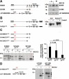
FIGURE 1.
FIGURE 1.
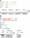
Loss of α-helical domains directs ER-targeted prion protein to proteasomal degradation in the cytosol. A, deletion of α-helical domains impairs ER import. Left panel, schematic presentation of the constructs. ER-SS, ER signal sequence; α, α-helical region; β, β-strand; CHO, N-linked glycosylation acceptor site; straight line, unstructured regions. Right panel, N2a cells were transiently transfected with the mutants depicted, and PrP present in the cell lysate (L) or in the cell culture medium (M) was analyzed by immunoblotting using the mAb 3F4. In addition, cells lysates were analyzed treated with or without the proteasomal inhibitor MG132 for 3 h prior to lysis (± MG132). B, an additional N-linked glycosylation acceptor site is functional. Left panel, schematic presentation of the constructs. GPI-SS, GPI signal sequence. The additional glycosylation site (CHO) at amino acid 31 in PrP/31CHO is marked in red. Right panel, N2a cells were transiently transfected with wild type PrP (PrP) or PrP/31CHO. Total cell lysates were either treated with the endoglycosidase H (+ EndoH) or left untreated (− EndoH), and PrP was detected by Western blotting. C and D, ER import efficiency correlates with the amount of α-helical domains. C, N2a cells transiently transfected with the constructs depicted in the upper panel were pulse-labeled (p) for 1 h with [35S]methionine and then chased (c) for 1 h in the presence (+ MG132) or absence (− MG132) of the proteasomal inhibitor MG132 (50 μm). PrP was immunoprecipitated using the mAb 3F4 and analyzed by SDS-PAGE. Quantification of three independent experiments is shown in the right panel. Data represent the ratio of glycosylated/unglycosylated PrP species present in the chase + MG132 (mean ± S.E.). p values were determined by Student's t test. D, α-helical domains are necessary and sufficient for ER import. N2a cells transiently transfected with the PrP mutants depicted in the left panel were metabolically labeled with [35S]methionine and then incubated in fresh medium for 1 h in the presence (+ MG132) or absence (− MG132) of MG132 (50 μm). PrP was immunoprecipitated using the mAb 3F4 and analyzed by SDS-PAGE. Open arrowheads represent unglycosylated PrP species; closed arrowheads represent glycosylated forms.
FIGURE 2.
FIGURE 2.
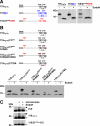
More efficient ER signal sequences cannot restore import of intrinsically disordered proteins. A, increase in polypeptide chain length could not promote translocation into the ER. Upper panel, schematic presentation of the proteins analyzed. The following domains were fused to PrP-115/31CHO: 115/31CHO+115, the unstructured domain of PrP (black); 115/31CHO+Tau, an unstructured domain of the Tau protein (green); and 115/31CHO+αsyn, an unstructured domain of α-synuclein (yellow). In addition we generated 31CHO+αsyn, a construct consisting of the ER signal sequence and the first 6 amino acids of PrP-115/31CHO, which mediates ER targeting and N-linked glycosylation, fused to the intrinsically disordered polypeptide derived from α-synuclein. Lower panel, N2a cells were transfected with the constructs depicted and grown for 3 h in the presence or absence of MG132 (± MG132). If indicated cell lysates were treated with EndoH (± EndoH). PrP was analyzed by Western blotting using the mAb 3F4. 31CHO+αsyn was analyzed using the anti-α-synuclein mAb42. B, efficient ER signal sequences do not restore ER import. The ER signal sequences from the yeast protein Cre5p (blue) or the rat growth hormone (GH; green) were fused to PrP or PrP-115/31CHO+115. Amino acid sequences of the ER-SS are depicted. Hydrophobic amino acids are marked in red. The panel shows a Kyte and Doolittle hydrophobicity plot of the signal sequences. Regions with values above 0 are hydrophobic in character. Lower panel, N2a cells were transiently transfected with the constructs indicated and analyzed by Western blot using the mAb 3F4. If indicated, cell lysates were treated with EndoH (± EndoH) prior to Western blotting.
FIGURE 3.
FIGURE 3.
ER import of unstructured domains is restored by increasing the content in α-helical domains. A and B, schematic presentation of the mutants analyzed. S-S, disulfide bond. The following domains were fused to PrP-115 or PrP-115/31CHO: 115α2α3, two α-helical domains of PrP (black); 115Dpl, two α-helical domains of Doppel (blue); and 115/31CHOGFR, two α-helical domains of GFR (red). A and B, N2a cells were transiently transfected with the mutants depicted. Cell lysates were either treated with EndoH (+ EndoH) or left untreated (− EndoH) and analyzed by Western blot using the mAb 3F4. C, α-helical but not unstructured domains restore in vitro translocation. PrP, 115α2α3, and 115/31CHO+115 were synthesized in vitro in the presence (+ microsomes) or absence (− microsomes) of ER-derived rough microsomes. If indicated, radioactively labeled products were treated with EndoH (± EndoH) before SDS-PAGE. Open arrowheads, unglycosylated PrP species; closed arrowheads, glycosylated PrP species.
FIGURE 4.
FIGURE 4.
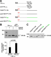
Polypeptides subjected to proteasomal degradation contain an uncleaved signal sequence. A, schematic presentation of the mutants analyzed. Two versions of the mutants 115α2α3, 115/31CHO+115, and 115/31CHO+Tau were generated, one version with the original ER-SS and one lacking the ER-SS (cyto forms). B, N2a cells were transiently transfected and incubated in the presence or absence of MG132 (30 μm; 3 h). In addition, cell lysates were either treated with EndoH (+ EndoH) or left untreated (− EndoH) prior to Western blotting using the mAb 3F4. Unglycosylated PrP with (+ ER-SS) and without (− ER-SS) the ER signal peptide are marked. Lower panel, quantification of at least three independent experiments. Plotted is the ratio of the amount of 115α2α3 with an uncleaved versus cleaved SS in EndoH-treated samples with or without proteasomal inhibition (± MG132) (mean ± S.E.). The p value was determined by Student's t test. C, N2a cells transfected with the mutants depicted were lysed, and proteins analyzed by Western blot using the mAb 3F4. The protein fraction with (+ ER-SS) and without (− ER-SS) the ER signal peptide is marked.
FIGURE 5.
FIGURE 5.
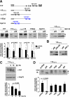
p58IPK promotes proteasomal degradation of ER-targeted polypeptides with extended unstructured domains at their N terminus. A, p58IPK promotes a preemptive/co-translocational quality control pathway. N2a cells were transiently co-transfected with the constructs indicated and p58IPK (p58) or a vector control (−). Right panel, cells co-transfected with p58IPK and PrP-115α2α3 were incubated in the presence or absence of MG132 (30 μm; 3 h). The lanes MG132+ are positioned directly next to the lanes MG132− although all lanes originate from one gel. B, p58IPK requires the ER signal peptide and J-domain to interfere with ER import of PrP-115α2α3. N2a cells were transiently co-transfected with PrP-115α2α3 and either p58IPK (p58), p58IPKΔSS (p58ΔSS), p58IPKΔJ (p58ΔJ), or a vector control (−). A and B, expression of PrP was analyzed by immunoblotting using the mAb 3F4; an open arrowhead marks the unglycosylated protein species; a closed arrowhead marks the glycosylated forms. C, reduced import under conditions of ER stress. N2a cells transfected with PrP-115α2α3 were incubated with thapsigargin (TG; 1 μm) for the time indicated and then analyzed by Western blot using the mAb 3F4 for detection of PrP and anti-Hsp70 mAb for detection of endogenous Hsp70 as loading control. D, overexpression of cytosolic Hsp70 or BiP does not interfere with ER import. N2a cells were transiently co-transfected with PrP-115α2α3 and the constructs indicated. Expression of PrP-115α2α3 was then analyzed by immunoblotting using the mAb 3F4; an open arrowhead marks the unglycosylated PrP species; a closed arrowhead marks the glycosylated forms. A, B, and C, quantifications were based on at least three independent experiments. Data were expressed as the ratio of glycosylated versus unglycosylated PrP (mean ± S.E.). Statistical analysis was performed using Student's t test. n.s., not significant.
FIGURE 6.
FIGURE 6.
A model for the co-translocational quality control of ER-targeted polypeptides containing extended unstructured domains. After targeting of the ribosome-nascent chain complex to the translocon via the N-terminal signal peptide (red), the translation of an extended unstructured domain delays productive translocation into the ER lumen. Instead the growing polypeptide chain is kept in a translocationally competent state inside or at the cytosolic side of the translocon (I). Depending on the folding state of the growing polypeptide chain two alternative pathways are conceivable. In case α-helical domains are synthesized, the polypeptide chain is efficiently imported and modified by the signal peptidase and the oligosaccharyltransferase (A). If the remaining part of the polypeptide is still devoid of α-helical domains productive ER import is not pursued, and the protein is disposed via the proteasome (B). The proteasome picture was adopted with permission from Walz et al. (54).

Similar articles

Cited by

References

    1. Pickart C. M. (2001) Annu. Rev. Biochem. 70, 503–533 - PubMed
    1. Hershko A., Ciechanover A. (1998) Annu. Rev. Biochem. 67, 425–479 - PubMed
    1. Ciechanover A., Orian A., Schwartz A. L. (2000) BioEssays 22, 442–451 - PubMed
    1. Nakatsukasa K., Brodsky J. L. (2008) Traffic 9, 861–870 - PMC - PubMed
    1. Ellgaard L., Helenius A. (2003) Nat. Rev. Mol. Cell Biol. 4, 181–191 - PubMed

Publication types

LinkOut - more resources