Review
. 2009:32:149-84.
doi: 10.1146/annurev.neuro.051508.135600.

The glial nature of embryonic and adult neural stem cells

Affiliations
Review

The glial nature of embryonic and adult neural stem cells

Arnold Kriegstein et al. Annu Rev Neurosci. 2009.

Abstract

Glial cells were long considered end products of neural differentiation, specialized supportive cells with an origin very different from that of neurons. New studies have shown that some glial cells--radial glia (RG) in development and specific subpopulations of astrocytes in adult mammals--function as primary progenitors or neural stem cells (NSCs). This is a fundamental departure from classical views separating neuronal and glial lineages early in development. Direct visualization of the behavior of NSCs and lineage-tracing studies reveal how neuronal lineages emerge. In development and in the adult brain, many neurons and glial cells are not the direct progeny of NSCs, but instead originate from transit amplifying, or intermediate, progenitor cells (IPCs). Within NSCs and IPCs, genetic programs unfold for generating the extraordinary diversity of cell types in the central nervous system. The timing in development and location of NSCs, a property tightly linked to their neuroepithelial origin, appear to be the key determinants of the types of neurons generated. Identification of NSCs and IPCs is critical to understand brain development and adult neurogenesis and to develop new strategies for brain repair.


Figures

Figure 1
Figure 1
Glial nature of neural stem cells (NSCs) in development and in the adult. Neuroepithelial cells in early development divide symmetrically to generate more neuroepithelial cells. Some neuroepithelial cells likely generate early neurons. As the developing brain epithelium thickens, neuroepithelial cells elongate and convert into radial glial (RG) cells. RG divide asymmetrically to generate neurons directly or indirectly through intermediate progenitor cells (nIPCs). Oligodendrocytes are also derived from RG through intermediate progenitor cells that generate oligodendrocytes (oIPCs). As the progeny from RG and IPCs move into the mantel for differentiation, the brain thickness, further elongating RG cells. Radial glia have apical-basal polarity: apically (down), RG contact the ventricle, where they project a single primary cilium; basally (up), RG contact the meninges, basal lamina, and blood vessels. At the end of embryonic development, most RG begin to detach from the apical side and convert into astrocytes while oIPC production continues. Production of astrocytes may also include some IPCs (see Figure 2) not illustrated here. A subpopulation of RG retain apical contact and continue functioning as NSCs in the neonate. These neonatal RG continue to generate neurons and oligodendrocytes through nIPCs and oIPCS; some convert into ependymal cells, whereas others convert into adult SVZ astrocytes (type B cells) that continue to function as NSCs in the adult. B cells maintain an epithelial organization with apical contact at the ventricle and basal endings in blood vessels. B cells continue to generate neurons and oligodendrocytes through (n and o) IPCs. This illustration depicts some of what is known for the developing and adult rodent brain. Timing and number of divisions likely vary from one species to another, but the general principles of NSC identity and lineages are likely to be preserved. Solid arrows are supported by experimental evidence; dashed arrows are hypothetical. Colors depict symmetric, asymmetric, or direct transformation. IPC, intermediate progenitor cell; MA, mantle; MZ, marginal zone; NE, neuroepithelium; nIPC, neurogenic progenitor cell; oIPC, oligodendrocytic progenitor cell; RG, radial glia; SVZ, subventricular zone; VZ, ventricular zone.
Figure 2
Figure 2
Lineage tree of NSCs. Purple to blue dots represent NSCs at different stages in development from neuroepithelial cells through early and late RG to adult NSCs (SVZ B cells and SGZ radial astrocytes). The derivation of embryonic progeny is depicted in the upper half, whereas the lower half shows lineages derived in the postnatal and adult brain. Solid lines indicate lineage conversions for which experimental evidence is available; dashed arrows are hypothetical. Intermediate progenitor cells (IPCs) for neurons (nIPCs), for oligodendrocytes (oIPCs), and for astrocytes (aIPCs) are indicated along each lineage leading to differentiated progeny. Note that in some instances this transit-amplifying step is bypassed. NSC, neural stem cell; RG, radial glia; SGZ: subgranular zone; SVZ, subventricular zone.
Figure 3
Figure 3
Three modes of neurogenesis during cortical development. RG in cortex generate neurons (a) directly through asymmetric division; (b) indirectly by generation of nIPCs and one round of amplification; or (c) indirectly again through nIPCs, but with two rounds of division and further amplification. This additional amplification stage may be fundamental to increase cortical size during evolution (see text). Subpopulations of nIPCs are likely to divide more than once in subcortical brain regions, but this has not yet been documented. For additional details, see Figure 1. CP, cortical plate; IZ, intermediate zone; MZ, marginal zone; nIPC, neurogenic intermediate progenitor cell; RG, radial glia; SVZ, subventricular zone; VZ, ventricular zone.
Figure 4
Figure 4
Regional specification of NSCs in the embryo and the adult brain. The left two panels show cross sections of the mouse forebrain from embryonic and adult brain showing four subdivisions of the VZ in different colors. On the right, RG in embryonic development (lower four panels) and type B cells in the adult brain (upper four panels) from the major forebrain subregions (septum, cortex, LGE, and MGE) are illustrated using the same colors as on the left. NSCs in the different subregions generate different types of neurons. Combinations of transcription factor expression (see text for additional details) define the different subdomains and the types of neurons produced. Representative camera lucida drawings of neuronal types produced within each subregion are illustrated. For example, neurons derived from cortical (green) RG in development give rise to large pyramidal neurons. In the adult, cortical progenitors give rise to olfactory bulb interneurons. NSCs in each subregion may give rise to an even wider diversity of cell types. Although in some cases (e.g., embryonic cortex) neuron progeny migrate radially, more frequently, immature neurons migrate tangentially for some distance before differentiating. Therefore, although mature neurons are shown deriving from distinct proliferative regions, we do not denote ultimate location, but rather that different neuronal phenotypes are derived from distinct pools of NSCs. LGE, lateral ganglionic eminence; MGE, medial ganglionic eminence; NSCs, neural stem cells; RG, radial glia; VZ, ventricular zone.
Figure 5
Figure 5
Schematic of progenitor types and lineages in the adult brain SVZ. NSCs in the wall of the lateral ventricles of adult rodents correspond to type B cells (SVZ astrocytes). These cells retain epithelial properties, including the extension of a thin apical process that ends on the ventricle and a basal process ending on blood vessels. B cells give rise to C cells, which correspond to nIPCs. B cells also generate oligodendrocytes through oIPCs. Dashed arrows illustrate hypothetical modes of division: blue for asymmetric and red for symmetric divisions. Investigators do not currently know how many times C cells divide. See Figure 1 for other information. nIPCs, neurogenic intermediate progenitor cells; NSCs, neural stem cells; oIPCs, oligodendrocytic intermediate progenitor cells; SVZ, subventricular zone.
Figure 6
Figure 6
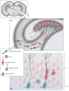
Schematic of progenitor types and lineages in the developing and adult brain dentate gyrus (DG) in the hippocampus. The subgranular zone (SGZ) is unique among forebrain germinal regions because it is detached from the walls of the ventricles. (a) Depicts a possible link between RG in the VZ facing the lateral ventricle (dentate neuroepithelium) and the developing SGZ. These RG migrate away from this region while they continue to generate nIPCs and early-born neurons. RG generate radial astrocytes, which become localized to the SGZ. These astrocytic cells have a prominent process (a, b) that traverses the dentate granule cell layer and branches in the deep molecular layer. (b) Radial astrocytes (also known as type I progenitors, see text) generate nIPCs (D cells or type II progenitors), which in turn generate young neurons. Young neurons remain tightly associated to radial processes of radial astrocytes before differentiating into granule cells. Dashed arrows indicate hypothetical symmetric (red) and asymmetric (blue) divisions. Black dashed arrows indicate hypothetical transformation. The inset in the upper left corner shows a cross section of the rodent forebrain to illustrate the location of the developing dentate gyrus in the hippocampus. nIPCs, neurogenic intermediate progenitor cells; RG, radial glia; SGZ, subgranular zone; VZ, ventricular zone.

Similar articles

Cited by

References

    1. Aaku-Saraste E, Oback B, Hellwig A, Huttner WB. Neuroepithelial cells downregulate their plasma membrane polarity prior to neural tube closure and neurogenesis. Mech Dev. 1997;69:71–81. - PubMed
    1. Aguirre A, Dupree JL, Mangin JM, Gallo V. A functional role for EGFR signaling in myelination and remyelination. Nat Neurosci. 2007;10:990–1002. - PubMed
    1. Altman J, Bayer SA. Horizontal compartmentation in the germinal matrices and intermediate zone of the embryonic rat cerebral cortex. Exp Neurol. 1990a;107:36–47. - PubMed
    1. Altman J, Bayer SA. Mosaic organization of the hippocampal neuroepithelium and the multiple germinal sources of dentate granule cells. J Comp Neurol. 1990b;301:325–42. - PubMed
    1. Altman J, Das GD. Autoradiographic and histological evidence of postnatal hippocampal neurogenesis in rats. J Comp Neurol. 1965;124:319–35. - PubMed

Publication types