SNX4 in complex with clathrin and dynein: implications for endosome movement
- PMID: 19529763
- PMCID: PMC2691479
- DOI: 10.1371/journal.pone.0005935
SNX4 in complex with clathrin and dynein: implications for endosome movement
Abstract
Background: Sorting nexins (SNXs) constitute a family of proteins classified by their phosphatidylinositol (PI) binding Phox homology (PX) domain. Some members regulate intracellular trafficking. We have here investigated mechanisms underlying SNX4 mediated endosome to Golgi transport.
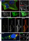
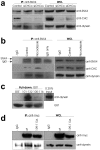
Methodology/principal findings: We show that SNX4 forms complexes with clathrin and dynein. The interactions were inhibited by wortmannin, a PI3-kinase inhibitor, suggesting that they form when SNX4 is associated with PI(3)P on endosomes. We further localized the clathrin interacting site on SNX4 to a clathrin box variant. A short peptide containing this motif was sufficient to pull down both clathrin and dynein. Knockdown studies demonstrated that clathrin is not required for the SNX4/dynein interaction. Moreover, clathrin knockdown led to increased Golgi transport of the toxin ricin, as well as redistribution of endosomes.
Conclusions/significance: We discuss the possibility of clathrin serving as a regulator of SNX4-dependent transport. Upon clathrin release, dynein may bind SNX4 and mediate retrograde movement.
Conflict of interest statement
Figures


Similar articles
-
Phosphoinositide-regulated retrograde transport of ricin: crosstalk between hVps34 and sorting nexins.Traffic. 2007 Mar;8(3):297-309. doi: 10.1111/j.1600-0854.2006.00527.x. Traffic. 2007. PMID: 17319803
-
SNX4 coordinates endosomal sorting of TfnR with dynein-mediated transport into the endocytic recycling compartment.Nat Cell Biol. 2007 Dec;9(12):1370-80. doi: 10.1038/ncb1656. Epub 2007 Nov 11. Nat Cell Biol. 2007. PMID: 17994011
-
Distinct complexes of yeast Snx4 family SNX-BARs mediate retrograde trafficking of Snc1 and Atg27.Traffic. 2017 Feb;18(2):134-144. doi: 10.1111/tra.12462. Epub 2017 Jan 16. Traffic. 2017. PMID: 28026081 Free PMC article.
-
Insights from yeast endosomes.Curr Opin Cell Biol. 2002 Aug;14(4):454-62. doi: 10.1016/s0955-0674(02)00352-6. Curr Opin Cell Biol. 2002. PMID: 12383796 Review.
-
Sorting nexins--unifying trends and new perspectives.Traffic. 2005 Feb;6(2):75-82. doi: 10.1111/j.1600-0854.2005.00260.x. Traffic. 2005. PMID: 15634208 Review.
Cited by
-
Clathrin is not required for SNX-BAR-retromer-mediated carrier formation.J Cell Sci. 2013 Jan 1;126(Pt 1):45-52. doi: 10.1242/jcs.112904. Epub 2012 Sep 26. J Cell Sci. 2013. PMID: 23015596 Free PMC article.
-
Regulation of endosomal clathrin and retromer-mediated endosome to Golgi retrograde transport by the J-domain protein RME-8.EMBO J. 2009 Nov 4;28(21):3290-302. doi: 10.1038/emboj.2009.272. Epub 2009 Sep 17. EMBO J. 2009. PMID: 19763082 Free PMC article.
-
A Daple-Akt feed-forward loop enhances noncanonical Wnt signals by compartmentalizing β-catenin.Mol Biol Cell. 2017 Dec 1;28(25):3709-3723. doi: 10.1091/mbc.E17-06-0405. Epub 2017 Oct 11. Mol Biol Cell. 2017. PMID: 29021338 Free PMC article.
-
Microtubule motors mediate endosomal sorting by maintaining functional domain organization.J Cell Sci. 2013 Jun 1;126(Pt 11):2493-501. doi: 10.1242/jcs.122317. Epub 2013 Apr 2. J Cell Sci. 2013. PMID: 23549789 Free PMC article.
-
CHC22 clathrin recruitment to the early secretory pathway requires two-site interaction with SNX5 and p115.EMBO J. 2024 Oct;43(19):4298-4323. doi: 10.1038/s44318-024-00198-y. Epub 2024 Aug 19. EMBO J. 2024. PMID: 39160272 Free PMC article.
References
Publication types
MeSH terms
Substances
LinkOut - more resources
Full Text Sources
Research Materials
Miscellaneous

