

Adenosine A(2A) receptor mediates microglial process retraction
- PMID: 19525944
- PMCID: PMC2712729
- DOI: 10.1038/nn.2341
Adenosine A(2A) receptor mediates microglial process retraction
Abstract
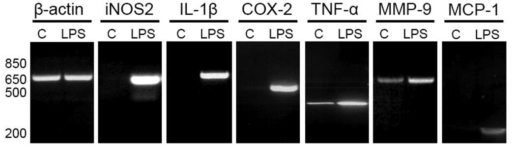
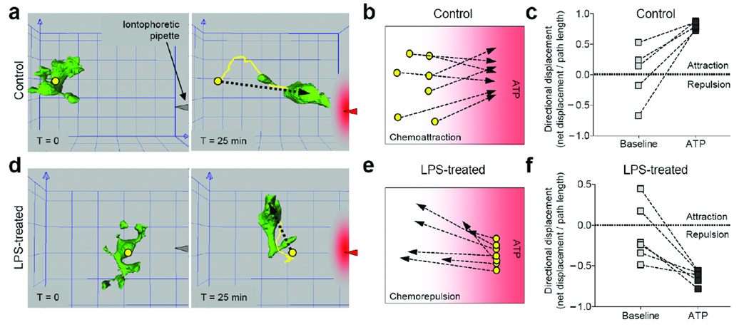
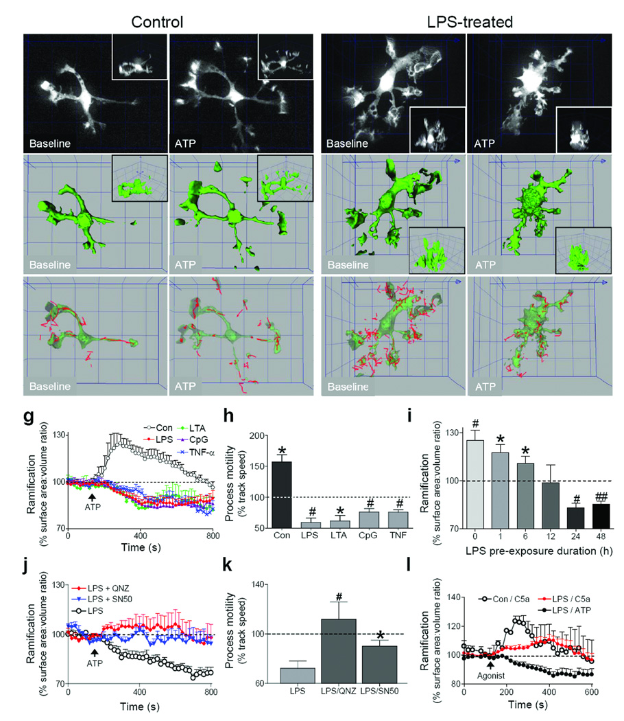
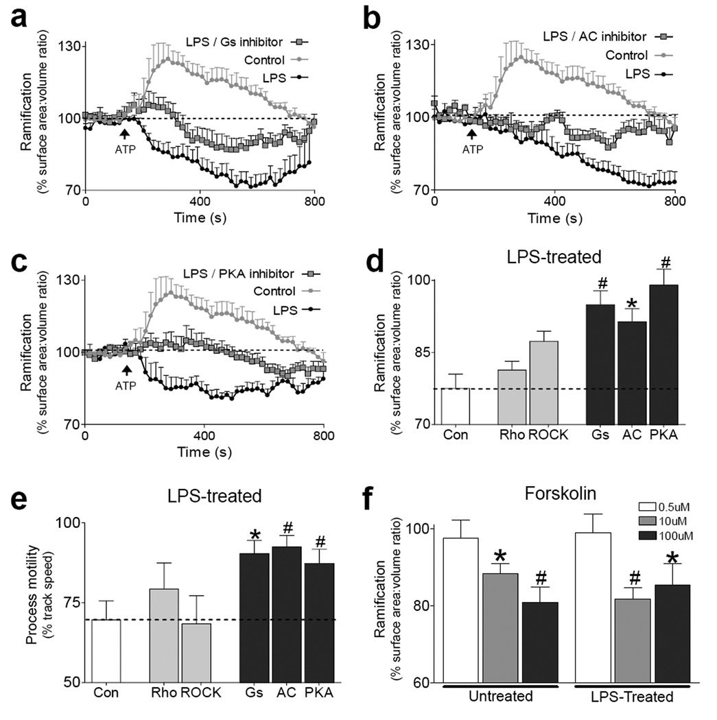
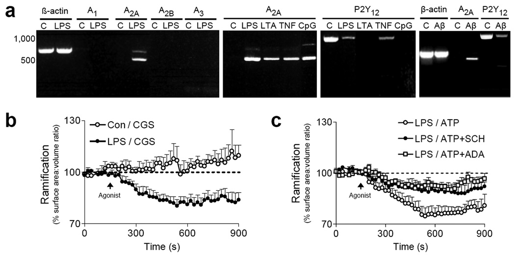
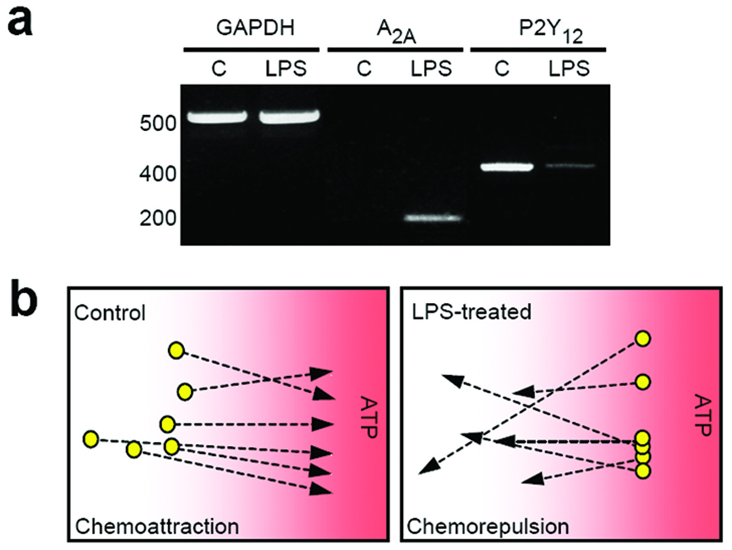
Cell motility drives many biological processes, including immune responses and embryonic development. In the brain, microglia are immune cells that survey and scavenge brain tissue using elaborate and motile cell processes. The motility of these processes is guided by the local release of chemoattractants. However, most microglial processes retract during prolonged brain injury or disease. This hallmark of brain inflammation remains unexplained. We identified a molecular pathway in mouse and human microglia that converted ATP-driven process extension into process retraction during inflammation. This chemotactic reversal was driven by upregulation of the A(2A) adenosine receptor coincident with P2Y(12) downregulation. Thus, A(2A) receptor stimulation by adenosine, a breakdown product of extracellular ATP, caused activated microglia to assume their characteristic amoeboid morphology during brain inflammation. Our results indicate that purine nucleotides provide an opportunity for context-dependent shifts in receptor signaling. Thus, we reveal an unexpected chemotactic switch that generates a hallmark feature of CNS inflammation.
Figures


Similar articles
-
Distinct P2Y Receptors Mediate Extension and Retraction of Microglial Processes in Epileptic and Peritumoral Human Tissue.J Neurosci. 2020 Feb 12;40(7):1373-1388. doi: 10.1523/JNEUROSCI.0218-19.2019. Epub 2020 Jan 2. J Neurosci. 2020. PMID: 31896671 Free PMC article.
-
Differential regulation of microglial motility by ATP/ADP and adenosine.Parkinsonism Relat Disord. 2009 Dec;15 Suppl 3(Suppl 3):S195-9. doi: 10.1016/S1353-8020(09)70813-2. Parkinsonism Relat Disord. 2009. PMID: 20082989 Free PMC article. Review.
-
ATP mediates rapid microglial response to local brain injury in vivo.Nat Neurosci. 2005 Jun;8(6):752-8. doi: 10.1038/nn1472. Epub 2005 May 15. Nat Neurosci. 2005. PMID: 15895084
-
The adenosine generating enzymes CD39/CD73 control microglial processes ramification in the mouse brain.PLoS One. 2017 Apr 4;12(4):e0175012. doi: 10.1371/journal.pone.0175012. eCollection 2017. PLoS One. 2017. PMID: 28376099 Free PMC article.
-
Purinergic signaling and microglia.Pflugers Arch. 2006 Aug;452(5):615-21. doi: 10.1007/s00424-006-0064-7. Epub 2006 Jun 21. Pflugers Arch. 2006. PMID: 16791619 Review.
Cited by
-
Purinergic Receptors of the Central Nervous System: Biology, PET Ligands, and Their Applications.Mol Imaging. 2020 Jan-Dec;19:1536012120927609. doi: 10.1177/1536012120927609. Mol Imaging. 2020. PMID: 32539522 Free PMC article. Review.
-
Alzheimer and Purinergic Signaling: Just a Matter of Inflammation?Cells. 2021 May 20;10(5):1267. doi: 10.3390/cells10051267. Cells. 2021. PMID: 34065393 Free PMC article. Review.
-
Established and emerging techniques for the study of microglia: visualization, depletion, and fate mapping.Front Cell Neurosci. 2024 Feb 15;18:1317125. doi: 10.3389/fncel.2024.1317125. eCollection 2024. Front Cell Neurosci. 2024. PMID: 38425429 Free PMC article. Review.
-
Age-Induced Spatial Memory Deficits in Rats Are Correlated with Specific Brain Region Alterations in Microglial Morphology and Gene Expression.J Neuroimmune Pharmacol. 2019 Jun;14(2):251-262. doi: 10.1007/s11481-018-9817-2. Epub 2018 Oct 20. J Neuroimmune Pharmacol. 2019. PMID: 30343448
-
Blockade of microglial adenosine A2A receptor suppresses elevated pressure-induced inflammation, oxidative stress, and cell death in retinal cells.Glia. 2019 May;67(5):896-914. doi: 10.1002/glia.23579. Epub 2019 Jan 22. Glia. 2019. PMID: 30667095 Free PMC article.
References
-
- Hanisch UK, Kettenmann H. Microglia: active sensor and versatile effector cells in the normal and pathologic brain. Nat Neurosci. 2007;10:1387–1394. - PubMed
-
- Kreutzberg GW. Microglia: a sensor for pathological events in the CNS. Trends Neurosci. 1996;19:312–318. - PubMed
-
- Nimmerjahn A, Kirchhoff F, Helmchen F. Resting microglial cells are highly dynamic surveillants of brain parenchyma in vivo. Science. 2005;308:1314–1318. - PubMed
-
- Davalos D, et al. ATP mediates rapid microglial response to local brain injury in vivo. Nat Neurosci. 2005;8:752–758. - PubMed
-
- Haynes SE, et al. The P2Y12 receptor regulates microglial activation by extracellular nucleotides. Nat Neurosci. 2006;9:1512–1519. - PubMed
Publication types
MeSH terms
Substances
Grants and funding
LinkOut - more resources
Full Text Sources
Other Literature Sources
Molecular Biology Databases
