. 2009 Jun 10;29(23):7459-73.
doi: 10.1523/JNEUROSCI.4872-08.2009.

Interaction of reelin with amyloid precursor protein promotes neurite outgrowth

Affiliations

Interaction of reelin with amyloid precursor protein promotes neurite outgrowth

Hyang-Sook Hoe et al. J Neurosci. .

Abstract

The processing of amyloid precursor protein (APP) to Abeta is an important event in the pathogenesis of Alzheimer's disease, but the physiological function of APP is not well understood. Our previous work has shown that APP processing and Abeta production are regulated by the extracellular matrix protein Reelin. In the present study, we examined whether Reelin interacts with APP, and the functional consequences of that interaction in vitro. Using coimmunoprecipitation, we found that Reelin interacted with APP through the central domain of Reelin (repeats 3-6) and the E1 extracellular domain of APP. Reelin increased cell surface levels of APP and decreased endocytosis of APP in hippocampal neurons in vitro. In vivo, Reelin levels were increased in brains of APP knock-out mice and decreased in APP-overexpressing mice. RNA interference knockdown of APP decreased neurite outgrowth in vitro and prevented Reelin from increasing neurite outgrowth. Knock-out of APP or Reelin decreased dendritic arborization in cortical neurons in vivo, and APP overexpression increased dendritic arborization. APP and Reelin have previously been shown to promote neurite outgrowth through interactions with integrins. We confirmed that APP interacted with alpha3beta1 integrin, and alpha3beta1 integrin altered APP trafficking and processing. Addition of an alpha3beta1 integrin antibody prevented APP and Reelin-induced neurite outgrowth. These findings demonstrate that Reelin interacts with APP, potentially having important effects on neurite development.


Figures

Figure 1.
Figure 1.
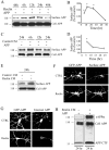
Reelin interacts with APP in primary cortical neurons and mouse brain. A, Mouse brain lysates (200 μg) were immunoprecipitated with 22C11 antibody against APP and probed with an anti-Reelin antibody (lanes 2 and 4). As a negative control, an irrelevant antibody (α-P-JNK) was included in the experiment (lanes 1 and 3). The asterisks denote antibody heavy and light chains. The bottom panels demonstrate total levels of APP and 300 kDa Reelin in the different cell lysates. B, Mouse brain lysates (200 μg) were immunoprecipitated with anti-Reelin antibody and probed with 22C11 antibody against APP (lane 2). C, Mouse brain lysates from wild-type and APP/APLP1 DKO were immunoprecipitated with anti-APP antibody and probed with anti-Reelin antibody. Interaction of APP with Reelin was detected in brain lysates from wild-type mice, but not from APP/APLP1 DKO mice. D, Primary cortical neuronal lysates were immunoprecipitated with anti-APP antibody and probed with the antibody against Reelin. Levels of APP or Reelin were measured in lysates from A–D. E, Cultured hippocampal neurons were fixed and immunostained at DIV 14 for APP and Reelin. Primary antibodies were detected with Alexa Flour 488 anti-rabbit antibody (in green; left panel) and Alexa Fluor 555 anti-mouse antibody (in red; middle panel). Immunolabeled neurons were imaged by confocal microscopy (63×). Colocalization of APP and Reelin appears as yellow in the right panel. F, Levels of both the 300 and 180 kDa forms of Reelin were detected using Western blot analyses with G10 antibody and compared between APP-deficient mice (n = 5) and wild-type mice (n = 5). Increased levels of the 300 and 180 kDa (top panel) were observed in the brain lysates from APP knock-out mice in comparison with wild-type mice for APP−/−. Additional Western blot analysis confirmed these observations for the 180 kDa (middle panel) and 300 kDa (data not shown). G, Levels of 300 and 180 kDa forms of Reelin were compared between APP Tg2576 mice (n = 3) and wild-type controls for APP Tg2576 mice (n = 3). H, Quantification of the levels of the Reelin 180 kDa band in four different Western blots (2 of which are shown in F and G) show that Reelin was increased in APP knock-out mice (n = 5; by 133%; *p < 0.01) and decreased in APP Tg 2576 mice (n = 3; by 87%; *p < 0.01). Error bars indicate SEM. I, Levels of APP, Reelin, and β1-integrin in mouse brain were examined by Western blots at various developmental stages (n = 3 at each age). APP was increased between P1 and P36, a critical period for development of neuronal processes and synaptogenesis. Levels of Reelin were also highest between P1 and P10.
Figure 2.
Figure 2.
The Reelin 3–6 domain interacts with the APP E1 domain. A, Schematic diagrams of Reelin constructs, with rectangles indicating signal peptides and circles indicating myc tags. B, Western blot analysis showing comparable expression levels of different Reelin constructs. C, COS7 cells were transfected with plasmids expressing different Reelin C-terminal constructs (indicated along the top of the panel) and APP. Cell lysates (200 μg) were immunoprecipitated with an anti-6E10 antibody and probed with an anti-myc antibody. Reelin constructs containing 3–6 domains coimmunoprecipitated with APP (constructs R1–R4), whereas the Reelin construct containing the 7–8 domains (construct R5) did not. The APP immunoblotting (bottom panel) showed similar expression levels of APP in all transfected cells. D, Schematic diagram of APP constructs. E, Western blot analysis with an anti-myc antibody exhibiting expression levels of individual APP constructs. F, COS7 cells were transfected with different APP constructs (indicated along the top of the film) and an N-terminal construct of Reelin (construct R6). Cell lysates (200 μg) were immunoprecipitated with anti-G10 and probed with anti-myc antibody. Reelin was immunoprecipitated with APP constructs containing the extracellular domain (constructs A1 and A4), but not with APP construct containing 430–770 aa (construct A2). G, COS7 cells were transfected with different APP constructs (indicated along the top of the film) and an N-terminal construct of Reelin (construct R6). Cell lysates (200 μg) were immunoprecipitated with anti-myc and probed with G10 antibody. Reelin was immunoprecipitated with full-length APP and the construct containing the extracellular domain of APP (constructs A1 and A4), but not with the construct containing intracellular domain of APP (construct A3). The Reelin immunoblot (bottom panel) showed comparable expression levels of Reelin in all transfected cells. H, Western blot with anti-myc antibody demonstrating expression levels of APP E1 and APP E2 domain. I, COS7 cells were cotransfected with N-terminal construct of Reelin (construct R6) and either APP E1 domain or APP E2 domain. Cell lysates (200 μg) were immunoprecipitated with G10 and probed with anti-myc antibody. Coimmunoprecipitation of Reelin was only detected when transfected with the construct containing the APP E1 domain (construct A5), but not with APP E2 domain (construct A6). The Reelin immunoblot (bottom panel) showed similar expression levels of Reelin in all transfection conditions. The asterisks shown in F, G, and I indicate IgG heavy and light chains.
Figure 3.
Figure 3.
Reelin regulates APP trafficking and processing. A, COS7 cells expressing APP were treated with Reelin-containing medium for indicated times (6, 12, 24, or 48 h). Cell surface proteins were biotinylated, isolated with avidin-conjugated beads, and immunoblotted with 6E10 antibody against APP. Total APP in cell lysates were measured in the bottom panel. B, Quantification of surface APP by Reelin in COS7 cells. Cell surface APP levels significantly increased after 6, 12, and 24 h of Reelin treatment (n = 3; by 39, 77, 69%; *p < 0.05). C, COS7 cells expressing APP were treated with control conditioned medium for indicated times (6, 12, or 24). Cell surface proteins were biotinylated, isolated with avidin-conjugated beads, and immunoblotted with 6E10 antibody against APP. Total APP in cell lysates were measured in the bottom panel. Error bars indicate SEM. D, Quantification of surface APP by control conditioned medium in COS7 cells. Cell surface APP levels were significantly increased after 6 h of control treatment (n = 4; by 36%; *p < 0.05), but not at 12 or 24 h. E, Cultured cortical neurons were treated with control or Reelin-containing medium for 24 h and analyzed for surface APP as in A. Cell surface proteins were labeled with biotin, isolated with avidin-conjugated beads, and immunoblotted with 22C11 antibody against APP. F, Cultured hippocampal neurons were transfected at DIV 12 with GFP-APP, treated with Reelin for 24 h, and stained with an antibody recognizing the N terminus of APP under impermeable conditions. Left panels, GFP-APP; right panels, surface APP (n = 8–10). G, Neurons were transfected with GFP-APP and treated with or without Reelin-containing medium for 24 h. After labeling cell surface APP, internalization was measured after 30 min with Alexa Fluor 555 anti-rabbit antibody (right) (n = 8–10). H, COS7 cells were transfected with APP and treated with control or Reelin-containing medium for 24 h. Secreted APP was measured in conditioned medium (15 μl) with antibody 6E10; APP CTF was detected from cell lysates (20 μg) with antibody c1/6.1.
Figure 4.
Figure 4.
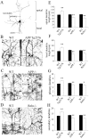
Interaction of APP with Reelin promotes neurite outgrowth. A, Neurons were infected with lentiviral constructs expressing GFP, human APP, or rat APP shRNA. After 24 h, cells were treated with control (top panels; n = 240, n = 191, n = 167) or Reelin (bottom panels; n = 218, n = 199, n = 199) and imaged after 24 h. B, C, Using Scholl analysis of cells in A, we graphed the average intersections per shell per neuron against the distance from the soma (in micrometers). D, E, The average actual neurite length for primary and secondary dendrites for each group from A. Results were pooled from 3 to 10 sets of cultures, and each culture included 20 random fields containing 5–8 cells/field. *p < 0.05. Error bars indicate SEM. F, Primary cortical neurons were infected with GFP, APP, APP shRNA, or APP and APP shRNA together and immunoblotted with c1/6.1, which recognizes both rat and human APP. shAPP only decreased rat APP levels (lane 4) but did not reduce human APP levels (lane 3). G, Neurons were infected with GFP (n = 87), APP (n = 69), APP shRNA (n = 55), or APP and APP shRNA together (n = 91), and then neurite lengths were measured. H, Averaged data include primary neurite lengths from each group. Results were pooled from 3 to 10 sets of cultures, and each culture included 20 random fields containing 5–8 cells per field. *p < 0.05.
Figure 5.
Figure 5.
Dendritic numbers and lengths are increased in APP transgenic mice and decreased in APP knock-out and Reeler mice. Mouse brains were Golgi stained and cortical neurons (layers II/III) imaged. A, Three-dimensional graphical tracing representing dendrite morphology. The numbers of primary and secondary dendrites were counted; the lengths of apical and basal dendrites were measured in micrometers. B–D, Representative Golgi impregnations for each condition. Scale bar, 100 μm. E, Averaged data include apical dendritic length from 3- to 4-week-old wild type, APP Tg2576, APP knock-out, and Reeler knock-out mice, analyzing 23–25 neurons from each. Error bars indicate SEM. F, Averaged data include basal dendritic length from each group, analyzing 23–25 neurons. G, Averaged data include number of primary dendrites from each group, analyzing 23–25 neurons. H, Averaged data include number of secondary dendrites from each group, analyzing 23–25 neurons. *p < 0.05; **p < 0.01.
Figure 6.
Figure 6.
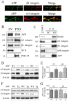
APP interacts with α3 and β1 integrin. A, Primary cortical neurons were fixed and immunostained at DIV 5 with c1/6.1 and anti-β1 or anti-α3 antibody. The antibodies were detected with Alexa Fluor 488 anti-mouse antibody (left panel) and Alexa Fluor 594 anti-rabbit antibody (middle panel). The neurons were observed under a confocal laser-scanning microscope (40×). Colocalization of APP and β1 integrin or α3 integrin is shown in the right panel. B, We performed immunoblot analysis of APP, α3 integrin, β1 integrin, synaptophysin, and PSD-95 in presynaptic vesicles (SV) and postsynaptic density (PSD) fractions. APP, α3 integrin, and β1 integrin were present in the presynaptic and postsynaptic fractions; PSD-95 was present in the postsynaptic fractions; synaptophysin was present presynaptically. C, Mouse brain lysates (300 μg) were immunoprecipitated with c1/6.1 and probed with G10, α3 integrin, β1 integrin, αM integrin, and APP. Interaction of APP with Reelin, α3 integrin, and β1 integrin was detected. D, Levels of α3 and β1 integrin were measured by Western blot analysis with α3 or β1 integrin antibody and compared between APP-deficient mice (n = 3) and wild-type mice (n = 3). E, Quantification of the levels of α3 and β1 integrins shown in D, demonstrating that both α3 and β1 integrins were significantly increased in APP knock-out mice, respectively (n = 3; by 59 and by 83%; *p < 0.05). Error bars indicate SEM. F, Protein levels of α3 and β1 integrins were analyzed and compared between APP Tg2576 mice (n = 3) and wild-type controls (n = 3). G, Quantification of the levels of α3 or β1 integrin shown in F, demonstrating that β1 integrin was significantly decreased in APP Tg2576 mice (n = 3; by 48%; *p < 0.01) but that α3 integrin was unaltered.
Figure 7.
Figure 7.
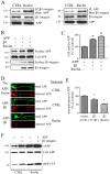
Reelin increases coprecipitation between APP and β1 integrin in primary neurons. A, Primary neuronal lysates (1 mg) were treated with control or Reelin-containing medium for 24 h and immunoprecipitated with a β1 integrin antibody and probed with c1/6.1 antibody (left panel). Inversely, primary neurons (1 mg) were immunoprecipitated with c1/6.1 and probed with anti-β1 integrin antibody (right panel). B, COS7 cells expressing APP or both APP and β1 integrin were treated with control or Reelin-containing medium for 24 h. Cell surface proteins were labeled with biotin, isolated with avidin beads, and immunoblotted with 6E10 for APP and anti-HA for β1 integrin (n = 3). C, Quantification of surface APP by Reelin in the presence of β1 integrin in COS7 cells. Cell surface levels of APP were significantly increased when COS7 cells were cotransfected with β1 integrin (n = 5; by 63%; *p < 0.05) and further by cotreatment with Reelin (n = 5; by 89%; *p < 0.05). Error bars indicate SEM. D, Primary hippocampal neurons were transfected with GFP-APP and vector or GFP-APP and β1 integrin and treated with control or Reelin-containing medium for 24 h. Internalized APP was labeled after 30 min with Alexa Fluor 555 anti-rabbit antibody. E, Quantitative analysis showed significant 29% (*p < 0.05; n = 6) and 47% (*p < 0.01; n = 6) decreases in APP internalization by β1 integrin or β1 integrin combined with Reelin, respectively. F, COS7 cells were transfected with APP and vector or APP and β1 integrin and treated with control or Reelin-containing medium for 24 h. Secreted APP was measured in conditioned media (15 μl) with antibody 6E10; APP CTF was detected from cell lysates (20 μg) with antibody c1/6.1 (n = 3). Reelin alone increased APP proteolytic fragments, and Reelin and β1 integrin together further increased them.
Figure 8.
Figure 8.
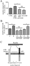
The effect of Reelin and APP on neurite outgrowth requires integrins. A, Hippocampal neurons were infected with lentiviral vectors expressing GFP (n = 163) or APP (n = 201) and treated with α3β1 antibody (n = 193). Average length of total dendrites was quantified. Results were pooled from 3 to 10 sets of cultures. Twenty random fields containing 5–8 neurons per field were taken from each culture (*p < 0.05). Error bars indicate SEM. B, Neurons were incubated in control (n = 100) or Reelin-containing medium (n = 100) and either anti-α3β1 antibody (control, n = 203; Reelin, n = 203) or anti-α5β1 antibody (control, n = 199; Reelin, n = 208) was added to the media. The length of dendritic neurites was quantified. The presence of the α3β1 integrin antibody significantly decreased neurite outgrowth and prevented the effect of Reelin on neurite outgrowth. C, A model of APP, Reelin, and α3β1 integrin interactions. Reelin repeats 1 and 2 interact with α3β1 integrin subunits, and the Reelin 3–6 domain interacts with APP E1 domain. The Reelin–α3β1 integrin and integrin–APP interaction may lead to reorganization of the cytoskeleton, or it may stabilize the actin cytoskeleton by inducing n-cofilin phosphorylation.

Similar articles

Cited by

References

    1. Alvarez VA, Ridenour DA, Sabatini BL. Retraction of synapses and dendritic spines induced by off-target effects of RNA interference. J Neurosci. 2006;26:7820–7825. - PMC - PubMed
    1. Andrade N, Komnenovic V, Blake SM, Jossin Y, Howell B, Goffinet A, Schneider WJ, Nimpf J. ApoER2/VLDL receptor and Dab1 in the rostral migratory stream function in postnatal neuronal migration independently of Reelin. Proc Natl Acad Sci U S A. 2007;104:8508–8513. - PMC - PubMed
    1. Andressen C, Arnhold S, Puschmann M, Bloch W, Hescheler J, Fässler R, Addicks K. Beta1 integrin deficiency impairs migration and differentiation of mouse embryonic stem cell derived neurons. Neurosci Lett. 1998;251:165–168. - PubMed
    1. Beffert U, Weeber EJ, Durudas A, Qiu S, Masiulis I, Sweatt JD, Li WP, Adelmann G, Frotscher M, Hammer RE, Herz J. Modulation of synaptic plasticity and memory by Reelin involves differential splicing of the lipoprotein receptor Apoer2. Neuron. 2005;47:567–579. - PubMed
    1. Botella-López A, Burgaya F, Gavín R, García-Ayllón MS, Gómez-Tortosa E, Peña-Casanova J, Ureña JM, Del Río JA, Blesa R, Soriano E, Sáez-Valero J. Reelin expression and glycosylation patterns are altered in Alzheimer's disease. Proc Natl Acad Sci U S A. 2006;103:5573–5578. - PMC - PubMed

Publication types

MeSH terms

LinkOut - more resources