. 2009 Jun 15;122(Pt 12):1970-8.
doi: 10.1242/jcs.044743.

Inactivation of the C. elegans lipin homolog leads to ER disorganization and to defects in the breakdown and reassembly of the nuclear envelope

Affiliations

Inactivation of the C. elegans lipin homolog leads to ER disorganization and to defects in the breakdown and reassembly of the nuclear envelope

Andy Golden et al. J Cell Sci. .

Abstract

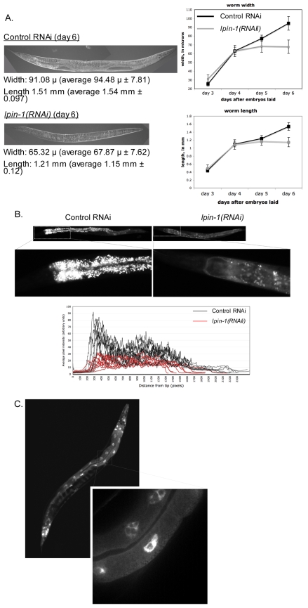
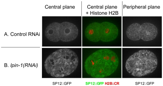
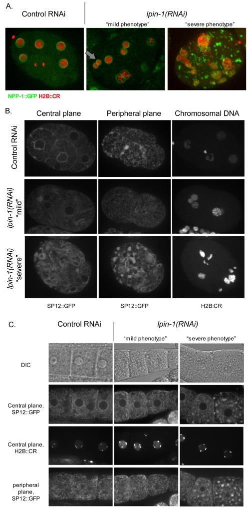
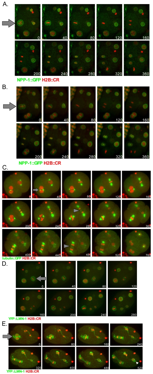
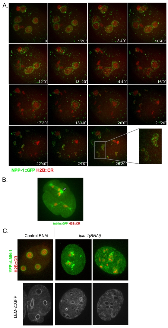
The nuclear envelope (NE) is a dynamic structure, undergoing periods of growth, breakdown and reassembly during the cell cycle. In yeast, altering lipid synthesis by inactivating the yeast homolog of lipin, a phosphatidic acid phosphohydrolase, leads to disorganization of the peripheral ER and abnormal nuclear shape. These results suggest that lipid metabolism contributes to NE dynamics; however, since yeast undergo closed mitosis, the relevance of these observations to higher eukaryotes is unclear. In mammals, lipin has been implicated in adipose tissue differentiation, insulin resistance, lipid storage and obesity, but the underlying cellular defects caused by altering lipin levels are not known. Here, we identify the Caenorhabditis elegans lipin homolog (LPIN-1) and examine its affect on NE dynamics. We find that downregulating LPIN-1 by RNAi results in the appearance of membrane sheets and other abnormal structures in the peripheral ER. Moreover, lpin-1 RNAi causes defects in NE breakdown, abnormal chromosome segregation and irregular nuclear morphology. These results uncover cellular processes affected by lipin in metazoa, and suggest that lipid synthesis has a role in NE dynamics.


Figures

Fig. 1.
Fig. 1.
RNAi of the C. elegans homolog of lipin results in reduced body size and defects in lipid storage. (A) Adult wild-type (N2) worms laid embryos for 1 hour on plates seeded with bacteria expressing either control RNAi or lpin-1(RNAi). The adult worms were then removed and the embryos were followed for the next 6 days. Each day, the length and width of at least 20 worms from the control plates (black line) or lpin-1(RNAi) plates (gray line) was determined as described in the Materials and Methods. A typical worm from each treatment (taken at day 6 after the embryos were laid) is shown. (B) Adult worms were placed on control or lpin-1(RNAi) plates as described above, except that the bacteria were mixed with Nile red (Ashrafi et al., 2003). Worms were imaged daily. Examples of a typical worm from each condition, taken on day 6, are shown. The bright white dots are lipid droplets that accumulated Nile red. The boxed areas are enlarged below. To quantify the Nile red fluorescence, average pixel intensity as a function of distance from the tip of the worm's pharynx (on the left of the graph) was determined for eight worms of each condition from day 6. Note that the average pixel intensity for control RNAi (black traces) are greater than the average pixel intensity for worms that hatched on lpin-1(RNAi), indicating the downregulation of LPIN-1 disrupted the accumulation of lipid droplets. (C) An adult worm expressing GFP::LPIN-1. A small region showing the pattern of GFP::LPIN-1 in the gut is enlarged.
Fig. 2.
Fig. 2.
lpin-1(RNAi) disrupts ER morphology. Embryos from L4-staged worms (strain OCF5) expressing SP12::GFP (green in the merged panel) and histone H2B::CR (red in the merged panel) that were grown on bacteria expressing either control dsRNA (panel A) or lpin-1 dsRNA (panel B) were imaged after 48 hours using confocal microscopy. Individual sections from either a central plane (left and middle columns) or a peripheral plane (right column) are shown.
Fig. 3.
Fig. 3.
lpin-1(RNAi) disrupts nuclear morphology. (A) L4-staged worms (strain OCF3) expressing NPP-1::GFP (green) and histone H2B::CR (red) were grown on bacteria expressing either control dsRNA (left) or lpin-1 dsRNA (middle and right) as described for Fig. 2, and embryos were imaged after 36-48 hours. Shown are merged images from multiple focal planes spanning each embryo. Note that in the case of embryos exhibiting a mild phenotype, some cells can have paired nuclei (gray arrow) whereas others can have a normal nuclear morphology. (B) Comparison of ER and nuclear morphologies following lpin-1(RNAi) treatment: worms expressing SP12::GFP and H2B::CR treated as described in A. For ER morphology (as detected by SP12::GFP), images were taken at a central plane (left column) or a peripheral plane (right column). To visualize the chromosomal DNA, multiple stacks traversing the embryo were merged, to visualize the total distribution of DNA. (C) L4-staged worms (strain OCF5) expressing SP12::GFP and histone H2B::CR grown on bacteria expressing either control dsRNA or lpin-1 dsRNA for 36-48 hours. Oocytes were imaged by confocal microscopy at either a central plane or a peripheral plane, as indicated. The designation of mild vs severe phenotypes was made based on the phenotype of the embryos from the same worms (not shown).
Fig. 4.
Fig. 4.
Nuclei exhibiting the lpin-1(RNAi)-induced mild phenotype give rise to paired nuclei following mitosis because of defects in NEBD. (A) L4-staged worms (strain OCF3) expressing NPP-1::GFP (green) and histone H2B::CR (red) grown on bacteria expressing control dsRNA for 48 hours. Live imaging of a four-cell-stage embryo from such a worm using confocal microscopy. Merged images of multiple focal planes from the indicated time point (in seconds) are shown. The nucleus labeled with an arrow is in prometaphase at time 0 seconds, in metaphase at 80 seconds, and in anaphase by 160 seconds. (B) L4-staged worms (strain OCF3) expressing NPP-1::GFP (green) and histone H2B::CR (red) were grown on bacteria expressing lpin-1 dsRNA for 36 hours and a four-cell embryo with cells exhibiting a mild phenotype (for example, see arrow) was imaged at the indicated time points, as described above. The timing of prometaphase, metaphase and anaphase is similar to that shown in A. (C) L4-staged worms (strain OCF2) expressing tubulin::GFP (green) and histone H2B::CR (red) were grown on bacteria expressing lpin-1 dsRNA for 36 hours and embryos were taken for live imaging as described above. In the example shown, a two-cell-stage embryo contains one cell with a normal nucleus (top right) and one cell with paired nuclei (bottom left). Chromosome segregation in the normal nucleus precedes that of the paired nuclei. In the case of the paired nuclei, the spindle poles align on both sides of the interface between the two DNA clusters before spindle elongation (arrow). Note the greater width of the spindle in the cell that has paired nuclei (arrowhead, time 480 seconds) compared with the spindle in the cell that has a single nucleus (arrowhead, time 280 seconds). (D,E) L4-staged worms (strain OCF4) expressing YFP::LMN-1 (green) and histone H2B::CR (red) were grown on bacteria expressing either control dsRNA (D) or lpin-1 dsRNA (E) for 24 hours at 24°C, and embryos from these worms were imaged by live microscopy as described above. In the case of the control RNAi, the nuclear lamina is at the nuclear periphery; it dissociates at the 160 second time point, during the early stages of anaphase. By contrast, in the case of lpin-1(RNAi) the lamina is present at the periphery of each of the paired nuclei, and is also hanging off the edges of the interface between the two nuclei (time point 520 seconds, arrowhead). As mitosis progresses, the nuclear lamina persists, and it is stretched as the distance between the segregating chromosomes increases. Note that the nuclear lamina appears to separate the DNA that had originated from each of the paired nuclei, presumably preventing their mixing.
Fig. 5.
Fig. 5.
Nuclei exhibiting the lpin-1(RNAi)-induced severe phenotype are defective in chromosome segregation and NE assembly. (A) L4-staged worms (strain OCF3) expressing NPP-1::GFP (green) and histone H2B::CR (red) were grown on bacteria expressing lpin-1 dsRNA for 48 hours. Live images of embryos were taken at the indicated time points. Note the unusual movement of the nuclei before chromosome segregation (time points 0 through 10'40”) and the abnormal movement of chromosomes during mitosis (from 17'20” to the end of the time course). Also note that the NPP-1::GFP foci in the cytoplasm disappear at the same time as those that are in the NE, around the nucleus (for example, time 20'00”). The last panel is an enlargement of the boxed area in time point 25'20”. (B) Image of an embryo expressing tubulin::GFP and histone H2B::CR (strain OCF2) from a worm treated with lpin-1(RNAi) as described above. The arrow indicates a spindle pole that nucleates two bundles of microtubules associated with two distinct DNA masses. (C) Worms expressing YFP::LMN-1 (green) and H2B::CR (red) (strain OCF4, top row), or LEM-2::GFP (bottom row), were treated with either control RNAi or lpin-1(RNAi), as described above. Shown are examples of embryos exhibiting severe phenotypes [in the case of lpin-1(RNAi)].

Similar articles

Cited by

References

    1. Anderson, D. J. and Hetzer, M. W. (2007). Nuclear envelope formation by chromatin-mediated reorganization of the endoplasmic reticulum. Nat. Cell Biol. 9, 1160-1166. - PubMed
    1. Anderson, D. J. and Hetzer, M. W. (2008). Reshaping of the endoplasmic reticulum limits the rate for nuclear envelope formation. J. Cell Biol. 182, 911-924. - PMC - PubMed
    1. Ashrafi, K., Chang, F. Y., Watts, J. L., Fraser, A. G., Kamath, R. S., Ahringer, J. and Ruvkun, G. (2003). Genome-wide RNAi analysis of Caenorhabditis elegans fat regulatory genes. Nature 421, 268-272. - PubMed
    1. Audhya, A., Desai, A. and Oegema, K. (2007). A role for Rab5 in structuring the endoplasmic reticulum. J. Cell Biol. 178, 43-56. - PMC - PubMed
    1. Campbell, J. L., Lorenz, A., Witkin, K. L., Hays, T., Loidl, J. and Cohen-Fix, O. (2006). Yeast nuclear envelope subdomains with distinct abilities to resist membrane expansion. Mol. Biol. Cell 17, 1768-1778. - PMC - PubMed

Publication types

MeSH terms

Substances