. 2009 Aug;83(16):7909-30.
doi: 10.1128/JVI.02541-08. Epub 2009 Jun 3.

3' cis-acting elements that contribute to the competence and efficiency of Japanese encephalitis virus genome replication: functional importance of sequence duplications, deletions, and substitutions

Affiliations

3' cis-acting elements that contribute to the competence and efficiency of Japanese encephalitis virus genome replication: functional importance of sequence duplications, deletions, and substitutions

Sang-Im Yun et al. J Virol. 2009 Aug.

Abstract

The positive-strand RNA genome of Japanese encephalitis virus (JEV) terminates in a highly conserved 3'-noncoding region (3'NCR) of six domains (V, X, I, II-1, II-2, and III in the 5'-to-3' direction). By manipulating the JEV genomic RNA, we have identified important roles for RNA elements present within the 574-nucleotide 3'NCR in viral replication. The two 3'-proximal domains (II-2 and III) were sufficient for RNA replication and virus production, whereas the remaining four (V, X, I, and II-1) were dispensable for RNA replication competence but required for maximal replication efficiency. Surprisingly, a lethal mutant lacking all of the 3'NCR except domain III regained viability through pseudoreversion by duplicating an 83-nucleotide sequence from the 3'-terminal region of the viral open reading frame. Also, two viable mutants displayed severe genetic instability; these two mutants rapidly developed 12 point mutations in domain II-2 in the mutant lacking domains V, X, I, and II-1 and showed the duplication of seven upstream sequences of various sizes at the junction between domains II-1 and II-2 in the mutant lacking domains V, X, and I. In all cases, the introduction of these spontaneous mutations led to an increase in RNA production that paralleled the level of protein accumulation and virus yield. Interestingly, the mutant lacking domains V, X, I, and II-1 was able to replicate in hamster BHK-21 and human neuroblastoma SH-SY5Y cells but not in mosquito C6/36 cells, indicating a cell type-specific restriction of its viral replication. Thus, our findings provide the basis for a detailed map of the 3' cis-acting elements in JEV genomic RNA, which play an essential role in viral replication. They also provide experimental evidence for the function of 3' direct repeat sequences and suggest possible mechanisms for the emergence of these sequences in the 3'NCR of JEV and perhaps in other flaviviruses.


Figures
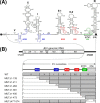
FIG. 1.
FIG. 1.
Schematic presentation of JEV 3′NCR mutants constructed in the context of a full-length infectious JEV cDNA molecular clone. (A) Detailed view of short CSs and predicted secondary structures within the 574-nt 3′NCR of JEV genomic RNA (30, 52, 55, 56, 68) from the translation stop codon (UAG; box outlined with a dotted line) of the single ORF to the 3′ end of the genome. Nucleotides are numbered from the first base of the 3′NCR, excluding the translation stop codon, toward its 3′ end. The 574-nt JEV 3′NCR was divided into six domains (55): V, nt 1 to 133; X, nt 134 to 211; I, nt 212 to 284; II-1, nt 285 to 381; II-2, nt 382 to 450; and III, nt 451 to 574, as ordered in the 5′ to 3′ direction. Highlighted in color are the five ∼20- to 30-nt CS or RCS motifs originally described by Hahn et al. (30): CS1, CS2, RCS2, CS3, and RCS3. Also indicated at the 3′ end of the genome are two well-documented RNA motifs, the 3′CYC (8-nt core sequence; box outlined with a solid line) embedded in the CS1 motif and the ∼90-nt 3′SL. (B) Schematic diagram of JEV 3′NCR deletion mutants. Shown at the top is the organization of the ∼11,000-nt JEV genomic RNA; viral proteins are indicated with the thick solid lines at both termini representing the 5′NCR and 3′NCR of the viral genome. Shown below is an expanded view of the six domains of the 574-nt JEV 3′NCR, in conjunction with the relative location of the five CSs (CSs or RCSs), 3′CYC, and 3′SL. Shown at the bottom are seven JEV 3′NCR deletion mutants, representing 5′- or 3′-truncated versions of various lengths within the 3′NCR, compared to full-length infectious JEV cDNA (WT). The designations of the seven mutants indicate the regions deleted from each of the individual constructs (shown by dotted lines).
FIG. 2.
FIG. 2.
Functional analysis of JEV 3′NCR mutants. Naïve BHK-21 cells were mock transfected (M) or transfected with 2 μg of the RNA transcripts synthesized from WT cDNA or each of the seven JEV 3′NCR mutant cDNAs. (A) Specific infectivity and representative focus (or plaque) morphology. The specific infectivities of RNA transcripts were determined by infectious center assays, as described in Materials and Methods. At 4 days after transfection, the cell monolayers were first immunostained with a mouse anti-JEV antiserum (representative foci) and then restained with crystal violet (representative plaques). RNA infectivities are given as FFU/microgram of RNA. The number of plus signs indicates the magnitude of the cytopathic effect produced by the corresponding mutant RNAs relative to that induced by the WT RNA; a minus sign indicates no cytopathic effect. N.D, not detectable. (B) Production of JEV RNA. Equal amounts of total cellular RNAs extracted at 6 and 22 h posttransfection (hpt) were used for real-time quantitative RT-PCR with a JEV-specific probe complementary to a sequence in the NS3 protein-coding region; a β-actin RNA-specific probe was used to normalize total RNA levels. Changes in JEV RNA levels in real-time quantitative RT-PCR assays relative to those detected at 6 hpt were calculated using the 2ΔΔCT method. (C) Accumulation of JEV proteins. Equal volumes of total cell lysates collected at the indicated time points were separated by SDS-polyacrylamide gel electrophoresis, and the levels of JEV proteins were visualized by immunoblotting with a mouse anti-JEV antiserum (anti-JEV). GAPDH, used as a loading and transfer control, was detected with a rabbit anti-GAPDH antiserum (anti-GAPDH). An asterisk indicates that ∼80% of RNA-transfected cells displayed a cytopathic effect. The positions of the viral proteins (E, NS1, and NS3) and a cleavage-related intermediate (open arrowhead) are shown on the left; molecular size markers (in kilodaltons) are shown on the right. (D) Yield of infectious virions. The amount of the infectious virus particles released into the culture supernatants was monitored by virus titration on naïve BHK-21 cells. The seven JEV 3′NCR mutants examined in our study were grouped into three classes (I to III) based on their phenotypes. See the text for detailed descriptions.
FIG. 3.
FIG. 3.
Recovery of MUTΔ1-450-derived pseudorevertants and their phenotypic characteristics. Undiluted culture supernatants of BHK-21 cells that had been transfected with 2 μg of the RNA synthesized from one of three mutant cDNAs (MUTΔ1-450, MUTΔ1-473, or MUTΔ477-574) were passaged six times in naïve BHK-21 cells. (A and B) Emergence of MUTΔ1-450-derived pseudorevertants. Culture medium (1 ml) collected at passage 0, 2, 4, and/or 6, as indicated, was used to infect naïve BHK-21 cells. (A) Equal amounts of the total cell lysates were analyzed for JEV protein expression at 72 h postinfection by immunoblotting with the mouse anti-JEV antiserum; the lysate from WT virus-infected cells at 24 h postinfection (WT*) was used as a reference to show the viral proteins E, NS1, and NS3 and a cleavage intermediate (open arrowhead). (B) To visualize representative foci (or plaques), cell monolayers at 4 days postinfection were immunostained first with the anti-JEV antiserum and then with crystal violet. The cytopathic effect produced by the individual mutant viruses relative to the WT level is indicated by plus or minus signs as described in the legend to Fig. 2. (C) Growth of MUTΔ1-450-derived pseudorevertants. Naïve BHK-21 cells were infected at an MOI of 0.1 with the WT or one of two MUTΔ1-450-originated pseudorevertants obtained at passages 2 and 6. Culture supernatants were harvested at the indicated hour postinfection (hpi) and used for virus titration on naïve BHK-21 cells. The data shown are from one of two independent experiments yielding similar results.
FIG. 4.
FIG. 4.
Identification of a novel 83-nt sequence duplication in the 3′-terminal region of the genomic RNAs extracted from MUTΔ1-450-derived pseudorevertants (MUTΔ1-450/Rev). (A) Relative location of a newly acquired 83-nt sequence (83-nt Ins; gray box) duplicated from the 3′-terminal region of the viral ORF in the 3′-terminal region of the MUTΔ1-450/Rev RNAs. (B) Nucleotide sequences of the 3′-terminal region of the original MUTΔ1-450 RNA (left) and its pseudorevertant MUTΔ1-450/Rev RNA (right). The uppercase sequences indicate the 3′-terminal sequences of the viral ORF; the lowercase sequences represent the entire 3′NCR of the corresponding RNAs. The 5′-end nucleotide of the 3′NCRs is numbered nt 451, and bases extending downstream are assigned consecutively according to the WT sequence. Also indicated are the translation stop codons UAG (open arrowheads), the 25-nt CS1 motif (curved lines), and the 8-nt core sequence of the 3′CYC motif (solid arrowheads). Highlighted only in MUTΔ1-450/Rev RNA is the newly discovered 83-nt sequence (double straight lines) originating from the corresponding 3′-terminal region (single straight lines) of the viral ORF.
FIG. 5.
FIG. 5.
Functional importance of the 83-nt sequence duplication in MUTΔ1-450 RNA replication. Naïve BHK-21 cells were mock transfected (M) or transfected with 2 μg of the synthetic RNA transcripts derived from the WT cDNA, the original MUTΔ1-450 cDNA, or its derivative MUTΔ1-450/83ntIns containing the 83-nt sequence duplication that was originally identified from MUTΔ1-450/Rev. (A) Specific infectivity, representative focus (or plaque) morphology, and cytopathic effect. At 4 days after transfection, the specific infectivity and representative focus morphology were assessed by immunostaining the cell monolayers with the anti-JEV antiserum; plaque morphology was then visualized by restaining with crystal violet. The cytopathic effect induced by MUTΔ1-450/83ntIns RNA replication is indicated relative to the WT level. N.D, not detectable. (B) Production of JEV RNA. The levels of JEV RNA production at 22 hpt relative to those at 6 hpt were evaluated by real-time quantitative RT-PCR using a JEV-specific probe. (C) Accumulation of JEV proteins. The levels of JEV protein accumulation at 24, 48, and 72 hpt were analyzed by immunoblotting with the anti-JEV antiserum (anti-JEV). GAPDH was used as a loading and transfer control (anti-GAPDH). An asterisk indicates that ∼80% of RNA-transfected cells displayed a cytopathic effect. The viral proteins E, NS1, and NS3 and a cleavage-related intermediate (open arrowhead) are shown on the left; on the right are molecular size markers in kilodaltons. (D) Production of infectious virions. Virus titers accumulated in the culture supernatants at the indicated time points were estimated on naïve BHK-21 cells. Experiments were carried out as described for Fig. 2.
FIG. 6.
FIG. 6.
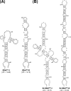
Predicted RNA secondary structures of the 83-nt sequence duplicated in the 3′-terminal region of the genomic RNAs of MUTΔ1-450-derived pseudorevertants. Shown are two pairs of four predicted stem-loop structures: one for a single copy of the 83-nt sequence (A) and the other for a tandem repeat of the 83-nt sequence (B). Also indicated are the initial thermodynamic free energies (ΔG, in kilocalories/mole) of all four potential structures, as determined by the RNA folding program mfold.
FIG. 7.
FIG. 7.
Characterization of recombinant viruses generated from five replication-competent JEV 3′NCR mutant cDNAs. (A and B) Viral growth and focus morphology. Subconfluent monolayers of BHK-21 cells were infected at an MOI of 0.1 with the WT or one of the five 3′NCR mutant viruses derived from the respective cDNAs, as indicated. (A) At the indicated hour postinfection (hpi), aliquots of culture supernatants were collected during a period of 4 days and used for virus titration on naïve BHK-21 cells. (B) The same monolayers were immunostained at 6 and 96 hpi (indicated by arrows) for infectious foci with the anti-JEV antiserum. The data shown are from one of two independent experiments, which produced similar results. M, mock infected. (C) Acquisition of spontaneous secondary mutations during three consecutive rounds of viral passage in BHK-21 cells. To examine the genetic stability of the five mutant viruses, each of the viruses collected from RNA-transfected cells (passage 0) was passaged three times in naïve BHK-21 cells at an MOI of 0.1. At the indicated passages, the 3′-terminal sequences of the replicating genomic RNAs were determined by 3′RACE, the cloning of the cDNA amplicons, and the sequencing of ∼20 independent clones containing the insert. Indicated is the presence (Yes) or absence (No) of secondary mutations for each mutant virus. N.T, not tested.
FIG. 8.
FIG. 8.
Identification of 12 point mutations acquired within the 3′-terminal region of the genomic RNAs of MUTΔ1-381-derived pseudorevertants. The original recombinant viral stock was generated by transfecting BHK-21 cells with RNA transcripts derived from MUTΔ1-381 cDNA (passage 0); this viral stock was serially passaged three times in naïve BHK-21 cells at an MOI of 0.1 (passages 1 to 3). The 3′-terminal sequences of the genomic RNAs at passages 0, 1, and 3 were determined as described in Materials and Methods. The relative locations (A) and nucleotide sequences (B) acquired at the 3′-terminal region of the genomic RNAs of MUTΔ1-381-derived pseudorevertants are shown. (A) Illustrated at the top is the organization of the genomic RNA derived from the MUTΔ1-381 cDNA; shown below is an expanded view of the 193-nt 3′NCR, including only the two 3′-proximal domains (II-2 and III), with the CS1 and CS2 motifs and the 3′SL indicated by gray boxes. (B) Twelve point mutations (single-nucleotide substitutions or deletions are in boldface type) acquired in MUTΔ1-381-derived pseudorevertants. Dots indicate the conserved nucleotide sequences in adapted pseudorevertants, and hyphens indicate the deleted nucleotide sequences. The number of clones containing each identified nucleotide sequence is given over the total number of clones containing the insert that were sequenced. Open arrowheads indicate the translation stop codon UAG. The 5′-end nucleotide of the 3′NCR is numbered nt 382, and downstream sequences are consecutively assigned according to the WT sequence.
FIG. 9.
FIG. 9.
Effects on RNA replication efficiency of 12 point mutations discovered in MUTΔ1-381-derived pseudorevertants. Naïve BHK-21 cells were mock transfected (Mock) or transfected with a synthetic RNA transcribed from the WT cDNA or one of the reconstructed MUTΔ1-381 derivative cDNAs, as indicated. (A) Representative focus morphology. Infectious foci were visualized at 4 days posttransfection by immunostaining with the anti-JEV antiserum. (B) Production of JEV RNA. The levels of JEV RNA production at 22 hpt relative to those at 6 hpt were estimated by real-time quantitative RT-PCR using a JEV-specific probe. (C) Accumulation of JEV proteins. The levels of JEV protein accumulation at 24 hpt were examined by immunoblotting with the anti-JEV antiserum (anti-JEV) or a rabbit antiserum specific for JEV NS1 (anti-NS1). The positions of the viral proteins E, NS1, and NS3 and a cleavage-related intermediate (open arrowhead) are shown on the left; molecular size markers are shown on the right. (D) Production of infectious virions. Virus yields in the culture supernatants at 24 hpt were determined by virus titration on naïve BHK-21 cells.
FIG. 10.
FIG. 10.
Identification of seven sequence insertions (duplications) of various sizes at the junction between domains II-1 and II-2 in the genomic RNAs of MUTΔ1-284-derived pseudorevertants. The original recombinant viral stock was obtained after transfection of BHK-21 cells with the RNA transcripts derived from MUTΔ1-284 cDNA (passage 0); this virus stock was then passaged three times in naïve BHK-21 cells at an MOI of 0.1 (passages 1 to 3). The 3′-terminal sequences of the genomic RNAs at passages 0, 1, and 3 were determined as described in Materials and Methods. (A) Relative locations of the seven sequence insertions (Ins1 to Ins7) duplicated from upstream sequences of domain II-2 in the genomic RNAs of MUTΔ1-284-derived pseudorevertants. At the top is a schematic presentation of the genomic RNA derived from the MUTΔ1-284 cDNA; given below is an expanded view of the 290-nt 3′NCR containing only the three 3′-proximal domains (II-1, II-2, and III), with the three CSs (CS1, CS2, and RCS2) and the 3′SL indicated. (B and C) Nucleotide sequences of the seven sequence insertions (Ins1 to Ins7) of various sizes. (B) The number of clones containing each identified nucleotide sequence/the total number of clones containing the insert that we examined is indicated. (C) The nucleotides of the seven sequence insertions (duplications) were aligned; the RCS2 motif is highlighted with a gray box. Open arrowheads indicate the translation stop codon UAG.
FIG. 11.
FIG. 11.
Effects of seven sequence insertions (duplications) discovered in MUTΔ1-284-derived pseudorevertants on RNA replication efficiency. Naïve BHK-21 cells were mock transfected (Mock) or transfected with the synthetic RNAs transcribed from WT cDNA or one of the reconstructed MUTΔ1-284 derivative cDNAs, as indicated. (A) Representative focus morphology. (B) Production of JEV RNA. (C) Accumulation of JEV proteins. (D) Production of infectious virions. Experiments were performed as described in the legend to Fig. 9.
FIG. 12.
FIG. 12.
Cell type-dependent replication of MUTΔ1-381-derived mutant virus. (A) Viral growth properties in human neuroblastoma SH-SY5Y and mosquito C6/36 cells. Monolayers of SH-SY5Y or C6/36 cells were infected at an MOI of 1 with the WT or one of the five 3′NCR mutant viruses derived from the respective cDNAs, as indicated. At various time points postinfection, aliquots of culture supernatants were collected and used for virus titration on naïve BHK-21 cells. Data were consistent in two independent experiments. (B) Viral protein accumulation in infected C6/36 cells. Monolayers of C6/36 cells were mock infected or infected at an MOI of 1 with WT, MUTΔ1-284-derived, or MUTΔ1-381-derived virus, or at an MOI of 10 with MUTΔ1-381-derived virus in parallel. At 48 and 144 h postinfection (hpi), the levels of JEV-specific proteins or JEV NS1 protein were analyzed by immunoblotting with the anti-JEV antiserum (anti-JEV) or a rabbit antiserum specific for JEV NS1 (anti-NS1), respectively. The positions of the viral proteins (E, NS1, and NS3) and a cleavage-related intermediate (open arrowhead) are indicated on the left. Molecular size markers (in kilodaltons) are shown on the right.

Similar articles

Cited by

References

    1. Ackermann, M., and R. Padmanabhan. 2001. De novo synthesis of RNA by the dengue virus RNA-dependent RNA polymerase exhibits temperature dependence at the initiation but not elongation phase. J. Biol. Chem. 27639926-39937. - PubMed
    1. Alvarez, D. E., A. L. De Lella Ezcurra, S. Fucito, and A. V. Gamarnik. 2005a. Role of RNA structures present at the 3′UTR of dengue virus on translation, RNA synthesis, and viral replication. Virology 339200-212. - PubMed
    1. Alvarez, D. E., M. F. Lodeiro, S. J. Luduena, L. I. Pietrasanta, and A. V. Gamarnik. 2005b. Long-range RNA-RNA interactions circularize the dengue virus genome. J. Virol. 796631-6643. - PMC - PubMed
    1. Alvarez, D. E., C. V. Filomatori, and A. V. Gamarnik. 2008. Functional analysis of dengue virus cyclization sequences located at the 5′ and 3′UTRs. Virology 375223-235. - PubMed
    1. Ball, L. A. 1997. Nodavirus RNA recombination. Semin. Virol. 895-100.

Publication types

MeSH terms

LinkOut - more resources