. 2009 Aug 1;332(1):166-76.
doi: 10.1016/j.ydbio.2009.05.566. Epub 2009 May 27.

Notch signaling is required for lateral induction of Jagged1 during FGF-induced lens fiber differentiation

Affiliations

Notch signaling is required for lateral induction of Jagged1 during FGF-induced lens fiber differentiation

Senthil S Saravanamuthu et al. Dev Biol. .

Abstract

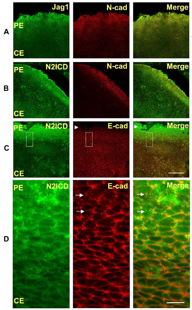
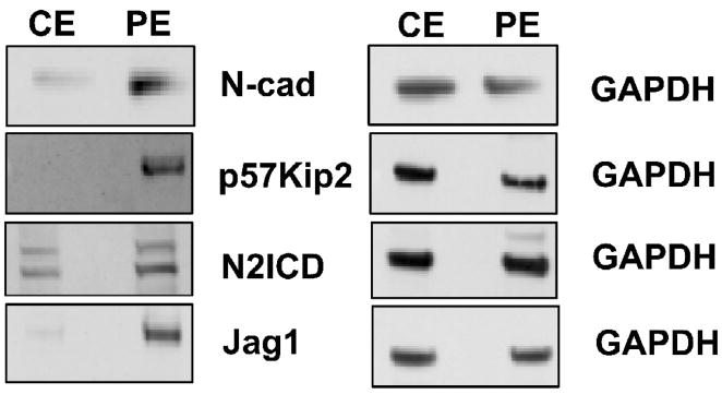
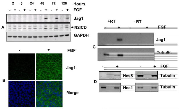
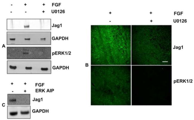
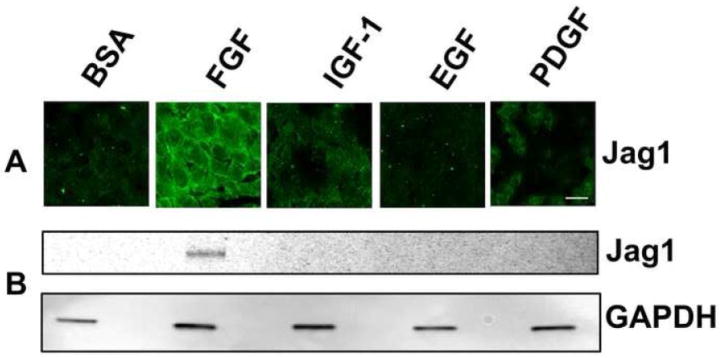
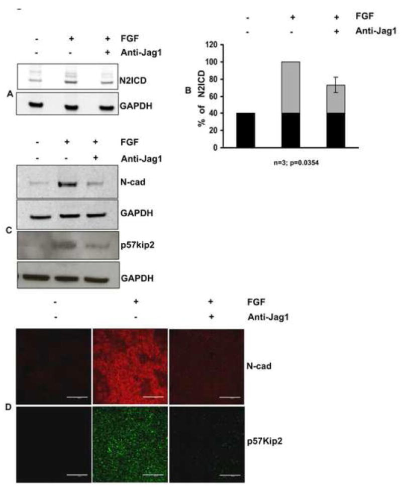
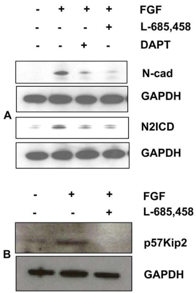
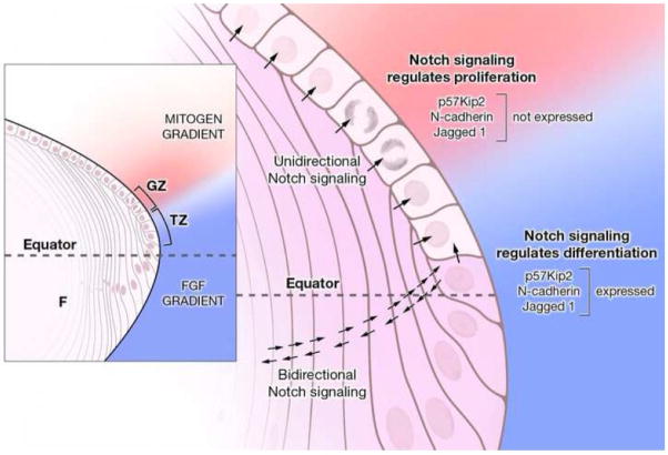
Previous studies of the developing lens have shown that Notch signaling regulates differentiation of lens fiber cells by maintaining a proliferating precursor pool in the anterior epithelium. However, whether Notch signaling is further required after the onset of fiber cell differentiation is not clear. This work investigates the role of Notch2 and Jagged1 (Jag1) in secondary fiber cell differentiation using rat lens epithelial explants undergoing FGF-2 dependent differentiation in vitro. FGF induced Jag1 expression and Notch2 signaling (as judged by the appearance of activated Notch2 Intracellular Domain (N2ICD)) within 12-24 h. These changes were correlated with induction of the Notch effector, Hes5, upregulation of N-cadherin (N-cad), and downregulation of E-cadherin (E-cad), a cadherin switch characteristic of fiber cell differentiation. Induction of Jag1 was efficiently blocked by U0126, a specific inhibitor of MAPK/ERK signaling, indicating a requirement for signaling through this pathway downstream of the FGF receptor. Other growth factors that activate MAPK/ERK signaling (EGF, PDGF, IGF) did not induce Jag1. Inhibition of Notch signaling using gamma secretase inhibitors DAPT and L-685,458 or anti-Jag1 antibody markedly decreased FGF-dependent expression of Jag1 demonstrating Notch-dependent lateral induction. In addition, inhibition of Notch signaling reduced expression of N-cad, and the cyclin dependent kinase inhibitor, p57Kip2, indicating a direct role for Notch signaling in secondary fiber cell differentiation. These results demonstrate that Notch-mediated lateral induction of Jag1 is an essential component of FGF-dependent lens fiber cell differentiation.


Figures

Fig. 1
Fig. 1. Jag1 and N2ICD are highly expressed in the peripheral lens epithelium
Representative immunofluorescence micrographs of whole mounts of freshly isolated postnatal day-2(PN2) rat lens epithelial explants stained with specific antibodies. (A,B) Jag1 (A) and N2ICD (B) expression colocalize with expression of N-cad, an early marker of differentiation, in the peripheral epithelium (PE). (C,D) The increase in the N2ICD corresponds to decreased immunostaining of Ecad (refer to arrowheads in panel C). Higher magnification (D), shown as an inset (dotted line box) in panel C, shows that the increased staining of N2ICD coincides with the appearance of intracellular vesicles (refer to arrows in panel D) that immunostain for E-cad, suggesting E-cad degradation (D). Scale bar on panel C represents 100 μm length and is applicable for all images of panels A, B and C. Scale bar on panel D represents 10 μm and is applicable for all images of panel D.
Fig. 2
Fig. 2
The protein profiles of microdissected peripheral epithelium (PE) and central epithelium (CE) with respect to N-cad, p57Kip2, Jag1 and N2ICD are compared in representative immunoblots. Near absence of N-cad, p57Kip2, and Jag1 in the CE demonstrates that microdissection of central explants efficiently separates cells of the CE from differentiating cells in the PE.
Fig. 3
Fig. 3. FGF induces Jag1 and activates Notch signaling
(A) Representative immunoblot of Jag1 and N2ICD from lens epithelial explants exposed to 100ng/mL FGF (+) or 0.1% BSA (−) for indicated times (hours). GAPDH has been used as a loading control. The two bands recognized by the N2ICD antibody likely represent sequential cleavage products of Notch2. Lower band represents the N2ICD (B) Representative immunofluorescence of Jag1 in the central region of rat lens epithelia cultured in the presence or absence of FGF for 4 days shows that FGF induces uniform expression of Jag1 in all cells in this region and demonstrates that Jag1(green) is localized along cell-cell boundaries. Nuclei have been stained with DAPI (blue). Scale bar is applicable to all images of panel B and represents 20 μm length (C) FGF induction of Jag1 mRNA was demonstrated by RT-PCR using total RNA isolated from CE explants cultured in the presence or absence of FGF (100 ng/mL) for 24 hours. Tubulin RT-PCR was performed as a positive control. Reactions lacking reverse transcriptase (RT) served as negative controls. Analyses of at least three different replicates provided similar results. (D) The ability of FGF to activate Notch signaling was examined by RT-PCR for Notch effectors Hes5 and Hes1, using total RNA isolated from CE explants cultured in the presence or absence of FGF (100 ng/mL) for 24 hours. Tubulin RT-PCR was performed as a positive control. Negative controls lacking reverse transcriptase (RT) showed no amplified products (not shown). Analyses of at least three different replicates provided similar results.
Fig. 4
Fig. 4. Jag1 induction is regulated by MAPK/ERK pathway
(A) Representative immunoblots of Jag1 and pERK1/2 expression in CE explants treated with the MEK inhibitor U0126. GAPDH expression is shown as loading control. (B) Representative whole mounts micrographs of rat lens CE explants cultured in the presence or absence of FGF and MAPK/ERK 1/2 pathway inhibitor U0126 (50 μM) for 48 hours and immunolabelled for Jag1 and pERK1/2. Scale bar is applicable to all images of panel B and represents 100 μm length. (C) Representative immunoblots of Jag1 expression in CE explants treated with the ERK Activation Inhibitor Peptide (ERK AIP). GAPDH expression is shown as loading control.
Fig. 5
Fig. 5. Jag1 expression is not induced by IGF-1, EGF, or PDGF
(A) Representative immunofluorescence micrographs of explants of central epithelium (CE) treated with BSA (control), FGF-2, IGF-1, EGF, or PDGF for 48 hours and immunostained for Jag1. Scale bar is applicable to all images of panel A and represents 10 μm length. (B) Corresponding immunoblots of lysates of treated explants from the same experiment immunoblotted for Jag1.
Fig. 6
Fig. 6. Blockade of Notch signaling with anti-Jag1 antibody reduces expression of p57Kip2 and N-cad
CE explants were cultured in the presence of FGF for 4 days in the presence or absence of anti- Jag1 antibody and analyzed for expression of N2ICD, p5Kip2, and N-cad. (A) Effectiveness of the blockade by anti-Jag1 was evaluated by immunoblotting for N2ICD. FGF induced an increase in N2ICD above the basal level (indicated by black in the bar graphs), as seen in control explants treated with only BSA. This FGF-dependent increase (indicated by gray areas of the bar graphs) was reduced by incubation with anti-Jag1. (B) Quantification of N2ICD expression by densitometric scanning of replicate immunoblots (n=3), demonstrated approximately a 50% decrease +/− 5.14 (s.e.m) (p=0.0354). (C) Immunoblotting lysates of the cultured explants confirmed the reduction in expression of p57Kip2 and N-cad. (D) Incubation with anti-Jag1 antibody sharply reduced immunofluorescence of p57Kip2 and N-cad in all cells of the explant. Scale bar is applicable to all images of panel D and represents 100 μm length.
Fig. 7
Fig. 7. Inhibition of Notch signaling with gamma secretase inhibitors reduces expression of N-cad and p57Kip2
(A) CE explants were cultured in the presence of FGF for 4 days in the presence or absence of the gamma secretase inhibitors, DAPT (50 μM) and L-685,458 (20 μM), and immunoblotted to determine expression of N2ICD and N-cad. Reduction of N2ICD with these pharmacological inhibitors was paralleled by reduction in N-cad expression, confirming the results obtained with anti-Jag1 antibody. (B) Lysates from CE explants cultured in the presence or absence of FGF and/or L-685,458 were immunoblotted for p57Kip2, representative immunoblot shows reduction in the expression of p57Kip2 during suppression of notch signaling by L-685,458.
Fig. 8
Fig. 8. Jag1-Notch signaling regulates Jag1 induction
(A) CE explants were cultured in the presence of FGF for 4 days in the presence or absence of anti- Jag1 antibody and analyzed for expression of Jag1. Immunoblotting demonstrated a marked reduction of Jag1 protein in the presence of anti-Jag1 antibody. (B) Quantification of Jag1expression by densitometric scanning of replicate immunoblots (n=3), demonstrated approximately a 70% decrease +/− 10.9 (s.e.m) (p=0.0238). (C) CE explants were cultured in the presence of FGF for 4 days in the presence or absence of the gamma secretase inhibitors, DAPT (50μM) and L-685,458 (20μM). Inhibition of Notch signaling with these pharmacological inhibitors confirmed that Notch signaling regulates induction of Jag1.
Fig. 9
Fig. 9. A dual role for Notch signaling in the lens
Epithelial cells in the germinative zone (GZ) and transition zone (TZ) are exposed to a cocktail of growth factors: FGF (blue gradient) diffusing from the vitreous humor and mitogenic growth factors (pink gradient) secreted into the aqueous humor by the ciliary body. Jag1 expression (shaded violet in the magnified view), which is restricted to fiber cells (F) and cells in the TZ in the early stages of differentiation, induces unidirectional Notch signaling (arrows) in the overlying epithelial cells and permits proliferation in the presence of appropriate mitogen concentration. Notch signaling in the GZ negatively regulates p57Kip2 (Jia et al., 2007; Rowan et al., 2008), thus maintaining a pool of precursor cells needed for secondary fiber cell production. As no differentiation occurs in this region, cells express E-cad, but not N-cad or Jag1. As the post-mitotic precursor cells migrate posteriorly they are exposed to increasing FGF concentration. At a critical, threshold concentration of FGF, an initial, Notch-independent induction of Jag1 occurs. This is rapidly amplified by lateral induction via positive feedback involving bidirectional Notch signaling between adjacent differentiating cells. A sharp boundary is thus established between non-differentiating and differentiating cells in the transition zone. In direct contrast to the role of Notch signaling in repressing p57Kip2 in the proliferating cells of the GZ, Notch signaling in the differentiating cells positively regulates p57Kip2. Moreover, Notch signaling regulates cadherin switching by augmenting N-cad expression, as E-cad is being lost from cell-cell junctions.

Similar articles

Cited by

References

    1. Ang SJ, Stump RJ, Lovicu FJ, McAvoy JW. Spatial and temporal expression of Wnt and Dickkopf genes during murine lens development. Gene Expr Patterns. 2004;4:289–95. - PubMed
    1. Bray S. Notch signalling in Drosophila: three ways to use a pathway. Semin Cell Dev Biol. 1998;9:591–7. - PubMed
    1. Bray SJ. Notch signalling: a simple pathway becomes complex. Nat Rev Mol Cell Biol. 2006;7:678–89. - PubMed
    1. Cain S, Martinez G, Kokkinos MI, Turner K, Richardson RJ, Abud HE, Huelsken J, Robinson ML, de Iongh RU. Differential requirement for beta-catenin in epithelial and fiber cells during lens development. Dev Biol. 2008;321:420–33. - PubMed
    1. Chandrasekher G, Sailaja D. Differential activation of phosphatidylinositol 3-kinase signaling during proliferation and differentiation of lens epithelial cells. Invest Ophthalmol Vis Sci. 2003;44:4400–11. - PubMed

Publication types

MeSH terms