

Down's syndrome suppression of tumour growth and the role of the calcineurin inhibitor DSCR1
- PMID: 19458618
- PMCID: PMC2724004
- DOI: 10.1038/nature08062
Down's syndrome suppression of tumour growth and the role of the calcineurin inhibitor DSCR1
Abstract
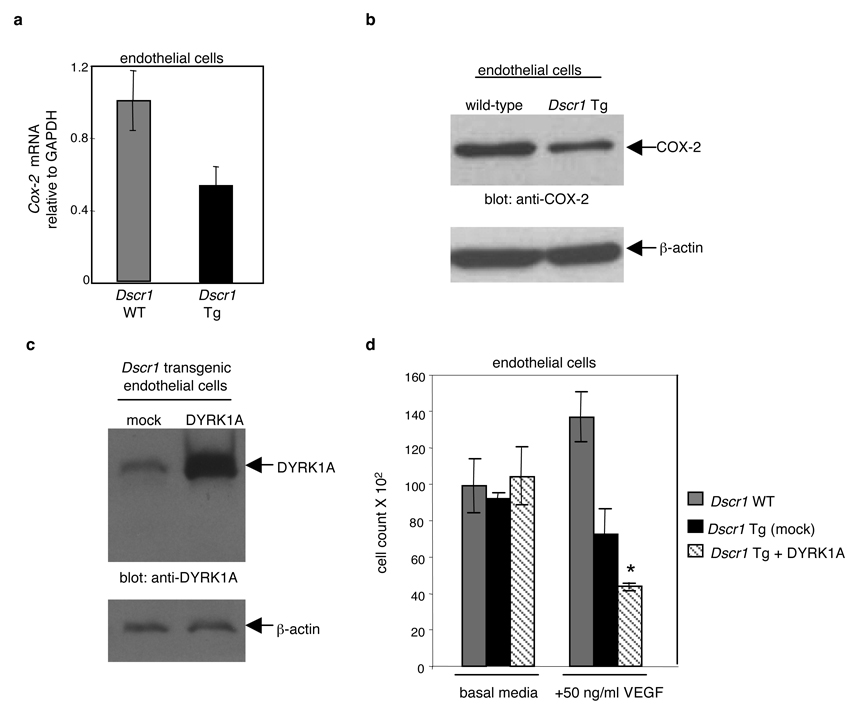
The incidence of many cancer types is significantly reduced in individuals with Down's syndrome, and it is thought that this broad cancer protection is conferred by the increased expression of one or more of the 231 supernumerary genes on the extra copy of chromosome 21. One such gene is Down's syndrome candidate region-1 (DSCR1, also known as RCAN1), which encodes a protein that suppresses vascular endothelial growth factor (VEGF)-mediated angiogenic signalling by the calcineurin pathway. Here we show that DSCR1 is increased in Down's syndrome tissues and in a mouse model of Down's syndrome. Furthermore, we show that the modest increase in expression afforded by a single extra transgenic copy of Dscr1 is sufficient to confer significant suppression of tumour growth in mice, and that such resistance is a consequence of a deficit in tumour angiogenesis arising from suppression of the calcineurin pathway. We also provide evidence that attenuation of calcineurin activity by DSCR1, together with another chromosome 21 gene Dyrk1a, may be sufficient to markedly diminish angiogenesis. These data provide a mechanism for the reduced cancer incidence in Down's syndrome and identify the calcineurin signalling pathway, and its regulators DSCR1 and DYRK1A, as potential therapeutic targets in cancers arising in all individuals.
Figures


Comment in
-
Down's syndrome patients are less likely to develop cancer.Clin Genet. 2010 Jul;78(1):35-7. doi: 10.1111/j.1399-0004.2010.01414_3.x. Clin Genet. 2010. PMID: 20597922 No abstract available.
Similar articles
-
NFAT dysregulation by increased dosage of DSCR1 and DYRK1A on chromosome 21.Nature. 2006 Jun 1;441(7093):595-600. doi: 10.1038/nature04678. Epub 2006 Mar 22. Nature. 2006. PMID: 16554754
-
A single extra copy of Dscr1 improves survival of mice developing spontaneous lung tumors through suppression of tumor angiogenesis.Cancer Lett. 2014 Jan 1;342(1):70-81. doi: 10.1016/j.canlet.2013.08.047. Epub 2013 Sep 16. Cancer Lett. 2014. PMID: 24051307
-
Increased dosage of DYRK1A and DSCR1 delays neuronal differentiation in neocortical progenitor cells.Genes Dev. 2013 Dec 15;27(24):2708-21. doi: 10.1101/gad.226381.113. Genes Dev. 2013. PMID: 24352425 Free PMC article.
-
[Molecular Mechanism Underlying Abnormal Differentiation of Neural Progenitor Cells in the Developing Down Syndrome Brain].Yakugaku Zasshi. 2017;137(7):795-800. doi: 10.1248/yakushi.16-00236-1. Yakugaku Zasshi. 2017. PMID: 28674289 Review. Japanese.
-
Multiple roles of the DSCR1 (Adapt78 or RCAN1) gene and its protein product calcipressin 1 (or RCAN1) in disease.Cell Mol Life Sci. 2005 Nov;62(21):2477-86. doi: 10.1007/s00018-005-5085-4. Cell Mol Life Sci. 2005. PMID: 16231093 Free PMC article. Review.
Cited by
-
Concise review: new paradigms for Down syndrome research using induced pluripotent stem cells: tackling complex human genetic disease.Stem Cells Transl Med. 2013 Mar;2(3):175-84. doi: 10.5966/sctm.2012-0117. Epub 2013 Feb 14. Stem Cells Transl Med. 2013. PMID: 23413375 Free PMC article. Review.
-
Secretion of Down Syndrome Critical Region 1 Isoform 4 in Ischemic Retinal Ganglion Cells Displays Anti-Angiogenic Properties Via NFATc1-Dependent Pathway.Mol Neurobiol. 2017 Oct;54(8):6556-6571. doi: 10.1007/s12035-016-0092-z. Epub 2016 Oct 12. Mol Neurobiol. 2017. PMID: 27734335
-
Loss of Down Syndrome Critical Region-1 Mediated-Hypercholesterolemia Accelerates Corneal Opacity Via Pathological Neovessel Formation.Arterioscler Thromb Vasc Biol. 2020 Oct;40(10):2425-2439. doi: 10.1161/ATVBAHA.120.315003. Epub 2020 Aug 13. Arterioscler Thromb Vasc Biol. 2020. PMID: 32787520 Free PMC article.
-
Reprogramming cellular identity for regenerative medicine.Cell. 2012 Mar 16;148(6):1110-22. doi: 10.1016/j.cell.2012.02.031. Cell. 2012. PMID: 22424223 Free PMC article.
-
The DYRK Family of Kinases in Cancer: Molecular Functions and Therapeutic Opportunities.Cancers (Basel). 2020 Jul 29;12(8):2106. doi: 10.3390/cancers12082106. Cancers (Basel). 2020. PMID: 32751160 Free PMC article. Review.
References
-
- Hasle H, Clemmensen IH, Mikkelsen M. Risks of leukaemia and solid tumors in individuals with Down’s syndrome. Lancet. 2000;355:165. - PubMed
-
- Satge D, et al. A lack of neuroblastoma in Down syndrome: a study from 11 European countries. Cancer Res. 1998;58:448. - PubMed
-
- Patja K, Pukkala E, Sund R, Iivanainen M, Kaski M. Cancer incidence of persons with Down syndrome in Finland: a population-based study. Int. J. Cancer. 2006;118:1769. - PubMed
-
- Yang Q, Rasmussen SA, Friedman JM. Mortality associated with Down's syndrome in the USA from 1983 to 1997: a population-based study. Lancet. 2002;359:1019. - PubMed
-
- Fuentes JJ, Pritchard MA, Estivill X. Genomic organization, alternative splicing, and expression patterns of the DSCR1 (Down syndrome candidate region 1) gene. Genomics. 1997;44:358. - PubMed
Publication types
MeSH terms
Substances
Grants and funding
LinkOut - more resources
Full Text Sources
Other Literature Sources
Medical
Molecular Biology Databases
