

Overexpression of catalase targeted to mitochondria attenuates murine cardiac aging
- PMID: 19451351
- PMCID: PMC2858759
- DOI: 10.1161/CIRCULATIONAHA.108.822403
Overexpression of catalase targeted to mitochondria attenuates murine cardiac aging
Abstract
Background: Age is a major risk for cardiovascular diseases. Although mitochondrial reactive oxygen species have been proposed as one of the causes of aging, their role in cardiac aging remains unclear. We have previously shown that overexpression of catalase targeted to mitochondria (mCAT) prolongs murine median lifespan by 17% to 21%.
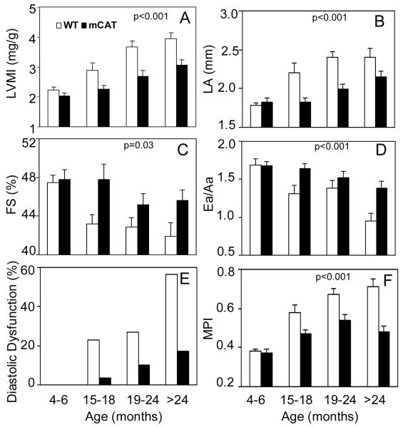
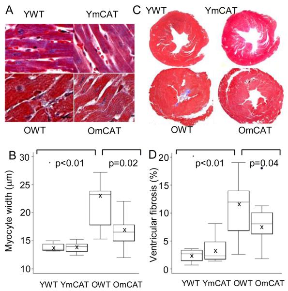
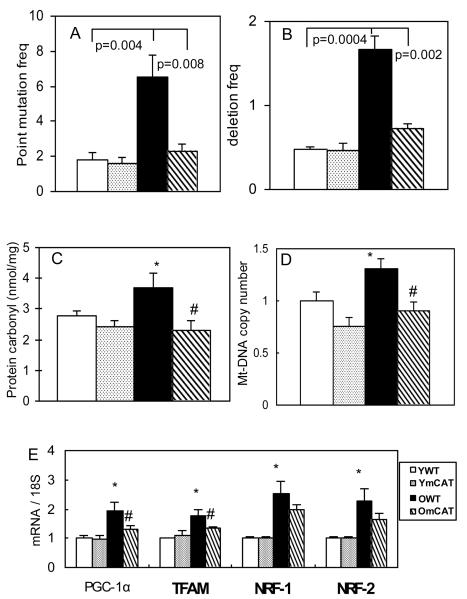
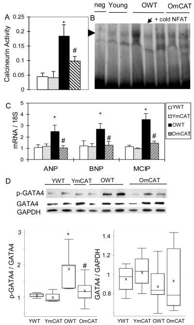
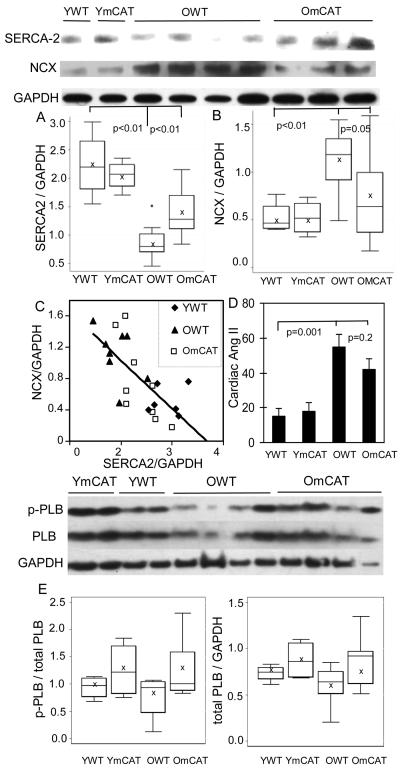
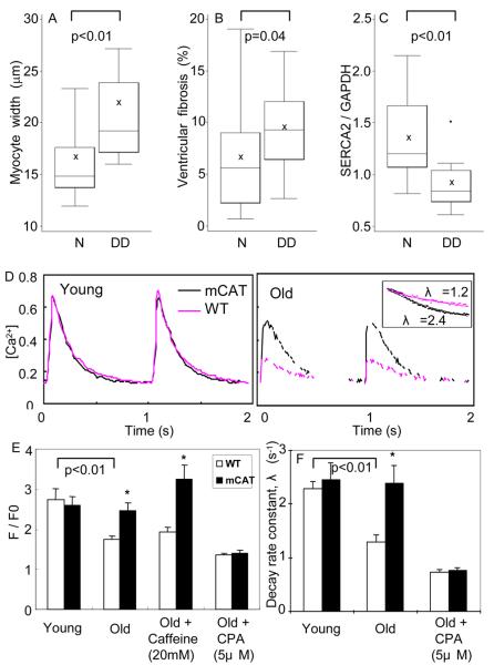
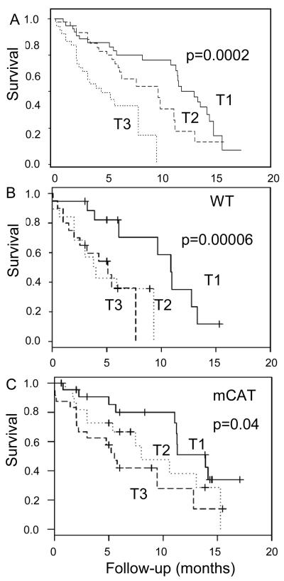
Methods and results: We used echocardiography to study cardiac function in aging cohorts of wild-type and mCAT mice. Changes found in wild-type mice recapitulate human aging: age-dependent increases in left ventricular mass index and left atrial dimension, worsening of the myocardial performance index, and a decline in diastolic function. Cardiac aging in mice is accompanied by accumulation of mitochondrial protein oxidation, increased mitochondrial DNA mutations and deletions and mitochondrial biogenesis, increased ventricular fibrosis, enlarged myocardial fiber size, decreased cardiac SERCA2 protein, and activation of the calcineurin-nuclear factor of activated T-cell pathway. All of these age-related changes were significantly attenuated in mCAT mice. Analysis of survival of 130 mice demonstrated that echocardiographic cardiac aging risk scores were significant predictors of mortality. The estimated attributable risk to mortality for these 2 parameters was 55%.
Conclusions: This study shows that cardiac aging in the mouse closely recapitulates human aging and demonstrates the critical role of mitochondrial reactive oxygen species in cardiac aging and the impact of cardiac aging on survival. These findings also support the potential application of mitochondrial antioxidants in reactive oxygen species-related cardiovascular diseases.
Figures
Similar articles
-
Cardiac aging in mice and humans: the role of mitochondrial oxidative stress.Trends Cardiovasc Med. 2009 Oct;19(7):213-20. doi: 10.1016/j.tcm.2009.12.004. Trends Cardiovasc Med. 2009. PMID: 20382344 Free PMC article. Review.
-
Age-dependent cardiomyopathy in mitochondrial mutator mice is attenuated by overexpression of catalase targeted to mitochondria.Aging Cell. 2010 Aug;9(4):536-44. doi: 10.1111/j.1474-9726.2010.00581.x. Epub 2010 Apr 29. Aging Cell. 2010. PMID: 20456298 Free PMC article.
-
Mitochondrial oxidative stress mediates angiotensin II-induced cardiac hypertrophy and Galphaq overexpression-induced heart failure.Circ Res. 2011 Apr 1;108(7):837-46. doi: 10.1161/CIRCRESAHA.110.232306. Epub 2011 Feb 10. Circ Res. 2011. PMID: 21311045 Free PMC article.
-
Hydrogen peroxide-mediated SERCA cysteine 674 oxidation contributes to impaired cardiac myocyte relaxation in senescent mouse heart.J Am Heart Assoc. 2013 Aug 20;2(4):e000184. doi: 10.1161/JAHA.113.000184. J Am Heart Assoc. 2013. PMID: 23963753 Free PMC article.
-
Oxidative stress and aging: catalase is a longevity determinant enzyme.Rejuvenation Res. 2005 Fall;8(3):138-40. doi: 10.1089/rej.2005.8.138. Rejuvenation Res. 2005. PMID: 16144468 Review.
Cited by
-
Regulation of signal transduction by reactive oxygen species in the cardiovascular system.Circ Res. 2015 Jan 30;116(3):531-49. doi: 10.1161/CIRCRESAHA.116.303584. Circ Res. 2015. PMID: 25634975 Free PMC article. Review.
-
Long-term treatment with Elamipretide enhances healthy aging phenotypes in mice.Aging Pathobiol Ther. 2022;4(3):76-83. doi: 10.31491/apt.2022.09.089. Epub 2022 Sep 30. Aging Pathobiol Ther. 2022. PMID: 36250163 Free PMC article.
-
Molecular basis of ventricular arrhythmogenicity in a Pgc-1α deficient murine model.Mol Genet Metab Rep. 2021 Apr 9;27:100753. doi: 10.1016/j.ymgmr.2021.100753. eCollection 2021 Jun. Mol Genet Metab Rep. 2021. PMID: 33898262 Free PMC article.
-
Overexpression of catalase targeted to mitochondria improves neurovascular coupling responses in aged mice.Geroscience. 2019 Oct;41(5):609-617. doi: 10.1007/s11357-019-00111-0. Epub 2019 Oct 23. Geroscience. 2019. PMID: 31643012 Free PMC article.
-
The role of inflammatory and fibrogenic pathways in heart failure associated with aging.Heart Fail Rev. 2010 Sep;15(5):415-22. doi: 10.1007/s10741-010-9161-y. Heart Fail Rev. 2010. PMID: 20213186 Free PMC article. Review.
References
-
- Lakatta EG, Levy D. Arterial and cardiac aging: major shareholders in cardiovascular disease enterprises: Part II: the aging heart in health: links to heart disease. Circulation. 2003;107:346–54. - PubMed
-
- Murphy SW. Diastolic Dysfunction. Curr Treat Options Cardiovasc Med. 2004;6:61–68. - PubMed
-
- Balaban RS, Nemoto S, Finkel T. Mitochondria, oxidants, and aging. Cell. 2005;120:483–95. - PubMed
-
- Vermulst M, Bielas JH, Kujoth GC, Ladiges WC, Rabinovitch PS, Prolla TA, Loeb LA. Mitochondrial point mutations do not limit the natural lifespan of mice. Nat Genet. 2007;39:540–3. - PubMed
Publication types
MeSH terms
Substances
Grants and funding
LinkOut - more resources
Full Text Sources
Other Literature Sources
Medical
Molecular Biology Databases
