Review
. 2009 Jun;212(Pt 11):1753-61.
doi: 10.1242/jeb.027284.

Regulation of luminal acidification in the male reproductive tract via cell-cell crosstalk

Affiliations
Review

Regulation of luminal acidification in the male reproductive tract via cell-cell crosstalk

Winnie W C Shum et al. J Exp Biol. 2009 Jun.

Abstract

In the epididymis, spermatozoa acquire their ability to become motile and to fertilize an egg. A luminal acidic pH and a low bicarbonate concentration help keep spermatozoa in a quiescent state during their maturation and storage in this organ. Net proton secretion is crucial to maintain the acidity of the luminal fluid in the epididymis. A sub-population of epithelial cells, the clear cells, express high levels of the proton-pumping V-ATPase in their apical membrane and are important contributors to luminal acidification. This review describes selected aspects of V-ATPase regulation in clear cells. The assembly of a particular set of V-ATPase subunit isoforms governs the targeting of the pump to the apical plasma membrane. Regulation of V-ATPase-dependent proton secretion occurs via recycling mechanisms. The bicarbonate-activated adenylyl cyclase is involved in the non-hormonal regulation of V-ATPase recycling, following activation of bicarbonate secretion by principal cells. The V-ATPase is also regulated in a paracrine manner by luminal angiotensin II by activation of the angiotensin II type 2 receptor (AGTR2), which is located in basal cells. Basal cells have the remarkable property of extending long and slender cytoplasmic projections that cross the tight junction barrier to monitor the luminal environment. Clear cells are activated by a nitric oxide signal that originates from basal cells. Thus, a complex interplay between the different cell types present in the epithelium leads to activation of the luminal acidifying capacity of the epididymis, a process that is crucial for sperm maturation and storage.


Figures

Fig. 1.
Fig. 1.
Schematic view of the epididymis. The epithelium lining the epididymis is composed of several cell types, including narrow, clear, principal and basal cells. Narrow and clear cells express high levels of the V-ATPase in their apical membrane and are important contributors to luminal acidification, especially in the distal region (cauda). Basal cells have the previously unrecognized property of sending narrow body projections that can contact the luminal side of the epithelium. Very few basal cells reaching the lumen were detected in the proximal regions including the initial segment and caput, but their number increased progressively in the corpus, to reach a maximum in the cauda.
Fig. 2.
Fig. 2.
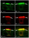
Immunolocalization of the a1 and a4 subunits of the V-ATPase, and comparison with the E subunit, a marker of all V-ATPase holoenzymes in clear cells. 5 μm sections of rat cauda epididymidis were stained for a1 (A; green) or a4 (B; green). The sections were double-labeled for E (C,D; red). a1 is located in sub-apical vesicles, where it colocalizes with E (yellow staining in the merged image shown in E), but it is absent from microvilli that are only labeled for the E subunit (red staining in E). a4 colocalizes with E in both sub-apical vesicles and apical microvilli (yellow-orange staining in the merged image shown in F). Scale bars, 5 μm. Reproduced from Pietrement et al. (Pietrement et al., 2006) with permission from Biology of Reproduction.
Fig. 3.
Fig. 3.
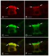
Immunolocalization of the B1 and B2 subunits of the V-ATPase, and comparison with the E subunit, a marker of all V-ATPase holoenzymes in clear cells. 5 μm sections of mouse cauda epididymidis were stained for B1 (A; red) or B2 (B; red). The sections were double-labeled for E (C,D; green). B1 colocalizes with E in both sub-apical vesicles and apical microvilli (yellow staining in the merged image in E). B2 is located in sub-apical vesicles, where it partially colocalizes with E (orange staining in the merged image in F), but it is absent from microvilli that are only labeled for the E subunit (green staining in F). Scale bars, 5 μm. Reproduced from Paunescu et al. (Paunescu et al., 2004) with permission from American Journal of Physiology – Cell Physiology.
Fig. 4.
Fig. 4.
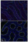
Relative numbers of clear cells in the rat caput (A) versus cauda (B) epididymidis. Rat epididymis was stained for the V-ATPase B1 subunit (green) to label clear cells, and NHERF1 (red), using antibodies that we have previously characterized (Pietrement et al., 2008). Nuclei and spermatozoa were stained with DAPI (blue). NHERF1 is located in the apical membrane of both principal cells and clear cells. B1-positive clear cells are much more numerous in the cauda (B) than in the caput (A) epididymidis. Scale bars, 50 μm.
Fig. 5.
Fig. 5.
Rat cauda epididymidis double-stained for CFTR (green) and the V-ATPase E subunit (red). Intense CFTR labeling is detected in the apical membrane of principal cells. Clear cells, identified by their positive labeling for the V-ATPase E subunit, do not express CFTR. The rabbit anti-CFTR antibody used here was purchased from Alomone Laboratory (Cat. no. ACL-006) and has been previously characterized in our laboratory (Pietrement et al., 2008). Sperm and nuclei were labeled with DAPI (blue). Scale bars, 15 μm. Lu, lumen.
Fig. 6.
Fig. 6.
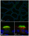
Rat cauda epididymidis perfused in vivo and stained for the V-ATPase B1 subunit (green). Nuclei were stained with DAPI (blue). (A) Numerous B1-positive clear cells were detected. Luminal spermatozoa are absent from these perfused tubules. (B) Higher magnification of a clear cell perfused with a control phosphate-buffered solution adjusted to pH 6.6 and containing the endocytic marker, HRP. Double-labeling for HRP (red) and the V-ATPase B1 subunit (green) was performed. The V-ATPase is distributed between sub-apical vesicles and short microvilli. The yellow staining indicates partial colocalization of the V-ATPase with HRP in endosomes. (C) Clear cell perfused with an `activation' buffer containing bicarbonate and cpt-cAMP. The V-ATPase is mainly located in longer microvilli (green) and no colocalization with HRP-labeled endosomes is detected (red). The staining was performed as previously characterized (Shum et al., 2008). Scale bars, 150 μm (A), 5 μm (B,C).
Fig. 7.
Fig. 7.
(A) Mouse caput epididymidis from a B1-EGFP transgenic mouse. Numerous EGFP-positive (green) clear cells are detected (see also Miller et al., 2005). Nuclei were stained with DAPI (blue). (B) RT-PCR detection of AGTR2 in clear cells isolated by FACS from B1-EGFP mouse epididymidis (GFP+) and in all other epididymal cell types (GFP–). AGTR2 was detected in the GFP-negative cell population, but not in the GFP-positive clear cells.
Fig. 8.
Fig. 8.
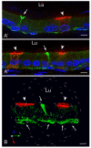
Expression of AGTR2 in basal cells. (A′,A″) Two examples of AGTR2 (green) and V-ATPase (red) labeling in rat cauda epididymidis. Arrows indicate AGTR2-positive basal cells, which send body projections towards the lumen. Arrowheads indicate nearby V-ATPase-positive clear cells. Nuclei were stained with DAPI (blue). (B) Three-dimensional (3D) reconstruction showing AGTR2-positive basal cells (green; arrows). One basal cell sends a projection between principal cells. Two clear cells, stained apically for the V-ATPase (red), are visible (arrowheads). The 3D mosaic was assembled from a stack of 0.1 μm interval optical Z sections obtained by laser scanning confocal microscopy. Lu, lumen. Scale bars, 5 μm. Reproduced from Shum et al. (Shum et al., 2008) with permission from Cell.
Fig. 9.
Fig. 9.
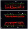
Basal cells reach the tight-junctions at the intersection between three epithelial cells. (A′,A″,A′″) Three different rotations of a three-dimensional reconstruction of an epididymis section stained for claudin-1 (red), a marker of basal cells, and the tight-junction protein ZO1 (green). Arrows indicate the tri-cellular corners where basal cells reach the tight-junctions. Scale bars, 10 μm. Reproduced from Shum et al. (Shum et al., 2008) with permission from Cell.
Fig. 10.
Fig. 10.
Basal cells cross the tight-junctions to reach the lumen (Lu). (A–D) Three-dimensional reconstructions of the apical region of basal cells from epididymis sections double stained for claudin-1 (red; a marker for basal cells) and ZO1 (green; a marker for tight junctions) showing different patterns of interaction. (A) No colocalization between claudin-1 and ZO1 (arrow); (B) partial colocalization of claudin-1 with ZO1 (yellow staining; arrows); (C) basal cell that penetrates the tight-junction (arrow); (D) basal cell forming a ZO1-stained tight junction (green) with adjacent cells (arrows). (E) Conventional microscopy image of the basal cell shown in D (arrow). A clear cell expressing apical V-ATPase (blue) is seen (arrowhead). The nuclei are also stained blue with DAPI. Scale bars, 5 μm. Reproduced from Shum et al. (Shum et al., 2008) with permission from Cell.
Fig. 11.
Fig. 11.
Schematic representation of cell–cell crosstalk in the epididymal epithelium. Basal cells extend a slender body projection toward the lumen, and form a new tight junction with adjacent epithelial cells. Luminal ANGII triggers the production of nitric oxide (NO) by activation of AGTR2 in basal cells. The NO then diffuses out of basal cells and acts locally on clear cells to produce cGMP by activation of the soluble guanylate cyclase (sGC), which is enriched in these cells. cGMP induces the accumulation of V-ATPase in microvilli, which results in the increase of proton secretion. Modified from Shum et al. (Shum et al., 2008) and reproduced with permission from Cell.

Similar articles

Cited by

References

    1. Acott, T. S. and Carr, D. W. (1984). Inhibition of bovine spermatozoa by caudal epididymal fluid: II. Interaction of pH and a quiescence factor. Biol. Reprod. 30, 926-935. - PubMed
    1. Bagnis, C., Marsolais, M., Biemesderfer, D., Laprade, R. and Breton, S. (2001). Na(+)/H(+)-exchange activity and immunolocalization of NHE3 in rat epididymis. Am. J. Physiol. Renal Physiol. 280, F426-F436. - PubMed
    1. Beaulieu, V., Da Silva, N., Pastor-Soler, N., Brown, C. R., Smith, P. J., Brown, D. and Breton, S. (2005). Modulation of the actin cytoskeleton via gelsolin regulates vacuolar H+ATPase (V-ATPase) recycling. J. Biol. Chem. 280, 8452-8463. - PubMed
    1. Beyenbach, K. W. and Wieczorek, H. (2006). The V-type H+ ATPase: molecular structure and function, physiological roles and regulation. J. Exp. Biol. 209, 577-589. - PubMed
    1. Blomqvist, S. R., Vidarsson, H., Soder, O. and Enerback, S. (2006). Epididymal expression of the forkhead transcription factor Foxi1 is required for male fertility. EMBO J. 25, 4131-4141. - PMC - PubMed

Publication types

Substances