. 2009 Jun 17;28(12):1732-44.
doi: 10.1038/emboj.2009.134. Epub 2009 May 14.

Modification of PATase by L/F-transferase generates a ClpS-dependent N-end rule substrate in Escherichia coli

Affiliations

Modification of PATase by L/F-transferase generates a ClpS-dependent N-end rule substrate in Escherichia coli

Robert L Ninnis et al. EMBO J. .

Abstract

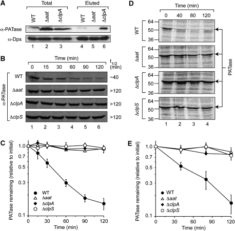
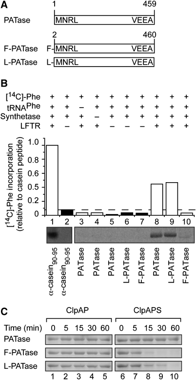
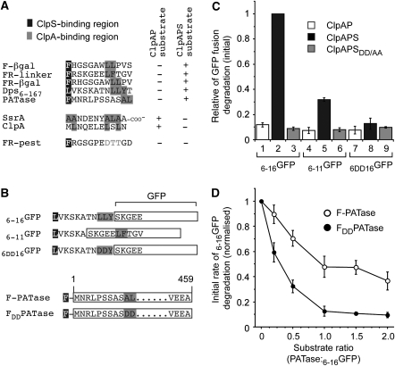
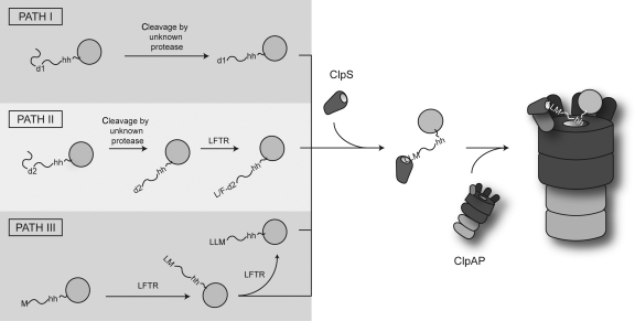
The N-end rule pathway is conserved from bacteria to man and determines the half-life of a protein based on its N-terminal amino acid. In Escherichia coli, model substrates bearing an N-degron are recognised by ClpS and degraded by ClpAP in an ATP-dependent manner. Here, we report the isolation of 23 ClpS-interacting proteins from E. coli. Our data show that at least one of these interacting proteins--putrescine aminotransferase (PATase)--is post-translationally modified to generate a primary N-degron. Remarkably, the N-terminal modification of PATase is generated by a new specificity of leucyl/phenylalanyl-tRNA-protein transferase (LFTR), in which various combinations of primary destabilising residues (Leu and Phe) are attached to the N-terminal Met. This modification (of PATase), by LFTR, is essential not only for its recognition by ClpS, but also determines the stability of the protein in vivo. Thus, the N-end rule pathway, through the ClpAPS-mediated turnover of PATase may have an important function in putrescine homeostasis. In addition, we have identified a new element within the N-degron, which is required for substrate delivery to ClpA.


Figures

Figure 1
Figure 1
N-degron dipeptides compete with model N-end rule substrates for binding to ClpS. (A) The ClpAPS-mediated degradation of FR-linker-GFP (1 μM) was monitored by fluorescence in the absence (filled circles) or presence of 25 μM (open diamonds), 75 μM (open triangles) and 150 μM (open squares) of FR. (B) The ClpAPS-mediated degradation of FR-linker-GFP was monitored in the absence of dipeptide (black bar), the presence of 150 μM N-degron dipeptide (white bars) or 150 μM non-N-end rule dipeptide (grey bars). The rate of FR-linker-GFP degradation by ClpAPS in the presence of dipeptide was determined relative to the rate in the absence of dipeptide. (C) The ClpAP-mediated degradation of GFP-ssrA (1 μM) was monitored either in the absence of dipeptide (black bar) or in the presence of increasing concentration of N-degron dipeptides (white bars). The rate of GFP-ssrA degradation by ClpAP in the presence of dipeptide was determined relative to the rate in the absence of dipeptide. (D) GFP-fusions FR-linker-GFP, FR-PEST-GFP and M-PEST-GFP were applied (T) to Ni-NTA agarose beads (lane 1), beads with immobilised ClpS (lane 3) and beads with immobilised ClpSDD/AA (lane 5). After a wash step, columns were treated with FR dipeptide and eluted samples (lanes 2, 4 and 6) were separated by SDS–PAGE and visualised by Coomassie blue staining; 4% of the total (T) and 10% of the eluted (E) samples were analysed.
Figure 2
Figure 2
Selected E. coli proteins are specifically eluted from immobilised ClpS using FR dipeptide. (A) Schematic diagram illustrating the procedure for isolation of specific ClpS-interacting proteins. (B) E. coli proteins were separated by Tricine SDS–PAGE after FR dipeptide elution from Ni-NTA agarose beads (lane 2), beads with immobilised ClpS (lane 4) or beads with immobilised ClpSDD/AA (lane 6). These profiles were compared with the control elution profiles, in the absence of extract, from immobilised ClpS (lane 3) or immobilised ClpSDD/AA (lane 5). (C, D) The 2D gel analysis of FR dipeptide-eluted E. coli proteins from Ni-NTA agarose beads containing (C) immobilised ClpS or (D) immobilised ClpSDD/AA.
Figure 3
Figure 3
Dps6−167 is a ClpS-dependent substrate of the ClpAP protease. (A) The ClpAP-mediated degradation of ClpS-interacting proteins was monitored in the presence of wild-type (lanes 1–5) or mutant ClpS (lanes 6–10). Proteins were separated by Tricine SDS–PAGE and visualised by Coomassie blue staining. (B) Interaction of ClpS with Dps, examined through coimmunoprecipitation. ClpS in the presence of Dps6−167 (lane 3), Dps2−167 (lane 4) or in the absence of Dps (lane 5) was subjected to coimmunoprecipitation with antibodies against Dps. Protein A Sepharose-eluted proteins separated by Tricine SDS–PAGE were analysed by immunoblotting (IB) as indicated. Dps6−167 only (lane 2) served as an IP control. (C) Time course of ClpAP-mediated degradation of Dps2−167 or Dps6−167 in the presence of wild-type (lanes 1–6) or mutant ClpS (lanes 7–12). Samples were separated by Tricine SDS–PAGE and proteins visualised by Coomassie blue staining. (D) Diagrammatic representation of the GFP fusions used in (E). (E) The ClpAP-dependent degradation of 2−12GFP (open symbols) and 6−16GFP (filled symbols) was monitored by fluorescence in the absence of additional components (squares), in the presence of ClpS (circles) or in the presence of ClpSDD/AA (diamonds).
Figure 4
Figure 4
Modified PATase is an N-end rule substrate. (A) The ClpAP-mediated degradation of ClpS-interacting proteins was monitored with time in the presence of wild-type (lanes 1–4) or mutant ClpS (lanes 5–8). Proteins were separated by SDS–PAGE, transferred to PVDF membrane and visualised by immunodecoration with anti-PATase antisera. (B) ClpS-interacting proteins were separated by 2D SDS–PAGE, PATase was excised from the gel and subjected to a GluC in-gel digest. The N-terminal peptides were identified by MALDI-TOF MS. (C) Schematic diagram showing the calculated mass of different N-terminal peptides of PATase (after digestion with GluC or trypsin). The observed amino-acid sequences of the tryptic peptides were determined by MS/MS fragmentation of the peptides (far right).
Figure 5
Figure 5
The degradation of PATase in vivo is dependent on the ClpAPS protease and modification by LFTR. (A) Extracts from E. coli strains indicated (lanes 1–3) were applied to Ni-NTA agarose beads containing immobilised ClpS. The ClpS-interacting proteins eluted using FR dipeptide are shown (lanes 4–6). After elution, proteins were separated by glycine SDS–PAGE and transferred to a PVDF membrane for immunodecoration with specific antisera as indicated. (B) The steady state turnover of PATase, in various E. coli strains, was analysed by immunoblotting after the addition of spectinomycin. (C) The relative amount of PATase was determined by quantification of the immunoreactive band from four independent experiments (using the GelEval software). Error bars represent the standard error of the mean determined from four independent experiments. (D) The half-life of newly synthesised PATase, in various E. coli strains, was analysed by pulse-chase IP of radiolabelled proteins using anti-PATase antisera. (E) The relative amount of newly synthesised PATase was determined by quantification of radiolabelled PATase from three independent experiments (using the GelEval software). Error bars represent the standard error of the mean determined from three independent experiments.
Figure 6
Figure 6
The N-terminal conjugation of PATase in vitro is dependent on LFTR and generates a ClpS-dependent N-end rule substrate of the ClpAP machine. (A) Diagrammatic representation of the various recombinant forms of PATase used in (B and C). (B) N-terminal conjugation of [14C]-Phe to various acceptor proteins (α-casein fragment (lanes 1 and 2), PATase (lanes 3–5 and 8) L-PATase (lanes 6 and 9) and F-PATase (lanes 7 and 10)) was performed in the presence (white bars) or absence (black bars) of LFTR. The level of PATase conjugation was determined by quantitation of the radiolabelled band (using the GelEval software), relative to conjugation of the control α-casein peptide (lane 1). (C) The ClpAP-mediated degradation of various recombinant forms of PATase was monitored in the absence (lanes 1–5) or presence of ClpS (lane 6–10).
Figure 7
Figure 7
N-end rule substrates contain a putative ClpA recognition motif downstream of the N-degron. (A) Sequence alignment of known ClpA and ClpS recognition motifs. The proposed ClpS-binding region is highlighted in black, known and proposed ClpA-binding regions are highlighted in grey. (B) Diagrammatic representation of the various GFP fusions and PATase mutants used in (C) or (D). (C) The ClpAPS-mediated degradation of GFP-fusion proteins 6−16GFP (lanes 1–3), 6−11GFP (lanes 4–6) and 6DD16GFP (lanes 7–9) was monitored in the absence of additional components (white bars), the presence of ClpS (black bars) or in the presence of ClpSDD/AA (grey bars). The rate of degradation of all GFP-fusion proteins was determined relative to ClpAP-mediated degradation of 6−16GFP in the presence of ClpS (lane 2). Error bars represent the standard error of the mean determined from quantification of three independent experiments. (D) The ClpAPS-mediated degradation of 6−16GFP was monitored in the presence of increasing concentrations of F-PATase (open circles) or FDD-PATase (filled circles). The rate of 6−16GFP degradation is relative to ClpAPS degradation in the absence of additional substrate. Error bars represent the standard error of the mean determined from quantification of three independent experiments.
Figure 8
Figure 8
Model for the generation of N-end rule substrates in E. coli. N-end rule proteins in E. coli contain an internal destabilising residue (d1 or d2), which after cleavage reveals a primary (e.g. Leu, Phe, Tyr, Trp; Path I) or secondary (e.g. Arg, Lys and possibly Met; Path II) destabilising residue at the N-terminus. Secondary destabilising residues are converted into primary destabilising residues by LFTR. In a newly proposed pathway (Path III), the initiating Met functions as a conditional secondary destabilising residue, which is converted by LFTR, into a primary destabilising residue. The addition of multiple primary destabilising residues to an N-terminal Leu (as observed for PATase) may only occur in the artificial setting of a cell lacking ClpA. It is nevertheless tempting to speculate that multiple modifications to the substrate N-terminus increase the linker length and may be required to optimise substrate delivery. All primary N-degrons (generated by Path I, II or III) bind to ClpS and are delivered to the ClpAP machine possibly through an interaction between the hydrophobic element (hh) within the substrate and the pore of ClpA.

Similar articles

Cited by

References

    1. Almiron M, Link AJ, Furlong D, Kolter R (1992) A novel DNA-binding protein with regulatory and protective roles in starved Escherichia coli. Genes Dev 6: 2646–2654 - PubMed
    1. Baba T, Ara T, Hasegawa M, Takai Y, Okumura Y, Baba M, Datsenko KA, Tomita M, Wanner BL, Mori H (2006) Construction of Escherichia coli K-12 in-frame, single-gene knockout mutants: the Keio collection. Mol Syst Biol 2: 2006 - PMC - PubMed
    1. Catanzariti AM, Soboleva TA, Jans DA, Board PG, Baker RT (2004) An efficient system for high-level expression and easy purification of authentic recombinant proteins. Protein Sci 13: 1331–1339 - PMC - PubMed
    1. Ceci P, Cellai S, Falvo E, Rivetti C, Rossi GL, Chiancone E (2004) DNA condensation and self-aggregation of Escherichia coli Dps are coupled phenomena related to the properties of the N-terminus. Nucleic Acids Res 32: 5935–5944 - PMC - PubMed
    1. Dougan DA, Mogk A, Bukau B (2002a) Protein folding and degradation in bacteria: to degrade or not to degrade? That is the question. Cell Mol Life Sci 59: 1607–1616 - PMC - PubMed

Publication types

MeSH terms