. 2009 Jul;83(14):7085-98.
doi: 10.1128/JVI.00437-09. Epub 2009 May 13.

The fowlpox virus BCL-2 homologue, FPV039, interacts with activated Bax and a discrete subset of BH3-only proteins to inhibit apoptosis

Affiliations

The fowlpox virus BCL-2 homologue, FPV039, interacts with activated Bax and a discrete subset of BH3-only proteins to inhibit apoptosis

Logan Banadyga et al. J Virol. 2009 Jul.

Abstract

Apoptosis is a potent immune barrier against viral infection, and many viruses, including poxviruses, encode proteins to overcome this defense. Interestingly, the avipoxviruses, which include fowlpox and canarypox virus, are the only poxviruses known to encode proteins with obvious Bcl-2 sequence homology. We previously characterized the fowlpox virus protein FPV039 as a Bcl-2-like antiapoptotic protein that inhibits apoptosis by interacting with and inactivating the proapoptotic cellular protein Bak. However, both Bak and Bax can independently trigger cell death. Thus, to effectively inhibit apoptosis, a number of viruses also inhibit Bax. Here we show that FPV039 inhibited apoptosis induced by Bax overexpression and prevented both the conformational activation of Bax and the subsequent formation of Bax oligomers at the mitochondria, two critical steps in the induction of apoptosis. Additionally, FPV039 interacted with activated Bax in the context of Bax overexpression and virus infection. Importantly, the ability of FPV039 to interact with active Bax and inhibit Bax activity was dependent on the structurally conserved BH3 domain of FPV039, even though this domain possesses little sequence homology to other BH3 domains. FPV039 also inhibited apoptosis induced by the BH3-only proteins, upstream activators of Bak and Bax, despite interacting detectably with only two: BimL and Bik. Collectively, our data suggest that FPV039 inhibits apoptosis by sequestering and inactivating multiple proapoptotic Bcl-2 proteins, including certain BH3-only proteins and both of the critical "gatekeepers" of apoptosis, Bak and Bax.


Figures
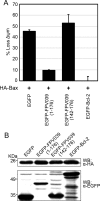
FIG. 1.
FIG. 1.
FPV039 inhibits Bax-induced apoptosis. (A) HeLa cells were cotransfected with either pEGFP, pEGFP-FPV039(1-176), pEGFP-FPV039(142-176), or pEGFP-Bcl-2 and pcDNA3-HA-Bax. Eighteen hours posttransfection, apoptosis was quantified by TMRE fluorescence in EGFP-positive cells using two-color flow cytometry. Standard deviations were calculated from three independent experiments. (B) Expression levels of the various transfected proteins. HeLa cells were transfected as above, and protein levels were determined by Western blotting (WB) of whole-cell lysates with either anti-EGFP or anti-HA antibodies.
FIG. 2.
FIG. 2.
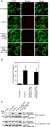
FPV039 inhibits Bax activity. (A) HeLa cells were infected with VV-EGFP, VVΔF1L, VVΔF1L-Flag-FPV039(1-176), or VVΔF1L-Flag-FPV039(Δ41-54) at an MOI of 10 for 24 h. Following fixation and permeabilization, Bax activation was visualized with anti-Bax6A7. (B) Microscopy analysis was quantified as the percentage of cells displaying Bax activation (mean ± SD). (C) HeLa cells were infected as for panel A. Cells were lysed using either 2% CHAPS or 1% Triton X-100, and activated Bax was immunoprecipitated (IP) with anti-Bax6A7 and Western blotted with anti-BaxN20.
FIG. 3.
FIG. 3.
FPV039 inhibits the conformational activation of Bax. Wild-type Jurkat cells (a to f), Jurkat cells devoid of Bak and Bax (g to k), or Jurkat cells overexpressing Bcl-2 (l to p) were infected with VV-EGFP, VVΔF1L, VVΔF1L-Flag-FPV039(1-176), or VVΔF1L-Flag-FPV039(Δ41-54) at an MOI of 5 for 6 h and then treated with 2 μM STS for 2 h to induce apoptosis. Bax activation was monitored by flow cytometry using anti-Bax6A7 or the isotype control antibody (NK1.1). Shaded histograms, untreated cells; open histograms, staurosporine-treated cells. Data are representative of at least three independent experiments.
FIG. 4.
FIG. 4.
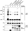
FPV039 inhibits Bax oligomerization. HeLa cells were cotransfected with pEGFP, pEGFP-FPV039(1-176), pEGFP-FPV039(Δ41-54), pEGFP-FPV039(1-94), or pEGFP-Bcl-2 and pcDNA3-HA-Bax. zVAD.fmk was added following transfection to prevent the activation of caspases. Cells were lysed in 2% CHAPS, and cell lysates were treated with 1 μM BMH and Western blotted (WB) with anti-HA to detect Bax oligomers (upper panel). A portion of the lysates (input) were treated with dimethyl sulfoxide alone and subjected to Western blotting with either anti-HA or anti-EGFP antibodies to determine expression levels of the transfected proteins (lower two panels). x1, Bax monomer; x2, Bax dimer; xN, higher-order Bax oligomers.
FIG. 5.
FIG. 5.
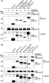
FPV039 interacts with Bax. (A) HEK293T cells were cotransfected with pEGFP, pEGFP-FPV039(1-176), pEGFP-FPV039(Δ41-54), pEGFP-FPV039(1-94), or pEGFP-Bcl-2 and pcDNA3-HA-Bax, followed by lysis with 2% CHAPS. Cell lysates were immunoprecipitated (IP) with anti-EGFP and Western blotted (WB) with either anti-BaxN20 or anti-EGFP to detect interaction. Whole-cell lysates were Western blotted with anti-BaxN20 or anti-EGFP to determine the expression levels of HA-Bax or the EGFP-tagged proteins, respectively. (B) HEK293T cells were cotransfected with pEGFP, pEGFP-F1L, pEGFP-M11L, pEGFP-FPV039(1-176), pEGFP-N1L, or pEGFP-Bcl-2 and pcDNA3-HA-Bax. Immunoprecipitations and Western blotting was performed as described for panel A. zVAD.fmk was added following transfection to prevent the activation of caspases.
FIG. 6.
FIG. 6.
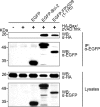
FPV039 retains an interaction with Bax in the absence of Bak. Bak−/− BMK cells were cotransfected with pEGFP, pEGFP-Bcl-2, or pEGFP-FPV039(1-176) and pcDNA3-HA-Bax, followed by lysis with 2% CHAPS. Cell lysates were immunoprecipitated (IP) with anti-EGFP and Western blotted (WB) with either anti-HA or anti-EGFP to detect interaction. Whole-cell lysates were Western blotted with anti-HA or anti-EGFP to determine the expression levels. zVAD.fmk was added following transfection to prevent the activation of caspases.
FIG. 7.
FIG. 7.
FPV039 interacts with active Bax in the context of virus infection. HeLa cells were mock infected or infected at an MOI of 5 with VVΔF1L, VV-Flag-F1L, or VVΔF1L-Flag-FPV039(1-176). Sixteen hours postinfection, cells were lysed in 2% CHAPS, and cell lysates were immunoprecipitated (IP) with either anti-Flag (A) or anti-Bax6A7 (B) and Western blotted (WB) with either anti-Flag or anti-Bax2D2 (A and B). Lysates were Western blotted with anti-Bax2D2 and anti-Flag (A) or anti-BaxN20 and anti-Flag (B). *, Flag-F1L (B).
FIG. 8.
FIG. 8.
FPV039 inhibits apoptosis induced by BH3-only proteins. HeLa cells were cotransfected with pEGFP and pEGFP-FPV039(1-176) along with one of the following BH3-only proteins and the relevant cellular Bcl-2 protein: pcDNA3-Flag-BimL and pEGFP-Bcl-2 (A), pcDNA3-Myc-Bik and pEGFP-Bcl-2 (B), pcDNA3-T7-Bmf and pEGFP-Bcl-2 (C), pXJ40-HA-Bad and pEGFP-Bcl-2 (D), pcDNA3-HA-Puma and pEGFP-Bcl-2 (E), pcDNA3-Bid-Flag and pEGFP-Bcl-XL (F), pcDNA3-tBid-Flag and pEGFP-Bcl-XL (G), or pcDNA3.1-Myc-Noxa and pEGFP-Mcl-1 (H). Eighteen hours posttransfection, apoptosis was quantified by TMRE fluorescence in EGFP-positive cells using two-color flow cytometry. Standard deviations were calculated from three independent experiments. Whole-cell lysates from parallel experiments were Western blotted with the following antibodies to determine expression levels of the BH3-only proteins: anti-Flag (A, F, and G), anti-Myc (B), anti-T7 (C), anti-HA (D and E), anti-Noxa (H). *, complete protection by EGFP-Bcl-2 or EGFP-Bcl-XL.
FIG. 9.
FIG. 9.
FPV039 interacts with BimL. HeLa cells were transfected with pEGFP, pEGFP-F1L, pEGFP-M11L, pEGFP-FPV039(1-176), or pEGFP-Bcl-2 and pcDNA3-Flag-BimL, followed by lysis with 2% CHAPS. Cell lysates were immunoprecipitated (IP) with anti-EGFP and Western blotted (WB) with either anti-Flag or anti-EGFP to detect interaction. Whole-cell lysates were Western blotted with anti-Flag or anti-EGFP to determine expression levels. zVAD.fmk was added following transfection to prevent the activation of caspases.
FIG. 10.
FIG. 10.
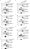
FPV039 interacts with a subset of BH3-only proteins. HEK293T cells were cotransfected with pEGFP and pEGFP-FPV039(1-176) along with one of the following BH3-only proteins and the relevant cellular Bcl-2 protein: pcDNA3-Myc-Bik and pEGFP-Bcl-2 (A), pcDNA3-T7-Bmf and pEGFP-Bcl-2 (B), pXJ40-HA-Bad and pEGFP-Bcl-2 (C), pcDNA3-HA-Puma and pEGFP-Bcl-2 (D), pcDNA3-Bid-Flag and pEGFP-Bcl-XL (E), pcDNA3-tBid-Flag and pEGFP-Bcl-XL (F), pcDNA3.1-Myc-Noxa and pEGFP-Mcl-1 (G). zVAD.fmk was added following transfection to prevent the activation of caspases. Cells were lysed with 2% CHAPS and immunoprecipitated (IP) with anti-EGFP. IP samples and whole-cell lysates were Western blotted (WB) with anti-EGFP and anti-Myc (A), anti-T7 (B), anti-HA (C and D), anti-Flag (E and F), or anti-Noxa (G).

Similar articles

Cited by

References

    1. Afonso, C. L., E. R. Tulman, Z. Lu, L. Zsak, G. F. Kutish, and D. L. Rock. 2000. The genome of fowlpox virus. J. Virol. 743815-3831. - PMC - PubMed
    1. Annis, M. G., E. L. Soucie, P. J. Dlugosz, J. A. Cruz-Aguado, L. Z. Penn, B. Leber, and D. W. Andrews. 2005. Bax forms multispanning monomers that oligomerize to permeabilize membranes during apoptosis. EMBO J. 242096-2103. - PMC - PubMed
    1. Antonsson, B., S. Montessuit, S. Lauper, R. Eskes, and J. C. Martinou. 2000. Bax oligomerization is required for channel-forming activity in liposomes and to trigger cytochrome c release from mitochondria. Biochem. J. 345271-278. - PMC - PubMed
    1. Antonsson, B., S. Montessuit, B. Sanchez, and J. C. Martinou. 2001. Bax is present as a high molecular weight oligomer/complex in the mitochondrial membrane of apoptotic cells. J. Biol. Chem. 27611615-11623. - PubMed
    1. Aoyagi, M., D. Zhai, C. Jin, A. E. Aleshin, B. Stec, J. C. Reed, and R. C. Liddington. 2007. Vaccinia virus N1L protein resembles a B cell lymphoma-2 (Bcl-2) family protein. Protein Sci. 16118-124. - PMC - PubMed

Publication types

MeSH terms

Substances