

White matter damage in frontotemporal dementia and Alzheimer's disease measured by diffusion MRI
- PMID: 19439421
- PMCID: PMC2732263
- DOI: 10.1093/brain/awp071
White matter damage in frontotemporal dementia and Alzheimer's disease measured by diffusion MRI
Abstract
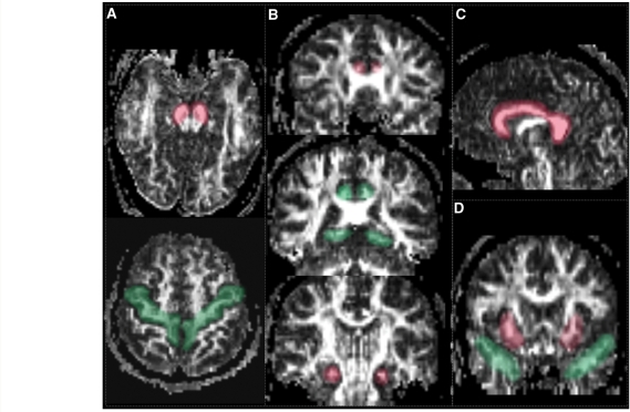
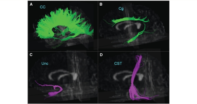
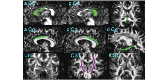
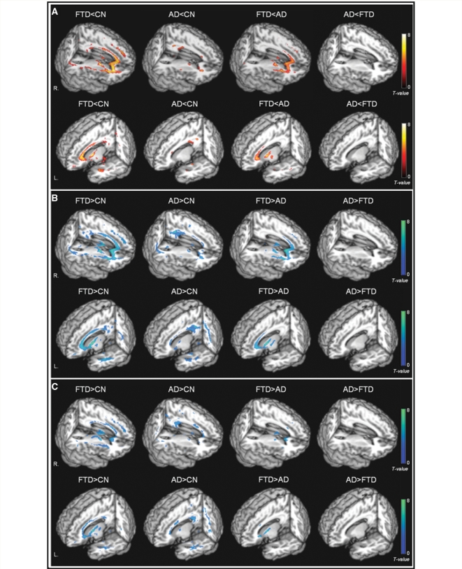
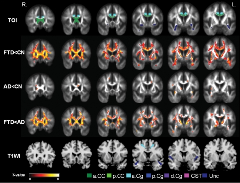
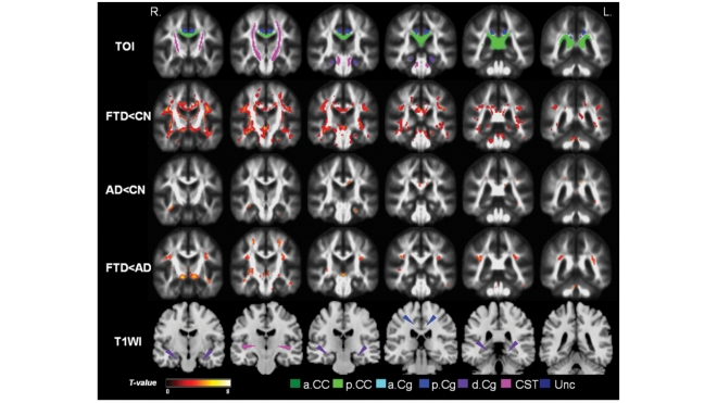
Frontotemporal dementia (FTD) and Alzheimer's disease are sometimes difficult to differentiate clinically because of overlapping symptoms. Using diffusion tensor imaging (DTI) measurements of fractional anisotropy (FA) can be useful in distinguishing the different patterns of white matter degradation between the two dementias. In this study, we performed MRI scans in a 4 Tesla MRI machine including T1-weighted structural images and diffusion tensor images in 18 patients with FTD, 18 patients with Alzheimer's disease and 19 cognitively normal (CN) controls. FA was measured selectively in specific fibre tracts (including corpus callosum, cingulum, uncinate and corticospinal tracts) as well as globally in a voxel-by-voxel analysis. Patients with FTD were associated with reductions of FA in frontal and temporal regions including the anterior corpus callosum (P < 0.001), bilateral anterior (left P < 0.001; right P = 0.005), descending (left P < 0.001; right P = 0.003) cingulum tracts, and uncinate tracts (left P < 0.001; right P = 0.005), compared to controls. Patients with Alzheimer's disease were associated with reductions of FA in parietal, temporal and frontal regions including the left anterior (P = 0.003) and posterior (P = 0.002) cingulum tracts, bilateral descending cingulum tracts (P < 0.001) and left uncinate tracts (P < 0.001) compared to controls. When compared with Alzheimer's disease, FTD was associated with greater reductions of FA in frontal brain regions, whereas no region in Alzheimer's disease showed greater reductions of FA when compared to FTD. In conclusion, the regional patterns of anisotropy reduction in FTD and Alzheimer's disease compared to controls suggest a characteristic distribution of white matter degradation in each disease. Moreover, the white matter degradation seems to be more prominent in FTD than in Alzheimer's disease. Taken together, the results suggest that white matter degradation measured with DTI may improve the diagnostic differentiation between FTD and Alzheimer's disease.
Figures
Similar articles
-
Different regional patterns of cortical thinning in Alzheimer's disease and frontotemporal dementia.Brain. 2007 Apr;130(Pt 4):1159-66. doi: 10.1093/brain/awm016. Epub 2007 Mar 12. Brain. 2007. PMID: 17353226 Free PMC article.
-
Longitudinal white matter change in frontotemporal dementia subtypes and sporadic late onset Alzheimer's disease.Neuroimage Clin. 2017 Sep 14;16:595-603. doi: 10.1016/j.nicl.2017.09.007. eCollection 2017. Neuroimage Clin. 2017. PMID: 28975068 Free PMC article.
-
An advanced white matter tract analysis in frontotemporal dementia and early-onset Alzheimer's disease.Brain Imaging Behav. 2016 Dec;10(4):1038-1053. doi: 10.1007/s11682-015-9458-5. Brain Imaging Behav. 2016. PMID: 26515192 Free PMC article.
-
The role of diffusion tensor imaging and fractional anisotropy in the evaluation of patients with idiopathic normal pressure hydrocephalus: a literature review.Neurosurg Focus. 2016 Sep;41(3):E12. doi: 10.3171/2016.6.FOCUS16192. Neurosurg Focus. 2016. PMID: 27581308 Review.
-
Diffusion tensor imaging in Alzheimer's disease and mild cognitive impairment.Behav Neurol. 2009;21(1):39-49. doi: 10.3233/BEN-2009-0234. Behav Neurol. 2009. PMID: 19847044 Free PMC article. Review.
Cited by
-
Simultaneous changes in gray matter volume and white matter fractional anisotropy in Alzheimer's disease revealed by multimodal CCA and joint ICA.Neuroscience. 2015 Aug 20;301:553-62. doi: 10.1016/j.neuroscience.2015.06.031. Epub 2015 Jun 23. Neuroscience. 2015. PMID: 26116521 Free PMC article.
-
Integrating retrogenesis theory to Alzheimer's disease pathology: insight from DTI-TBSS investigation of the white matter microstructural integrity.Biomed Res Int. 2015;2015:291658. doi: 10.1155/2015/291658. Epub 2015 Jan 20. Biomed Res Int. 2015. PMID: 25685779 Free PMC article. Review.
-
Differential diagnosis of neurodegenerative diseases using structural MRI data.Neuroimage Clin. 2016 Mar 5;11:435-449. doi: 10.1016/j.nicl.2016.02.019. eCollection 2016. Neuroimage Clin. 2016. PMID: 27104138 Free PMC article.
-
Magnetic Resonance Imaging in Tauopathy Animal Models.Front Aging Neurosci. 2022 Jan 25;13:791679. doi: 10.3389/fnagi.2021.791679. eCollection 2021. Front Aging Neurosci. 2022. PMID: 35145392 Free PMC article. Review.
-
Diffusion kurtosis imaging allows the early detection and longitudinal follow-up of amyloid-β-induced pathology.Alzheimers Res Ther. 2018 Jan 9;10(1):1. doi: 10.1186/s13195-017-0329-8. Alzheimers Res Ther. 2018. PMID: 29370870 Free PMC article.
References
-
- Anderson VC, Litvack ZN, Kaye JA. Magnetic resonance approaches to brain aging and Alzheimer disease-associated neuropathology [Review] Top Magn Reson Imaging. 2005;16:439–52. - PubMed
-
- Aoki S, Iwata NK, Masutani Y, Yoshida M, Abe O, Ugawa Y, et al. Quantitative evaluation of the pyramidal tract segmented by diffusion tensor tractography: feasibility study to patients with amyotrophic lateral sclerosis. Radiat Med. 2005;23:195–9. - PubMed
-
- Bartenstein P, Minoshima S, Hirsch C, Buch K, Willoch F, Mösch D, et al. Quantitative assessment of cerebral blood flow in patients with Alzheimer's disease by SPECT. J Nucl Med. 1997;38:1095–101. - PubMed
-
- Bartzokis G. Age-related myelin breakdown: a developmental model of cognitive decline and Alzheimer's disease [Review] Neurobiol Aging. 2004;25:5–18. - PubMed
-
- Borroni B, Brambati SM, Agosti C, Gipponi S, Bellelli G, Gasparotti R, et al. Evidence of white matter changes on diffusion tensor imaging in frontotemporal dementia. Arch Neurol. 2007;64:246–51. - PubMed
Publication types
MeSH terms
Grants and funding
LinkOut - more resources
Full Text Sources
Other Literature Sources
Medical
