. 2009;4(5):e5515.
doi: 10.1371/journal.pone.0005515. Epub 2009 May 13.

Abberant alpha-synuclein confers toxicity to neurons in part through inhibition of chaperone-mediated autophagy

Affiliations

Abberant alpha-synuclein confers toxicity to neurons in part through inhibition of chaperone-mediated autophagy

Maria Xilouri et al. PLoS One. 2009.

Abstract

Background: The mechanisms through which aberrant alpha-synuclein (ASYN) leads to neuronal death in Parkinson's disease (PD) are uncertain. In isolated liver lysosomes, mutant ASYNs impair Chaperone Mediated Autophagy (CMA), a targeted lysosomal degradation pathway; however, whether this occurs in a cellular context, and whether it mediates ASYN toxicity, is unknown. We have investigated presently the effects of WT or mutant ASYN on the lysosomal pathways of CMA and macroautophagy in neuronal cells and assessed their impact on ASYN-mediated toxicity.

Methods and findings: Novel inducible SH-SY5Y and PC12 cell lines expressing human WT and A53T ASYN, as well as two mutant forms that lack the CMA-targeting motif were generated. Such forms were also expressed in primary cortical neurons, using adenoviral transduction. In each case, effects on long-lived protein degradation, LC3 II levels (as a macroautophagy index), and cell death and survival were assessed. In both PC12 and SH-SY5Y cycling cells, induction of A53T ASYN evoked a significant decrease in lysosomal degradation, largely due to CMA impairment. In neuronally differentiated SH-SH5Y cells, both WT and A53T ASYN induction resulted in gradual toxicity, which was partly dependent on CMA impairment and compensatory macroautophagy induction. In primary neurons both WT and A53T ASYN were toxic, but only in the case of A53T ASYN did CMA dysfunction and compensatory macroautophagy induction occur and participate in death.

Conclusions: Expression of mutant A53T, and, in some cases, WT ASYN in neuronal cells leads to CMA dysfunction, and this in turn leads to compensatory induction of macroautophagy. Inhibition of these lysosomal effects mitigates ASYN toxicity. Therefore, CMA dysfunction mediates aberrant ASYN toxicity, and may be a target for therapeutic intervention in PD and related disorders. Furthermore, macroautophagy induction in the context of ASYN over-expression, in contrast to other settings, appears to be a detrimental response, leading to neuronal death.


Conflict of interest statement

Competing Interests: The authors have declared that no competing interests exist.

Figures

Figure 1
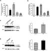
Figure 1. Over-expression of A53T ASYN in PC12 cells results in lysosomal dysfunction due to CMA impairment.
(A) Generation of stable inducible Tet-Off PC12 cell lines over-expressing human A53T and ΔDQ/A53T ASYN. Cells were cultured in the presence (+) or absence (−) of dox (2 µg/ml) for 4 days and assayed for ASYN expression with the C20 polyclonal Ab. ERK Ab is used as a loading control. (B, C) PC12 cells stably transfected with WT or mutant ASYNs (A53T, ΔDQ/A53T) or control bgal were labeled with [3H] leucine for 48 hrs (2 µCi/ml). Cells were treated with or without NH4Cl (25 mM) or 3MA (10 mM) and degraded proteins were assayed 14 hrs later. Rate of total (B) (inhibitable by NH4Cl) and (C) of macroautophagic (inhibitable by 3MA) long lived protein degradation in PC12 cell lines expressing bgal, WT, A53T or ΔDQ/A53T ASYN. For each line, protein degradation was assessed in the presence or absence of dox, and the results are reported as percentage degradation in the absence compared to the presence of dox (induced vs. non-induced). All presented data are the mean of 3 independent experiments and within each experiment triplicate samples per condition were assessed. (***p<0.001, one way ANOVA followed by the Student-Newman-Keuls' test, comparing between cells expressing all forms of ASYN and bgal controls; ## p<0.01, comparing between cells expressing A53T and ΔDQ/A53T ASYN).
Figure 2
Figure 2. Over-expression of A53T ASYN impairs lysosomal function due to CMA targeting in proliferating SH-SY5Y cells.
(A) Generation of stable inducible Tet-Off SH-SY5Y cell lines over-expressing human A53T and ΔDQ/A53T ASYN. The cells were cultured in the presence (+) or absence (−) of dox (3 µg/ml) for 4 days and assayed for ASYN expression with the C20 polyclonal Ab. ERK Ab is used as a loading control. (B, C) SH-SY5Y cells stably transfected with WT or mutant ASYNs (A53T, ΔDQ/A53T) or control bgal were labeled with [3H] leucine for 48 hrs (2 µCi/ml). Cells were treated with or without Baf (500 nM) or 3MA (10 mM) in serum free medium and degraded proteins were assayed 14 hrs later. Rate of total (B) (inhibitable by Baf) and (C) of macroautophagic (inhibitable by 3MA) long lived protein degradation in SH-SY5Y cells expressing bgal, WT, A53T or ΔDQ/A53T ASYN. For each line, protein degradation was assessed in the presence or absence of dox, and the results are reported as percentage degradation in the absence compared to the presence of dox (induced vs. non-induced). All presented data are the mean of 3 independent experiments and within each experiment triplicate samples per condition were assessed. (***p<0.001, one way ANOVA followed by the Student-Newman-Keuls' test, comparing between cells expressing all forms of ASYN and bgal controls; ### p<0.001, comparing between cells expressing A53T and ΔDQ/A53T ASYN).
Figure 3
Figure 3. Over-expression of ASYNs impairs lysosomal function in differentiated SH-SY5Y cells, dependent on CMA targeting.
(A, B) SH-SY5Y cells expressing WT or mutant ASYNs were differentiated in 20 µM RA for 5 days in the presence or absence of dox and then treated as in Figure 2. Rate of total (A) and (B) macroautophagy-dependent long lived protein degradation in differentiated SH-SY5Y cells expressing ASYN (WT, ΔDQ/WT, A53T or ΔDQ/A53T). Bgal expressing cells are used as controls. For each line, protein degradation was assessed in the presence or absence of dox, and the results are reported as percentage degradation in the absence compared to the presence of dox (induced/non-induced). All presented data are the mean of 4 independent experiments and within each experiment triplicate samples per condition were assessed. (***p<0.001, one way ANOVA followed by the Student-Newman-Keuls' test, comparing between cells expressing all forms of ASYN and control bgal; ### p<0.001, comparing between cells expressing WT and ΔDQ/WT ASYN). (C, D) Western blotting analysis of differentiated SH-SY5Y cells (7 days, +/−dox) expressing A53T, ΔDQ/A53T, WT or ΔDQ/WT ASYN. ERK Ab is used as a loading control. Representative immunoblots of LC3 II are presented in the left panels and quantification of LC3 II levels after ASYN induction (−dox compared to +dox) is shown in the right panels. All results are expressed as the ratio of OD values to the corresponding controls and data are presented as mean of ±S.E. of 3 independent experiments [*p<0.05, Student's t-test comparing between cells expressing A53T and ΔDQ/A53T (C), or between cells expressing WT and ΔDQ/WT ASYN (D)].
Figure 4
Figure 4. ASYN-induction is toxic to differentiated SH-SH5Y cells due to CMA blockade and macroautophagy induction.
(A, B) ASYN (WT, ΔDQ/WT, A53T or ΔDQ/A53T)-expressing SH-SH5Y cells, were differentiated in RA (+/−dox). Samples were collected at 5, 7, 9, 12, 14 days after RA addition and survival was assessed by counting the number of intact nuclei. Rate of survival, presented in each case as the percentage of the (−) over the (+) dox condition, is shown for WT/ΔDQ and WT ASYN (A), and for A53T/ΔDQ and A53T ASYN (B) cells. All presented data are the mean of 3 independent experiments. Within each experiment triplicate samples per condition were assessed. (*p<0.05, **p<0.01, ***p<0.001, Student's t-test comparing WT or A53T ASYN-expressing cells and their corresponding ΔDQ mutants). (C, D) Suppression of macroautophagy with 3MA (C) or with ATG 5 siRNA (D), rescues differentiated SH-SY5Y cells from ASYN-induced death. (C) ASYN (WT, ΔDQ/WT, A53T)-expressing SH-SY5Y cells, were differentiated (+/−dox) for 5 days before 3MA addition. 36 hrs later, survival was assessed as in A, B. (*p<0.05, student's t-test, comparing WT or A53T-expressing cells+/−3MA). (D) WT or A53T ASYN cells and bgal cells were differentiated for 5 days (+/−dox) and transfected with scrambled (scr) or ATG 5 siRNA together with EGFP. Cell death was assessed 72 hrs later by counting the percentage of EGFP-positive transfected cells that were also Ethidium Homodimer-positive. At least 100 EGFP-positive cells were counted per well per condition. The data are presented as mean±SE of 3 independent experiments. Within each experiment triplicate samples per condition were assessed (### p<0.001, one way ANOVA followed by the Student-Newman-Keuls' test, comparing WT or A53T ASYN cells+/−dox; *p<0.05, **p<0.01, comparing WT or A53T ASYN-induced cells (−dox) transfected with ATG 5/EGFP to the control scr/EGFP transfected cells).
Figure 5
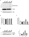
Figure 5. Over-expression of A53T ASYN in primary cortical neurons alters lysosomal function dependent on CMA targeting.
(A) Five day-old cortical cultures were transduced with adenoviruses (MOI 150) expressing human ASYN (WT, ΔDQ/WT, A53T or ΔDQ/A53T) or EGFP (control virus) as described in Materials and Methods. ASYN expression was assessed 96 hrs later by performing immunoblotting with the C20 polyclonal Ab. ERK Ab is used as a loading control. A representative immunoblot of ASYN expression is shown. (B, C) Rate of total (inhibitable by NH4Cl) and of macroautophagic (inhibitable by 3MA) long lived protein degradation in rat cortical cultures, 96 hrs after transduction with adenoviruses expressing WT or mutant ASYNs. EGFP transduced neurons are used as controls. All presented data are the mean of 4 independent experiments and within each experiment triplicate samples per condition were assessed. (D) Cortical neurons were treated as in B, lysed and assessed by western immunoblotting for LC3 II levels. ERK Ab is used as a loading control. Representative immunoblot of LC3 II is presented in the left panel and quantification of LC3 II levels after WT or mutant ASYN transduction is shown in the right panel. All results are expressed as the ratio of OD values to the corresponding controls and all data are presented as mean of ±S.E. of 4 independent experiments (* p<0.05, one way ANOVA followed by the Student-Newman-Keuls' test, comparing between cultures expressing various forms of ASYN and control EGFP; # p<0.05, comparing between cultures transduced with A53T and ΔDQ/A53T ASYN).
Figure 6
Figure 6. Over-expression of human ASYN (WT, mutants) is toxic to rat cortical neurons.
(A) Five days-old cortical cultures were transduced with adenoviruses (MOI 150) expressing human ASYN (WT, ΔDQ/WT, A53T or ΔDQ/A53T) or EGFP (control virus) as described in Materials and Methods. 96 hrs later cells were lysed with a nuclear-sparing buffer and intact nuclei were counted in a hemacytometer. (B) Cortical cultures were treated as in A with slight modifications. 72 hrs post-infection, 3MA (10 mM) was added and intact nuclei were counted 24 hrs later. All data are presented as mean±SE of independent experiments and within each experiment triplicate samples per condition were assessed. (*p<0.05, **p<0.01, one way ANOVA followed by the Student-Newman-Keuls' test, comparing between cultures expressing various forms of ASYN and control EGFP; # p<0.05 comparing between cultures transduced with A53T and ΔDQ/A53T ASYN or between A53T ASYN transduced neurons+/−3MA).
Figure 7
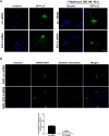
Figure 7. ATG 5 down-regulation decreases human A53T ASYN-induced death in rat cortical neurons.
(A) Five day-old cortical cultures were transfected with GFP-LC3 cDNA together with the scrambled (scr) or the ATG 5 siRNA. 24 hrs later, rapamycin was added (500 nM, 48 hrs) and formation of GFP-LC3 vacuoles (dots) was determined by fluorescent microscopy. Representative fluorescent microscopic pictures showing the formation of GFP-LC3 vacuoles (indicated by the arrow) in cultures transfected with the GFP-LC3 construct together with the scr siRNA after rapamycin addition are shown. Vacuoles (dots) failed to be observed in rapamycin-treated cultures transfected with the GFP-LC3 construct together with the ATG 5 siRNA. (B) Five day-old cortical cultures were transduced with A53T ASYN adenovirus and 24 hrs later were transiently transfected with the scr or ATG siRNA along with an EGFP construct to monitor transfection. Cell death was assessed 72 hrs later by counting the percentage of EGFP-positive transfected cells that were positive for Ethidium Homodimer stain (dying cells). Representative pictures are shown in the upper panel and quantification of the percentage of scr or ATG5/EGFP positive cells that were also stained with Ethidium Homodimer is shown in the bottom panel. At least 100 EGFP-positive cells were counted per condition. The data are presented as mean±SE of 3 independent experiments (**p<0.01, Student's t-test, comparing A53T ASYN transduced neurons transfected with the scr or the ATG 5 siRNA. Bars: (A) 10 µM; (B) 50 µM.
Figure 8
Figure 8. Reduction of dopamine levels improves lysosomal dysfunction and survival of differentiated SH-SY5Y WT ASYN expressing cells.
(A, B) SH-SY5Y cells expressing WT or ΔDQ/WT ASYN were differentiated in RA (20 µM) in the presence (+) or absence (−) of dox (3 µg/ml) for 5 days before addition of alpha-mehtyl-p-tyrosine (AMPT, 1 mM). Next, cells were labeled with [3H] leucine for 48 hrs (2 µCi/ml) and degraded proteins were assayed 14 hrs later. (A) Rate of total long lived protein degradation in differentiated SH-SY5Y cell lines expressing WT ASYN+/−AMPT. ΔDQ/WT ASYN expressing cells are used as control. All data are presented as the relative percentage of long-lived protein degradation in the (−) relative to the (+) dox setting for each condition (B) Cells expressing WT or ΔDQ/WT ASYN were differentiated as in A for 5 days and AMPT was added for subsequent 3 days. Survival was assessed by counting the number of intact nuclei and is presented as (−) relative to (+) dox condition. All presented data are the mean of 3 independent experiments, and within each experiment triplicate samples per condition were assessed. (*p<0.05, **p<0.01, ***p<0.001, one-way ANOVA followed by the Student-Newman-Keuls' test, comparing between cells expressing WT ASYN+/−AMPT, and between cells expressing WT and ΔDQ/WT ASYN).
Figure 9
Figure 9. Schematic diagram of the lysosomal effects and relevant cell death pathways induced by aberrant ASYN.
Aberrant ASYN induces in all settings CMA dysfunction. When this leads to compensatory macroautophagy induction (as assessed by autophagosome formation/accumulation), autophagic cell death, inhibited by macroautophagy inhibitors, occurs. In certain settings (A53T ASYN in neuronally differentiated SH-SY5Y cells), lysosomal dysfunction occurs independent of CMA targeting and may contribute to death (dotted arrows).

Similar articles

Cited by

References

    1. Spillantini MG, Crowther RA, Jakes R, Hasegawa M, Goedert M. alpha-Synuclein in filamentous inclusions of Lewy bodies from Parkinson's disease and dementia with lewy bodies. Proc Natl Acad Sci U S A. 1998;95:6469–6473. - PMC - PubMed
    1. Dauer W, Przedborski S. Parkinson's disease: mechanisms and models. Neuron. 2003;39:889–909. - PubMed
    1. Giasson BI, Duda JE, Murray IV, Chen Q, Souza JM, et al. Oxidative damage linked to neurodegeneration by selective alpha-synuclein nitration in synucleinopathy lesions. Science. 2000;290:985–989. - PubMed
    1. Polymeropoulos MH, Lavedan C, Leroy E, Ide SE, Dehejia A, et al. Mutation in the alpha-synuclein gene identified in families with Parkinson's disease. Science. 1997;276:2045–2047. - PubMed
    1. Kruger R, Kuhn W, Muller T, Woitalla D, Graeber M, et al. Ala30Pro mutation in the gene encoding alpha-synuclein in Parkinson's disease. Nat Genet. 1998;18:106–108. - PubMed

Publication types