. 2009 Jun 1;122(Pt 11):1778-87.
doi: 10.1242/jcs.040956. Epub 2009 May 12.

Alpha3beta1 integrin in epidermis promotes wound angiogenesis and keratinocyte-to-endothelial-cell crosstalk through the induction of MRP3

Affiliations

Alpha3beta1 integrin in epidermis promotes wound angiogenesis and keratinocyte-to-endothelial-cell crosstalk through the induction of MRP3

Kara Mitchell et al. J Cell Sci. .

Abstract

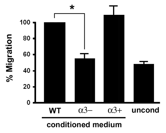
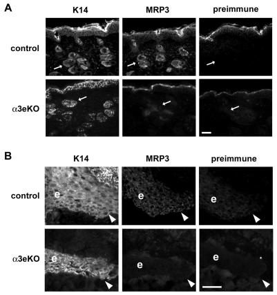
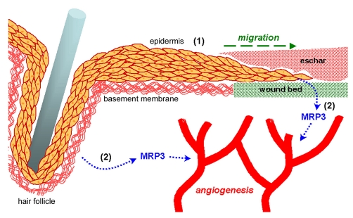
During cutaneous wound healing, epidermal keratinocytes play essential roles in the secretion of factors that promote angiogenesis. However, specific cues in the wound microenvironment that trigger the production of pro-angiogenic factors by keratinocytes, and the cellular receptors that mediate this response, remain unclear. In this study, we exploited a model of conditional integrin knockout to demonstrate impaired wound angiogenesis in mice that lack alpha3beta1 integrin in epidermis. In addition, we used genetic and shRNA approaches to determine that alpha3beta1-integrin deficiency in keratinocytes leads to reduced mRNA and protein expression of the pro-angiogenic factor mitogen-regulated protein 3 (MRP3; also known as PRL2C4), and to demonstrate that this regulation provides a mechanism of keratinocyte-to-endothelial-cell crosstalk that promotes endothelial-cell migration. Finally, we showed that the impaired wound angiogenesis in epidermis-specific alpha3-integrin-knockout mice is correlated with reduced expression of MRP3 in wounded epidermis. These findings identify a novel role for alpha3beta1 integrin in promoting wound angiogenesis through a mechanism of crosstalk from epidermal to endothelial cells, and they implicate MRP3 in this integrin-dependent crosstalk. Such a mechanism represents a novel paradigm for integrin-mediated regulation of wound angiogenesis that extends beyond traditional roles for integrins in cell adhesion and migration.


Figures

Fig. 1.
Fig. 1.
α3-integrin expression is absent from epidermis of α3eKO mice. (A) Schematic representation of the strategy used to generate the targeted floxed Itga3 allele (see Materials and Methods for details). Frt and LoxP sites are indicated by red and blue triangles, respectively. NeoR, neomycin-resistance gene. (B,C) H&E histological sections from back skin of adult Itga3flx/– mice that either lack (B, control) or express (C, α3eKO) K14-Cre. Scale bar: 50 μm. (D-I) Frozen skin sections from adult control mice (Itga3flx/flx; D,F,H) or α3eKO mice (K14-Cre:Itga3flx/flx; E,G,I) were stained by immunofluorescence with anti-keratin 14 (K14), anti-α3-integrin (α3), or anti-LN-332 (LN). Arrowheads in D-G point to basal keratinocytes; arrows in H,I point to basement membrane; e, epidermis; d, dermis; f, hair follicle. Note that specific α3-integrin staining is absent from the basal cell layer of α3eKO epidermis [G; asterisks in F,G indicate non-specific staining in both control and α3eKO epidermis, which was also seen with the corresponding pre-immune serum (not shown)]. Scale bar: 25 μm.
Fig. 2.
Fig. 2.
α3 integrin is absent from wound keratinocytes in α3eKO mice. (A,B) H&E-stained histological sections from 5-day excision wounds showing the epidermis migrating over the wound bed. Sections were from control mice (Itga3flx/–; A) or α3eKO mice (K14-Cre:Itga3flx/–; B); dotted lines outline the migrating epidermal tongue. Scale bar: 50 μm. (C-J) A series of adjacent cryosections from 5-day excision wounds of control mice (Itga3flx/flx; C,E,G,I) or α3eKO mice (K14-Cre:Itga3flx/flx; D,F,H,J) were stained by immunofluorescence with anti-keratin 14 (C,D), anti-α3-integrin (E,F), corresponding pre-immune serum (G,H), or anti-LN-332 (I,J). Arrowheads point to the leading edge of migrating epidermis. Scale bar: 50 μm. e, epidermis; d, dermis; s, eschar; wb, wound bed.
Fig. 3.
Fig. 3.
Wound angiogenesis is reduced in mice with α3β1-integrin-deficient epidermis. (A) CD31/PECAM-1 immunostaining in the dermis in frozen skin sections away from (distal) or within (wound bed) 5-day wounds of control (Itga3flx/–) or α3eKO (K14-Cre:Itga3flx/–) mice. Arrows point to examples of CD31-positive blood vessels. Scale bar: 50 μm. (B) Quantification of blood-vessel density. Three 20× fields were collected from within each wound bed (see C), and total area of CD31-staining was determined per field. Average blood-vessel area was then determined for each wound; each wound was from a separate 6.5-week-old mouse. Graph shows CD31-positive area for control mice and α3eKO mice. Data are mean ±s.e.m.; five mice were analyzed for each genotype; Student's t-test, *P<0.01. (C) Schematic illustration of a wound, showing approximate positions of fields (boxes, not to scale) that were imaged for analysis of blood-vessel density in panel B. d, distal; e, epidermis; s, eschar; wb, wound bed.
Fig. 4.
Fig. 4.
α3β1-integrin deficiency in keratinocytes leads to reduced secretion of soluble factors that induce endothelial-cell migration. Transwell migration assays were performed to compare relative cell migration of HUVECs in response to treatment with conditioned culture medium from wild-type keratinocytes (WT), Itga3-null keratinocytes (α3–), or the latter cells rescued with human α3 integrin (α3+). Bottom chambers of transwells contained keratinocyte-conditioned medium, or unconditioned medium (uncond) as a control to establish baseline migration. Graph shows average HUVEC migration as a percentage of that in cells treated with medium from wild-type cells; mean ±s.e.m.; n=3; *P<0.023, Student's t-test.
Fig. 5.
Fig. 5.
α3β1 integrin regulates Mrp3 mRNA and protein expression in cultured keratinocytes. (A) α3β1-integrin-expressing (α3+) or α3-integrin-deficient (α3–) keratinocytes were cultured for 24 hours on LN-332 ECM in serum-free medium, and total RNA was isolated. Semi-quantitative RT-PCR was performed to assess mRNA levels for Mrp3, Mmp9, Vegf (bands correspond to 120 kD and 160 kD isoforms) and β-actin. Results shown are representative of three independent experiments. (B) Keratinocytes were cultured as in A, and total cell lysate, or equivalent proportions of conditioned culture media, were assessed for MRP3 protein levels by immunoblot. Cells assayed included wild type (WT), Itga3-null (α3–) or the latter cells stably transfected with human α3-integrin subunit (α3+). Note that MRP3 migrates as a diffuse band, probably due to heterogeneity in extent of glycosylation (Corbacho et al., 2002). The asterisk indicates a non-specific band, which was also detected with corresponding pre-immune serum (not shown). Immunoblot for anti-keratin 14 (K14) was performed to control for loading differences. (C) Quantification of MRP3 protein levels in cell lysates such as those from B. MRP3 protein levels are indicated as a proportion of levels in wild-type cells after normalization to keratin 14. Data are the mean ±s.e.m.; n=3; *P<0.001, Student's t-test.
Fig. 6.
Fig. 6.
MRP3 expression is required in keratinocytes for the α3β1-integrin-dependent induction of endothelial-cell migration. α3β1-integrin-expressing keratinocytes were transduced with lentivirus expressing non-targeting shRNA (ctrl) or three different shRNAs that target murine Mrp3 (1-3), and stable populations were selected in puromycin. To confirm stable suppression of Mrp3 mRNA and MRP3 protein, transduced cells were cultured on collagen for 24 hours in serum-free medium, then (A) total RNA was isolated for RT-PCR analysis of Mrp3 mRNA, or β-actin mRNA as a control, and (B) conditioned culture medium was collected for immunoblot analysis of secreted MRP3 protein. (C) Transwell migration assays were performed as in Fig. 4 to compare HUVEC migratory responses to conditioned culture medium from keratinocytes that express the control shRNA (ctrl) or each of the Mrp3-targeting shRNAs (1-3); treatment with unconditioned medium was included to establish baseline migration. Graph shows relative HUVEC migration normalized to basal migration of cells treated with unconditioned medium (uncond). Data are mean±s.e.m.; n≥4 over two independent experiments; *P<0.05 compared to cells expressing control shRNA; one-way ANOVA followed by a Dunnett's post-hoc test.
Fig. 7.
Fig. 7.
Reduced expression of MRP3 in wounded epidermis of α3eKO mice. (A) Adjacent cryosections from skin that is proximal to 5-day wounds (approximately 1000 μm), were obtained from 6.5-week-old control mice (Itga3flx/–) or α3eKO mice (K14-Cre:Itga3flx/–) and stained by immunofluorescence with anti-keratin 14 (K14) to label follicular and interfollicular epidermis, or with anti-MRP3 or corresponding pre-immune serum, as indicated. Arrows point to hair follicles. (B) Adjacent cryosections of the wound edge, showing the migrating epidermal tongue (e), were collected from wounds of the same mice assessed in A and stained with anti-keratin 14, anti-MRP3 or pre-immune serum. Arrowheads point to the leading edge of migrating epidermis. Scale bars: 50 μm.
Fig. 8.
Fig. 8.
Model depicting α3β1-integrin-mediated functions of epidermis that contribute to wound healing. In vitro and in vivo studies indicate distinct and separable roles for α3β1 integrin in the epidermis during wound healing: (1) complex roles in epidermal migration and regeneration (see text for details and references), and (2) production of pro-angiogenic factors, such as MRP3, that promote wound angiogenesis (current study). α3β1-integrin-dependent MRP3 expression is indicated in keratinocytes within a wound-proximal hair follicle, as well as in epidermis that is migrating into the wound beneath the eschar and over the provisional ECM of the wound bed (shown in green). The basement membrane (shown in red) is indicated beneath the adjacent epidermis and around the hair follicle.

Similar articles

Cited by

References

    1. Bengtson, N. W. and Linzer, D. I. (2000). Inhibition of tumor growth by the antiangiogenic placental hormone, proliferin-related protein. Mol. Endocrinol. 14, 1934-1943. - PubMed
    1. Brakebusch, C., Grose, R., Quondamatteo, F., Ramirez, A., Jorcano, J. L., Pirro, A., Svensson, M., Herken, R., Sasaki, T., Timpl, R. et al. (2000). Skin and hair follicle integrity is crucially dependent on beta 1 integrin expression on keratinocytes. EMBO J. 19, 3990-4003. - PMC - PubMed
    1. Castilho, R. M., Squarize, C. H., Patel, V., Millar, S. E., Zheng, Y., Molinolo, A. and Gutkind, J. S. (2007). Requirement of Rac1 distinguishes follicular from interfollicular epithelial stem cells. Oncogene 26, 5078-5085. - PubMed
    1. Chattopadhyay, N., Wang, Z., Ashman, L. K., Brady-Kalnay, S. M. and Kreidberg, J. A. (2003). alpha3beta1 integrin-CD151, a component of the cadherin-catenin complex, regulates PTPmu expression and cell-cell adhesion. J. Cell Biol. 163, 1351-1362. - PMC - PubMed
    1. Chmielowiec, J., Borowiak, M., Morkel, M., Stradal, T., Munz, B., Werner, S., Wehland, J., Birchmeier, C. and Birchmeier, W. (2007). c-Met is essential for wound healing in the skin. J. Cell Biol. 177, 151-162. - PMC - PubMed

Publication types

Substances

LinkOut - more resources