

Protective actions of sex steroid hormones in Alzheimer's disease
- PMID: 19427328
- PMCID: PMC2728624
- DOI: 10.1016/j.yfrne.2009.04.015
Protective actions of sex steroid hormones in Alzheimer's disease
Abstract
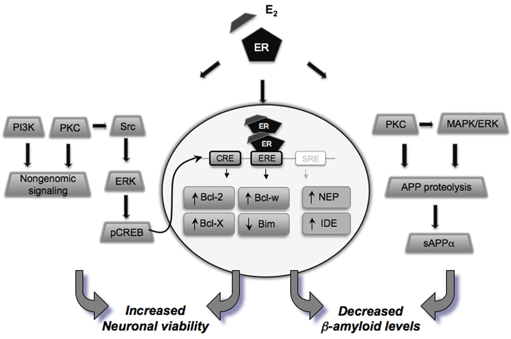
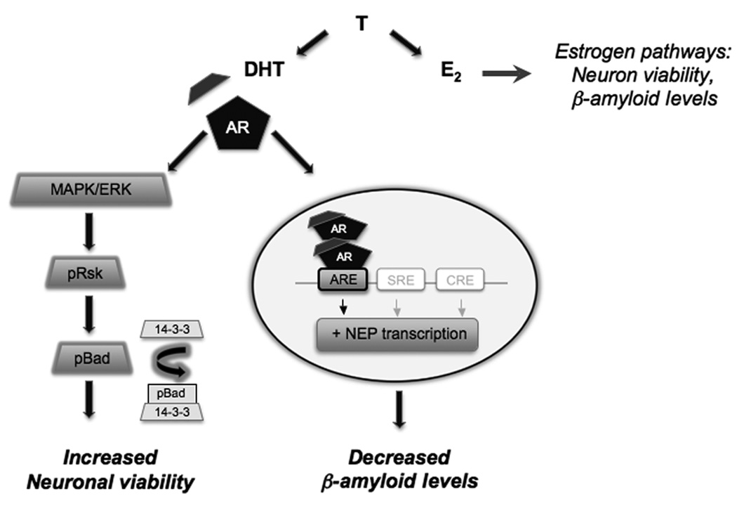
Risk for Alzheimer's disease (AD) is associated with age-related loss of sex steroid hormones in both women and men. In post-menopausal women, the precipitous depletion of estrogens and progestogens is hypothesized to increase susceptibility to AD pathogenesis, a concept largely supported by epidemiological evidence but refuted by some clinical findings. Experimental evidence suggests that estrogens have numerous neuroprotective actions relevant to prevention of AD, in particular promotion of neuron viability and reduction of beta-amyloid accumulation, a critical factor in the initiation and progression of AD. Recent findings suggest neural responsiveness to estrogen can diminish with age, reducing neuroprotective actions of estrogen and, consequently, potentially limiting the utility of hormone therapies in aged women. In addition, estrogen neuroprotective actions are also modulated by progestogens. Specifically, continuous progestogen exposure is associated with inhibition of estrogen actions whereas cyclic delivery of progestogens may enhance neural benefits of estrogen. In recent years, emerging literature has begun to elucidate a parallel relationship of sex steroid hormones and AD risk in men. Normal age-related testosterone loss in men is associated with increased risk to several diseases including AD. Like estrogen, testosterone has been established as an endogenous neuroprotective factor that not only increases neuronal resilience against AD-related insults, but also reduces beta-amyloid accumulation. Androgen neuroprotective effects are mediated both directly by activation of androgen pathways and indirectly by aromatization to estradiol and initiation of protective estrogen signaling mechanisms. The successful use of hormone therapies in aging men and women to delay, prevent, and or treat AD will require additional research to optimize key parameters of hormone therapy and may benefit from the continuing development of selective estrogen and androgen receptor modulators.
Figures
Similar articles
-
The potential use of hormone-based therapeutics for the treatment of Alzheimer's disease.Curr Alzheimer Res. 2012 Jan;9(1):18-34. doi: 10.2174/156720512799015109. Curr Alzheimer Res. 2012. PMID: 22329650 Review.
-
Sex hormones, aging, and Alzheimer's disease.Front Biosci (Elite Ed). 2012 Jan 1;4(3):976-97. doi: 10.2741/E434. Front Biosci (Elite Ed). 2012. PMID: 22201929 Free PMC article. Review.
-
Gender, sex steroid hormones, and Alzheimer's disease.Horm Behav. 2013 Feb;63(2):301-7. doi: 10.1016/j.yhbeh.2012.04.006. Epub 2012 Apr 19. Horm Behav. 2013. PMID: 22554955 Free PMC article. Review.
-
Effects of sex hormones on Alzheimer's disease-associated β-amyloid oligomer formation in vitro.Exp Neurol. 2011 Apr;228(2):298-302. doi: 10.1016/j.expneurol.2011.01.011. Epub 2011 Jan 31. Exp Neurol. 2011. PMID: 21281631
-
Select estrogens within the complex formulation of conjugated equine estrogens (Premarin) are protective against neurodegenerative insults: implications for a composition of estrogen therapy to promote neuronal function and prevent Alzheimer's disease.BMC Neurosci. 2006 Mar 13;7:24. doi: 10.1186/1471-2202-7-24. BMC Neurosci. 2006. PMID: 16533397 Free PMC article.
Cited by
-
Sex-specific risk factors and clinical dementia outcomes for white matter hyperintensities in a large South Korean cohort.Alzheimers Res Ther. 2024 Nov 1;16(1):243. doi: 10.1186/s13195-024-01598-2. Alzheimers Res Ther. 2024. PMID: 39482724 Free PMC article.
-
Neuroactive hormones and personal growth: associations in Chilean adolescents (ages 12-25) with ovulatory dysfunction.Front Psychol. 2024 Aug 21;15:1433437. doi: 10.3389/fpsyg.2024.1433437. eCollection 2024. Front Psychol. 2024. PMID: 39233885 Free PMC article.
-
Treadmill Exercise Reverses the Change of Dendritic Morphology and Activates BNDF-mTOR Signaling Pathway in the Hippocampus and Cerebral Cortex of Ovariectomized Mice.J Mol Neurosci. 2021 Sep;71(9):1849-1862. doi: 10.1007/s12031-021-01848-0. Epub 2021 May 26. J Mol Neurosci. 2021. PMID: 34041687
-
Evaluation of the effects of testosterone and luteinizing hormone on regulation of β-amyloid in male 3xTg-AD mice.Brain Res. 2012 Jul 23;1466:137-45. doi: 10.1016/j.brainres.2012.05.011. Epub 2012 May 14. Brain Res. 2012. PMID: 22587890 Free PMC article.
-
Interactions between inflammation, sex steroids, and Alzheimer's disease risk factors.Front Neuroendocrinol. 2016 Oct;43:60-82. doi: 10.1016/j.yfrne.2016.09.001. Epub 2016 Sep 17. Front Neuroendocrinol. 2016. PMID: 27651175 Free PMC article. Review.
References
-
- Effects of estrogen or estrogen/progestin regimens on heart disease risk factors in postmenopausal women. The Postmenopausal Estrogen/Progestin Interventions (PEPI) Trial. The Writing Group for the PEPI Trial. Jama. 1995;273:199–208. - PubMed
-
- Ahlbom E, Prins GS, Ceccatelli S. Testosterone protects cerebellar granule cells from oxidative stress-induced cell death through a receptor mediated mechanism. Brain Research. 2001;892:255–262. - PubMed
-
- Alexander GM, Swerdloff RS, Wang C, Davidson T, McDonald V, Steiner B, Hines M. Androgen-behavior correlations in hypogonadal men and eugonadal men. II.Cognitive abilities. Horm Behav. 1998;33:85–94. - PubMed
Publication types
MeSH terms
Substances
Grants and funding
LinkOut - more resources
Full Text Sources
Other Literature Sources
Medical
