. 2009 Jul 10;390(2):262-77.
doi: 10.1016/j.jmb.2009.04.068. Epub 2009 May 5.

Thermostabilization of the neurotensin receptor NTS1

Affiliations

Thermostabilization of the neurotensin receptor NTS1

Yoko Shibata et al. J Mol Biol. .

Abstract

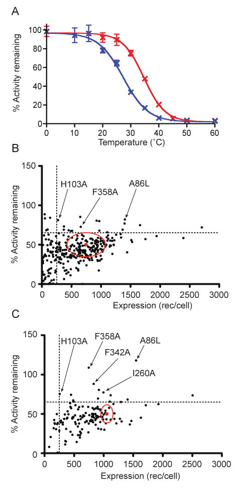
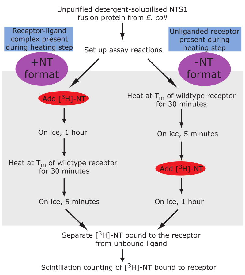
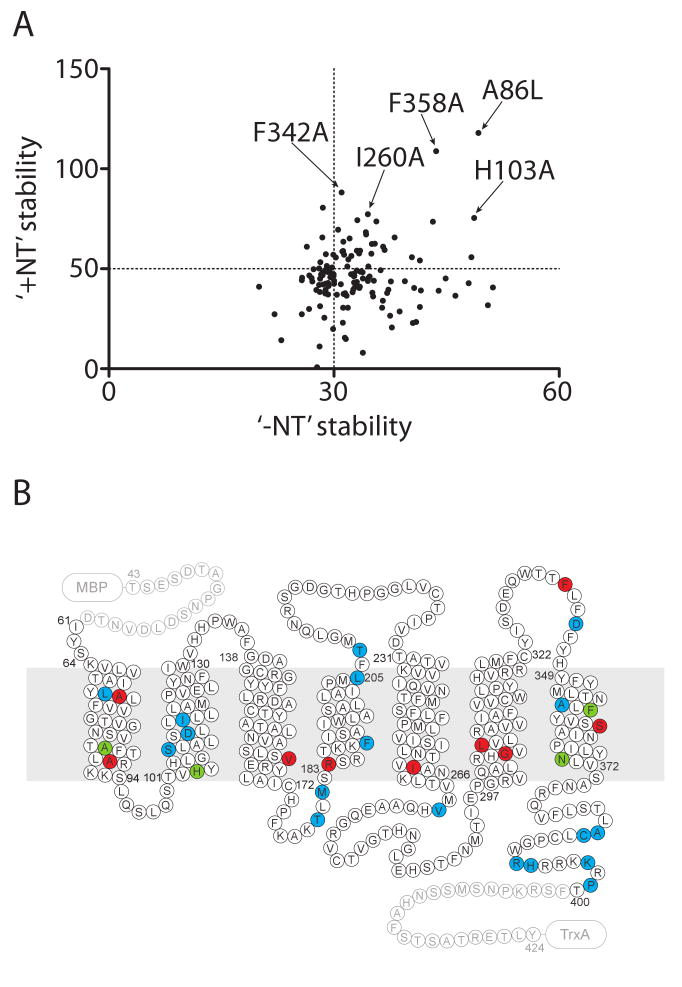
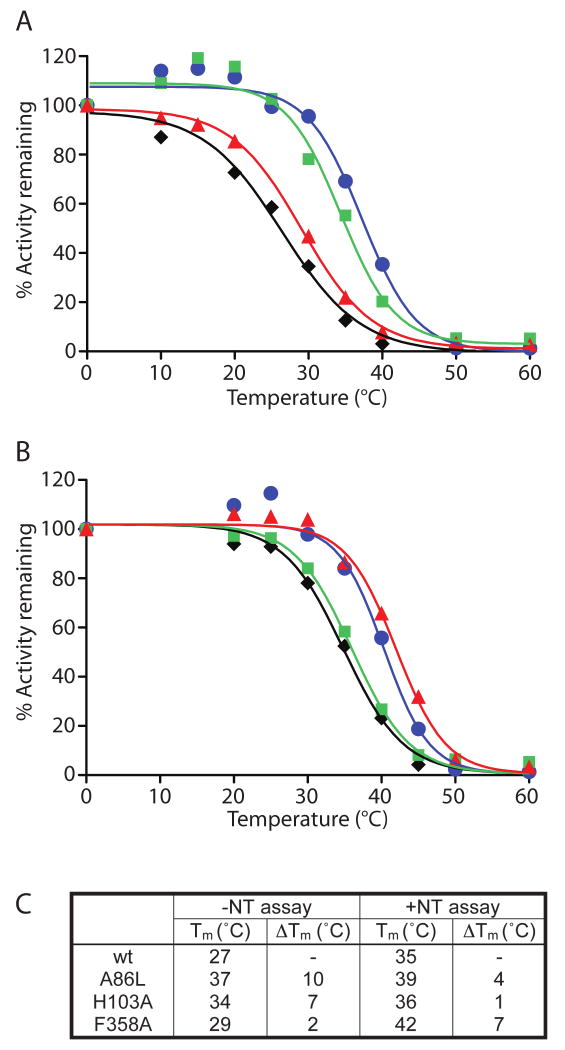
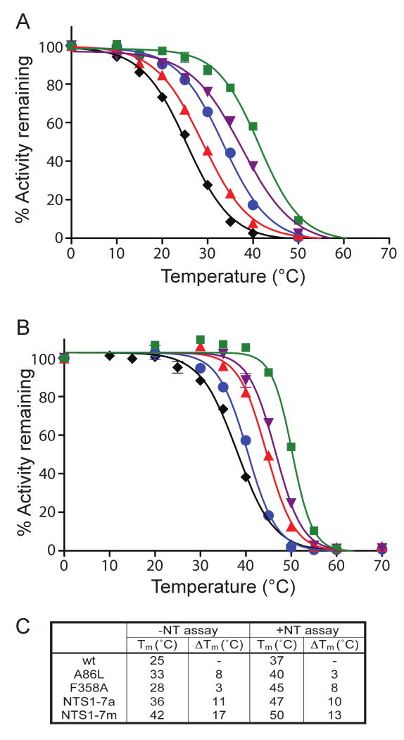
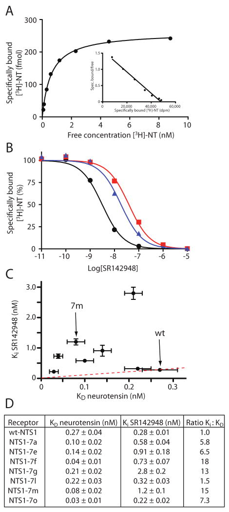
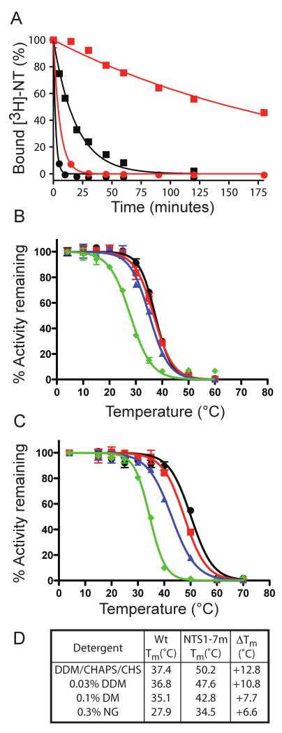
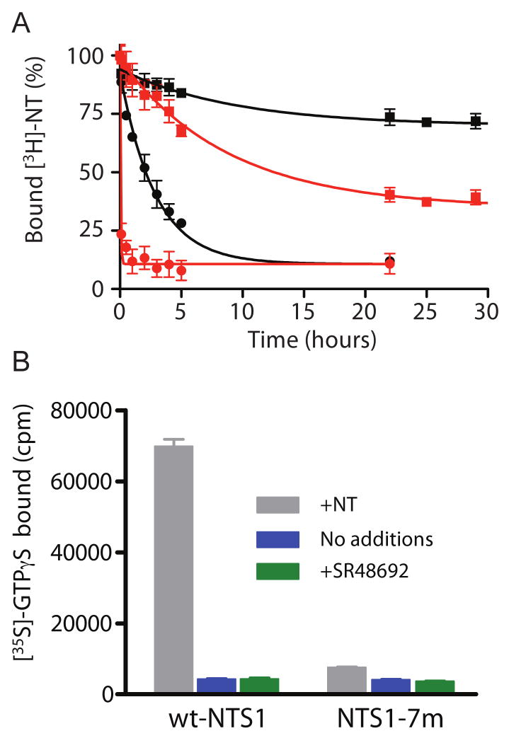
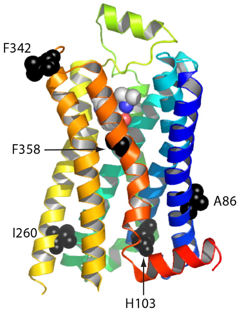
Structural studies on G-protein-coupled receptors have been hampered for many years by their instability in detergent solution and by the number of potential conformations that receptors can adopt. Recently, the structures of the beta(1) and beta(2) adrenergic receptors and the adenosine A(2a) receptor were determined in the antagonist-bound state, a receptor conformation that is thought to be more stable than the agonist-bound state. In contrast to these receptors, the neurotensin (NT) receptor NTS1 is much less stable in detergent solution. We have therefore used a systematic mutational approach coupled with activity assays to identify receptor mutants suitable for crystallization, both alone and in complex with the peptide agonist NT. The best receptor mutant NTS1-7m contained four point mutations. It showed increased stability compared to the wild-type receptor, in the absence of ligand, after solubilization with a variety of detergents. In addition, NTS1-7m bound to NT was more stable than unliganded NTS1-7m. Of the four thermostabilizing mutations, only one residue (A86L) is predicted to be in the lipid environment. In contrast, I260A appears to be buried within the transmembrane helix bundle, F342A may form a distant part of the putative ligand-binding site, whereas F358A is likely to be in a region that is important for receptor activation. NTS1-7m binds NT with a similar affinity for the wild-type receptor. However, agonist dissociation was slower, and NTS1-7m activated G-proteins poorly. The affinity of NTS1-7m for the antagonist SR48692 was also lower than that of the wild-type receptor. Thus, we have successfully stabilized NTS1 in an agonist-binding conformation that does not efficiently couple to G-proteins.


Figures

Figure 1
Figure 1
Thermal stability and expression levels of NTS1 single Ala/Leu mutants. (A) The thermal stability of wt-NTS1 in the unliganded state (blue) and with neurotensin bound (red) was assessed by determining the apparent Tm value from the mid-point of the curves; apparent Tm of unliganded wt-NTS1, 24±2°C; apparent Tm of neurotensin-bound wt-NTS1, 37±2°C. (B-C) Individual mutants of NTS1, each containing a single alanine mutation (if the original amino acid was alanine then it was mutated to leucine) are summarized for its expression level in E. coli (number of functional receptors/cell), its thermal stability in the absence of neurotensin (B), and in the presence of neurotensin (C). Thermal stability was measured after incubating each detergent-solubilized mutant at 24 °C (B) or 37 °C (C) for 30 minutes, and the percentage of activity remaining after incubation was determined with respect to its own unheated control. All the stability data are normalized against the wt-NTS1 stability for each set of experiment (wt=50 %). The mean wt-NTS1 expression level and stability (red dot) and standard errors (red oval) are shown in the plots. The dotted lines show the cut-off values for the stabilized mutants (65 % activity remaining, 250 receptors/cell).
Figure 2
Figure 2
Schematic of the +NT and −NT thermostability assays.
Figure 3
Figure 3
Comparison of unliganded-state and agonist-bound state stabilities of NTS1 single Ala/Leu mutants. (A) The stability of each mutant is shown in both the unliganded state (-NT stability) and agonist-bound state (+NT stability). Mutations combined to optimally stabilise NTS1 are indicated. The intersection of the dotted lines in the plot corresponds to the position of wt-NTS1. (B) The locations of 31 stabilizing mutations are shown in the snake plot; positions of stabilising mutations are shown for the unliganded receptor (blue), neurotensin-bound receptor (red) or both (green).
Figure 4
Figure 4
Denaturation profiles of the three best NTS1 single Ala/Leu mutants in the absence and presence of neurotensin. Denaturation curves of the three best thermostable single mutants of NTS1, A86L, H103A, and F358A, were determined by heating the solubilized mutants at elevated temperatures for 30 minutes, either in the absence of neurotensin (A) or in the presence of 12 nM [3H]-NT (B). NTS1 mutants shown are: wt (black diamonds), A86L (blue circles), H103A (green squares), and F358A (red triangles). (C) Table summarizing the apparent Tm values determined by non-linear regression of the above curves; constraint of upper lower boundaries was not used. The estimated error from repeated experiments is ±2 °C. Activity remaining was normalised to 100% based upon the amount of binding measured in the samples incubated on ice.
Figure 5
Figure 5
Denaturation profiles of NTS1 multiple mutants in the absence or presence of neurotensin. Denaturation curves of four examples of the best thermostable mutants, NTS1-7a (A86L/F358A), NTS1-7m (A86L/I260A/F342A/F358A), A86L and F358A were compared to wt-NTS1. The solubilized receptors were heated for 30 minutes, either in the absence (A) or presence (B) of neurotensin: wt-NTS1 (black diamonds), A86L (blue closed circles), F358A (red triangles), NTS1-7a (purple diamonds), and NTS1-7m (green squares). (C) Table summarizing the apparent Tm values determined from the above curves. The estimated error from repeated experiments is ±2 °C. Activity remaining was normalised to 100% based upon the amount of binding measured in the samples incubated on ice.
Figure 6
Figure 6
Agonist and antagonist binding to NTS1 mutants. (A) Saturation binding curve of a representative [3H]-NT binding experiment with NTS1-7m in intact E. coli cells. The Scatchard plot is shown as an inset (one-site fit, KD 0.34±0.03 nM). (B) Competition assays were performed using intact E. coli cells expressing either wt-NTS1 or the NTS1-mutants. Increasing quantities of antagonist SR142948 were incubated with the cells in the presence of 5 nM agonist [3H]-NT. Competition curves for wt-NTS1 (black circles), NTS1-7a (blue triangles) and NTS1-7m (red squares) are shown. Ki values were determined by non-linear regression analyses using KD values for NT-binding determined from the saturation binding curves (D). (C) The correlation between the KD(NT) and Ki(SR142948) for each mutant is shown as a scatter plot with results shown of a representative experiment, with error bars representing the SEM of data fitting. The red dashed line represents the ratio between the Ki and KD values for wt-NTS1. (D) Table summarizing the apparent KD values for [3H]-NT binding, Ki values for SR142948 and the ratio Ki:KD for each of the mutants tested. KD and Ki determinations were performed simultaneously for each mutant in duplicate. SEMs are from one representative experiment and arise from data fitting.
Figure 7
Figure 7
NTS1-7m shows improved thermal stability as well as stability in short-chain detergents compared to wt-NTS1. (A) The rates of thermal inactivation of solubilised wt-NTS1 (circles) and NTS1-7m (squares) in DDM/CHAPS/CHS were compared by heating the samples at 45°C either in the presence (red lines) or absence (black lines) of [3H]-NT. Half-lives were determined from the curves by non-linear regression of the single-exponential curve after constraining the values for Y=0 to 100 % and the plateau=0 %: unliganded wt-NTS1, 1.3 minutes; neurotensin-bound wt-NTS1, 5.7 minutes; unliganded NTS1-7m, 13.4 minutes; neurotensin-bound NTS1-7m, 220 minutes. (B-D) Thermostability of wt-NTS1 and NTS1-7m in various detergents. Receptors were solubilised in DDM/CHAPS/CHS, bound to Ni2+-NTA beads and then washed and eluted with buffer containing either DDM/CHAPS/CHS (black circles), 0.03 % DDM (red squares), 0.1 % DM (blue triangles) or 0.3 % NG green diamonds). Thermostability assays were performed in the presence of NT (B: wt-NTS1, C: NTS1-7m). The activity remaining was normalised against the unheated control in each detergent condition (100%), although the recovery yields were different in each case (see main text). The apparent Tm values (D) were determined from the curves by non-linear regression.
Figure 8
Figure 8
Rate of dissociation of NT and activation of G protein by wt-NTS1 and NTS1-7m. (A) The dissociation rates of [3H]-NT from wt-NTS1 (circles) and NTS1-7m (squares) were determined by quantifying the amount of [3H]-NT remaining bound to the receptors (total NT concentration in the assay, 2 nM) upon addition of 50 μM unlabeled NT on ice in the presence (red) or absence (black) of NaCl. The rate of [3H]-NT dissociation were determined by non-linear regression with single exponential decay. (B) Recombinant receptors in urea-washed insect cell membranes were tested for their ability to stimulate G protein using a GDP/[35S]-GTPγS exchange assay. All assays contained purified recombinant Gαqβ1γ1, [35S]-GTPγS and insect cell membranes containing either wt-NTS1 or NTS1-7m. Receptors were incubated with no additional ligands (blue bars), with neurotensin (grey bars) or with the antagonist SR48692 (green bars). The amount of [35S]-GTPγS bound to the G protein complex was determined as described in the text.
Figure 9
Figure 9
Positions of the thermostabilising mutations in NTS1-7m. The structure of the β1-adrenergic receptor (PDB 2vt4) is shown in rainbow coloration (N-terminus in blue, C-terminus in red) with the bound antagonist cyanopindolol shown as a space-filling model. The equivalent positions (via primary amino-acid sequence alignment) of five thermostabilising mutations of NTS1 are shown with the side chains as space-filling models (black) with the labels corresponding the amino acid residues in NTS1.

Similar articles

Cited by

References

    1. Warne T, Serrano-Vega MJ, Tate CG, Schertler GF. Development and crystallization of a minimal thermostabilized G protein-coupled receptor. Protein Exp Purif. 2009;65:204–213. - PubMed
    1. Deisenhofer J, Epp O, Miki K, Huber R, Michel H. Structure of the protein subunits in the photosynthetic reaction centre of Rhodopseudomonas viridis at 3Å resolution. Nature. 1985;318:618–624. - PubMed
    1. Weiss MS, Schulz GE. Structure of porin refined at 1.8 Å resolution. J Mol Biol. 1992;227:493–509. - PubMed
    1. Tsukihara T, Aoyama H, Yamashita E, Tomizaki T, Yamaguchi H, Shinzawa-Itoh K, Nakashima R, Yaono R, Yoshikawa S. The whole structure of the 13-subunit oxidized cytochrome c oxidase at 2.8 Å. Science. 1996;272:1136–44. - PubMed
    1. Xia D, Yu CA, Kim H, Xia JZ, Kachurin AM, Zhang L, Yu L, Deisenhofer J. Crystal structure of the cytochrome bc1 complex from bovine heart mitochondria. Science. 1997;277:60–66. - PubMed

Publication types