. 2009 May 4;185(3):459-73.
doi: 10.1083/jcb.200810029.

The nucleoporins Nup170p and Nup157p are essential for nuclear pore complex assembly

Affiliations

The nucleoporins Nup170p and Nup157p are essential for nuclear pore complex assembly

Tadashi Makio et al. J Cell Biol. .

Abstract

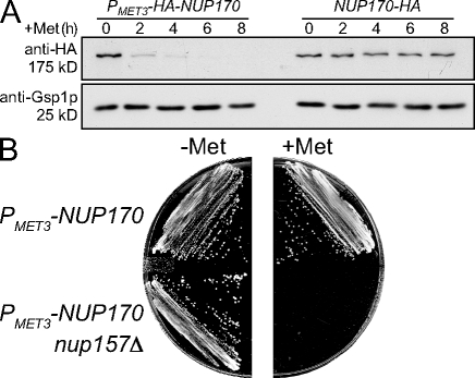
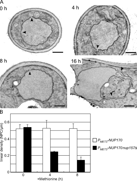
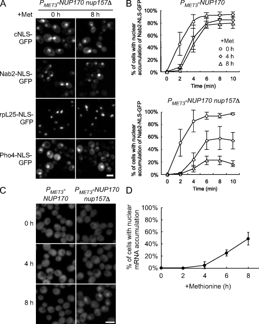
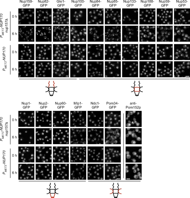
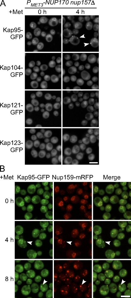
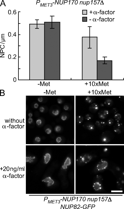
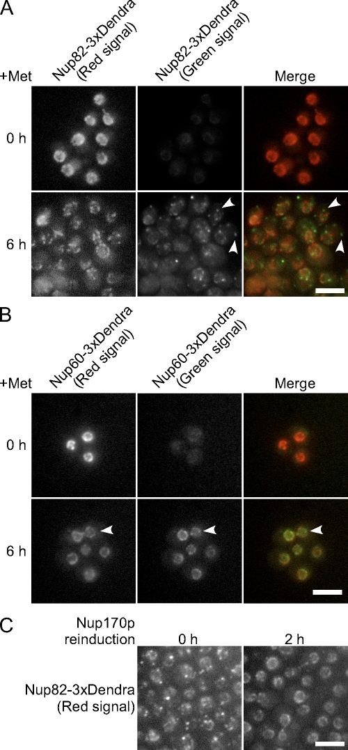
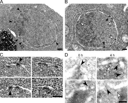
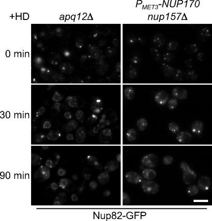
We have established that two homologous nucleoporins, Nup170p and Nup157p, play an essential role in the formation of nuclear pore complexes (NPCs) in Saccharomyces cerevisiae. By regulating their synthesis, we showed that the loss of these nucleoporins triggers a decrease in NPCs caused by a halt in new NPC assembly. Preexisting NPCs are ultimately lost by dilution as cells grow, causing the inhibition of nuclear transport and the loss of viability. Significantly, the loss of Nup170p/Nup157p had distinct effects on the assembly of different architectural components of the NPC. Nucleoporins (nups) positioned on the cytoplasmic face of the NPC rapidly accumulated in cytoplasmic foci. These nup complexes could be recruited into new NPCs after reinitiation of Nup170p synthesis, and may represent a physiological intermediate. Loss of Nup170p/Nup157p also caused core and nucleoplasmically positioned nups to accumulate in NPC-like structures adjacent to the inner nuclear membrane, which suggests that these nucleoporins are required for formation of the pore membrane and the incorporation of cytoplasmic nups into forming NPCs.


Figures

Figure 1.
Figure 1.
Depletion of Nup170p in the nup157Δ background causes a defect in cell growth. (A) The strains TMY1098 (PMET3-HA-NUP170) and TMY1269 (NUP170-HA) were grown in medium lacking methionine. Methionine was added to induce NUP170 shut-off and incubated for the indicated times. Samples were analyzed by Western blotting using anti-HA and anti-Gsp1p (load control) antibodies. (B) The yeast strains TMY1098 (PMET3-HA-NUP170) and TMY1126 (PMET3-HA-NUP170 nup157Δ) were streaked on CM (−Met) and CM (+Met) plates, and grown for 2 d at 30°C.
Figure 2.
Figure 2.
Depletion of Nup170p (nup157Δ) leads to a progressive decrease in the density of NPCs. The strain TMY1126 was grown in media lacking methionine. Methionine was added to repress NUP170 expression, and cells were processed for examination by TEM. (A) Typical TEM images of TMY1126 at the indicated times after methionine addition. Arrowheads indicate gaps in the NE of a size consistent with NPCs. Note that by 16 h, the NE becomes distorted, with this section showing a large invagination of the cytoplasm. Bars, 0.5 µm. (B) The number of NPCs was counted in each cell section and divided by the length of the NE to calculate a linear density of NPCs. A minimum of 30 cells was examined in each experiment, and standard deviations were estimated from three independent experiments. White bar, TMY1098; shaded bar, TMY1126.
Figure 3.
Figure 3.
Decrease in NPC density caused by the depletion of Nup170p (nup157Δ) is accompanied by a decrease in the rate of protein import and mRNA export. (A) TMY1126 cells harboring plasmids encoding the cNLS-GFP, Nab2-NLS-GFP, rpL25-NLS-GFP, and Pho4-NLS-GFP reporters were transferred to media containing methionine for the indicated times to repress NUP170 expression, then examined using an epifluorescence microscope. Bar, 5 µm. (B) Analysis of Nab2-NLS–mediated import. Strains of TMY1098 and TMY1126 harboring a Nab2-NLS-GFP plasmid were grown in media containing methionine for the indicated times. A relative measure of in vivo import rates of the Nab2-NLS-GFP reporter was examined. Standard deviations estimated from at least four independent measurements are shown as error bars. (C) NUP170 expression was repressed in the TMY1098 and TMY1126 strains, and, at the indicated times, the localization of poly-A mRNA was examined by FISH analysis using an oligo (dT50) probe. Bar, 5 µm. (D) For TMY1126 (PMET3-NUP170 nup157Δ), the number of cells that showed a nuclear accumulation of poly-A mRNA at each time point was determined. A minimum of 200 cells was counted in each experiment and standard deviations were estimated from two independent experiments.
Figure 4.
Figure 4.
The loss of Nup170p and Nup157p has distinct effects on the localization of NPC components. (A) The endogenous gene encoding the indicated nup was tagged with GFP in the TMY1098 and TMY1126 strains, and the fusion proteins were examined at the indicated times after repression of NUP170 expression using an epifluorescence microscope. Pom152p localization was analyzed by indirect immunofluorescence using an anti-Pom152p antibody (mAb118C3). Note, the brighter Ndc1-GFP foci of seen at the NE at 0 h likely reflect the shared spindle pole body association of Ndc1p (Chial et al., 1998). Diagrams adjacent to the images depict the general localization of each nup within the NPC (red). Nup100p is present on both faces of the NPC but is biased to the cytoplasm. Bars, 5 µm.
Figure 5.
Figure 5.
Cytoplasmic foci contain multiple nups. Strains expressing Nup159-mRFP and either Nup188-GFP, Nup2-GFP, or Ndc1-GFP within the PMET3-NUP170 nup157Δ background were examined at the indicated times after repression of NUP170 expression using a confocal fluorescence microscope. It should be noted that the foci of Ndc1-GFP and Nup188-GFP generally colocalize with those of Nup159-mRFP (arrows) but not vice versa (arrowheads). Bars, 5 µm.
Figure 6.
Figure 6.
Kap95p binds cytoplasmic foci in cells lacking Nup170p and Nup157p. (A) Strains producing GFP-tagged Kap95p, Kap104p, Kap121p, or Kap123p in the PMET3-NUP170 nup157Δ background were transferred to media containing methionine to repress NUP170 expression. Kap-GFP localization was examined using an epifluorescence microscope at the indicated times. At 4 h after NUP170 repression (nup157Δ), Kap95-GFP was visible at cytoplasmic foci (arrowheads). (B) A PMET3-NUP170 nup157Δ strain producing Kap95-GFP and Nup159-mRFP was grown in media containing methionine for the indicated times. Images were acquired using a confocal microscope. As indicated by the arrowheads, the Nup159-mRFP foci overlap with those of the Kap95-GFP. Bars, 5 µm.
Figure 7.
Figure 7.
Cell cycle arrest induced by α-factor inhibits the decrease in NE-associated Nup82-GFP after the repression of NUP170 (nup157Δ). (A) The strain TMY1203 (PMET3-NUP170 nup157Δ) was transferred to media lacking (dark gray bars) or containing (light gray bars) α-factor in the presence (+Met) or absence (−Met) of methionine for 6 h. The linear density of NPCs was calculated as in Fig. 2. A minimum of 30 cells was examined in each experiment, and the standard deviation was estimated from two independent experiments. (B) Nup82-GFP localization. The localization of Nup82-GFP in TMY1209 (PMET3-NUP170 nup157Δ NUP82-GFP) cells was examined by epifluorescence microscopy after 6 h with or without α-factor and in the presence (+Met) or absence (−Met) of methionine. Bar, 5 µm.
Figure 8.
Figure 8.
Nup170p and Nup157p are required for the incorporation of newly synthesized nups into NPCs. The strains KWY2216 (NUP82-Dendra PMET3-NUP170 nup157Δ; A) and KWY2215 (NUP60-Dendra PMET3-NUP170 nup157Δ; B) were transferred to media containing methionine to repress NUP170 expression. At the same time, UV light was applied to the cells to induce photoconversion of Dendra (green to red). Cells were imaged at 0 and 6 h after photoconversion. Nup82-Dendra produced after photoconversion (green) appears at foci generally distinct from existing NPCs (A, arrowheads). NE-associated Nup60-Dendra-green foci that were distinct from Nup60-Dendra-red were also detected (B, arrowheads). (C) KWY2216 cells were switched to media containing methionine; then, at 5 h after NUP170 repression, UV light was applied to the cells to induce photoconversion of Dendra to the red fluorophore. Cells were transferred to the media lacking methionine for reinduction of NUP170. At 0 and 2 h after induction, images of the Nup82-Dendra (red) were captured using an epifluorescence microscope. Bars, 5 µm.
Figure 9.
Figure 9.
NPC-like structures accumulate at the inner nuclear membrane after the loss of Nup170p and Nup157p. (A–C) The strains TMY1098 (PMET3-NUP170; A) and TMY1126 (PMET3-NUP170 nup157Δ; B and C) were harvested at 4 h after NUP170 repression. The cells were then examined by TEM. Black arrowheads indicate the mature NPCs. White arrowheads indicate structures attached on the inner nuclear membrane with similar size and staining properties of NPCs. (D) TMY1126 cells were grown in media containing methionine for the indicated time points. Cells were then harvested and analyzed by immunoelectron microscopy using the mAb414 antibody. The nucleoplasm (N) and the cytoplasm (C) are indicated. Black arrowheads indicate gold particles bound to NPCs as defined by discontinuities in NE lumen. White arrowheads indicate gold particles attached to the inner nuclear membrane. Bars, 0.2 µm.
Figure 10.
Figure 10.
Hexane-1, 6-diol suppresses the NPC assembly defect of the apq12Δ strain but not that of cells lacking Nup170p and Nup157p. apq12Δ (TMY1290) and PMET3-NUP170 nup157Δ (TMY1135) strains expressing Nup82-GFP were grown in YPD overnight at 23°C, and for 6 h in media containing methionine to induce NPC assembly defects and accumulation of Nup82-GFP foci. Hexane-1, 6-diol (+HD; final 3% wt/vol) was then added to the media, and samples were taken at the indicated times. Images of Nup82-GFP were acquired using an epifluorescence microscope. Bar, 5 µm.

Comment in

Similar articles

Cited by

References

    1. Aitchison J.D., Rout M.P., Marelli M., Blobel G., Wozniak R.W. 1995. Two novel related yeast nucleoporins Nup170p and Nup157p: complementation with the vertebrate homologue Nup155p and functional interactions with the yeast nuclear pore-membrane protein Pom152p.J. Cell Biol. 131:1133–1148 - PMC - PubMed
    1. Alber F., Dokudovskaya S., Veenhoff L.M., Zhang W., Kipper J., Devos D., Suprapto A., Karni-Schmidt O., Williams R., Chait B.T., et al. 2007a. Determining the architectures of macromolecular assemblies.Nature. 450:683–694 - PubMed
    1. Alber F., Dokudovskaya S., Veenhoff L.M., Zhang W., Kipper J., Devos D., Suprapto A., Karni-Schmidt O., Williams R., Chait B.T., et al. 2007b. The molecular architecture of the nuclear pore complex.Nature. 450:695–701 - PubMed
    1. Amberg D.C., Goldstein A.L., Cole C.N. 1992. Isolation and characterization of RAT1: an essential gene of Saccharomyces cerevisiae required for the efficient nucleocytoplasmic trafficking of mRNA.Genes Dev. 6:1173–1189 - PubMed
    1. Antonin W., Franz C., Haselmann U., Antony C., Mattaj I.W. 2005. The integral membrane nucleoporin pom121 functionally links nuclear pore complex assembly and nuclear envelope formation.Mol. Cell. 17:83–92 - PubMed

Publication types

MeSH terms

Substances