. 2009 Jul;83(14):7202-9.
doi: 10.1128/JVI.00076-09. Epub 2009 Apr 29.

X-box binding protein 1 contributes to induction of the Kaposi's sarcoma-associated herpesvirus lytic cycle under hypoxic conditions

Affiliations

X-box binding protein 1 contributes to induction of the Kaposi's sarcoma-associated herpesvirus lytic cycle under hypoxic conditions

Lucy Dalton-Griffin et al. J Virol. 2009 Jul.

Abstract

Kaposi's sarcoma-associated herpesvirus (KSHV), like other herpesviruses, has two stages to its life cycle: latency and lytic replication. KSHV is required for development of Kaposi's sarcoma, a tumor of endothelial origin, and is associated with the B-cell tumor primary effusion lymphoma (PEL) and the plasmablastic variant of multicentric Castleman's disease, all of which are characterized by predominantly latent KSHV infection. Recently, we and others have shown that the activated form of transcription factor X-box binding protein 1 (XBP-1) is a physiological trigger of KSHV lytic reactivation in PEL. Here, we show that XBP-1s transactivates the ORF50/RTA promoter though an ACGT core containing the XBP-1 response element, an element previously identified as a weakly active hypoxia response element (HRE). Hypoxia induces the KSHV lytic cycle, and active HREs that respond to hypoxia-inducible factor 1alpha are present in the ORF50/RTA promoter. Hypoxia also induces active XBP-1s, and here, we show that both transcription factors contribute to the induction of RTA expression, leading to the production of infectious KSHV under hypoxic conditions.


Figures

FIG. 1.
FIG. 1.
The KSHV ORF50 promoter contains a functional XRE. (A) Schematic representation of the ORF50 promoter showing the location of the dominant HRE (HRE2) as well as the newly identified XRE, previously identified as the putative HRE4 element (4). Numbers in brackets indicate the response element start sites relative to the KSHV genome sequence (NC_003409). The wild-type sequences of both HRE2 and XRE are shown, with the core ACGT sequence element highlighted in gray. The mutated sequence of the XRE is indicated underneath, with mutated nucleotides indicated by •. (B) HEK 293T cells were transfected with the DsRed reporter plasmid with the wild-type (p50redi; black bars) or the mutant (pMUT50redi; white bars) ORF50 promoter sequence, together with pIG (control), RTA-expressing (pCMV-RTA), or XBP-1s-expressing (pXBP-sIG) plasmids. The percentages of DsRed-positive cells were quantified by flow cytometry at 48 h posttransfection. Percentages of DsRed-positive cells are expressed relative to the level for empty-vector control pIG.
FIG. 2.
FIG. 2.
Acute hypoxia stabilizes HIF-1α and activates XBP-1 in PEL. (A) Western blot analysis of whole-cell lysates of the PEL cell line rKSHV.219 JSC-1 cultured in normoxia (N; 21%O2) or exposed to hypoxia (H; 3% O2) 48 or 72 h with anti-HIF-1α antibody (BD transduction laboratories). HIF-1α is stabilized in rKSHV.219 JSC-1 in response to hypoxia at 48 and 72 h, compared to the level for normoxia, and β-actin acts as a loading control. (B) RT-PCR amplification across the XBP-1 intron produces a 249-bp amplicon from XBP-1u mRNA and a 223-base amplicon from XBP-s mRNA. PstI digests only the XBP-1u amplicon, resulting in two bands, whereas XBP-1s results in a single band (50). RT-PCR amplification from the total mRNA of the rKSHV.219 JSC-1 cell line cultured under normoxic conditions (N; 21%O2) or exposed to hypoxia (H; <3% O2) for 48 or 72 h shows that XBP-1s is produced after 72 h of hypoxia but is absent in cells under normoxic conditions. In the 72-h-hypoxia sample, a slower-migrating, non-PstI-digestible PCR hybrid between the XBP-1s and XBP-1u products is visible, similar to what was previously described (50).
FIG. 3.
FIG. 3.
Acute hypoxia leads to accumulation of KSHV RTA, induces ORF29a/b lytic gene expression, and produces progeny virions, as measured by ORF37. (A) Western blot analysis of whole-cell lysates of the PEL cell line rKSHV.219 JSC-1 cultured in normoxia (N; 21%O2) or exposed to hypoxia (H; <3% O2) for 48 or 72 h with rabbit anti-RTA. RTA is induced by hypoxia at 48 h and further increased at 72 h of exposure, compared to cells under normoxic conditions. β-Actin acts as a loading control. (B) Total cellular RNA was isolated from rKSHV.219 JSC-1 cultured under normoxic or hypoxic conditions for 48 and 72 h. With RT-PCR, cDNA was amplified with gene-specific primers for the spliced lytic transcript, ORF29a/b. The 300-bp product is seen only in samples exposed to hypoxia. (C) qPCR for ORF37 from KSHV virion DNA extracted from the supernatants of JSC-1 and HBL-6 cells cultured for 72 h in normoxia (N; 21%O2; black bars) or hypoxia (H; <3% O2; white bars). Copy numbers were determined using an ORF37 standard curve and normalized for total DNA input. Hypoxia leads to a significant increase in virus copies present in the supernatant. Western blots for HIF-1α and RTA of cells corresponding to the supernatants at 48 h show that samples are HIF-1α positive at 48 h and that RTA is induced.
FIG. 4.
FIG. 4.
The KSHV RTA promoter contains response elements for XBP-1 and HIF-1α and can respond to both. (A) Schematic of the predicted HREs in the KSHV RTA promoter region (adapted from reference 4). (B) Schematic of reporter plasmids pRpluc1-3087+s containing a 3-kb sequence upstream of the RTA transcriptional start site and a 1-kb splicing sequence of RTA that drives the expression of firefly luciferase (FL) (4) as well as truncated promoters named pRpluc1115-1327 (HRE2 only) and pRpluc1-550 XRE (HRE4 only). (B) A fixed amount of the reporter plasmids was transfected into HEK 293T cells, together with pIG (empty vector control; ↓), RTA-expressing (pCMV-RTA; black bars), XBP-1s-expressing (pXBP-sIG; striped bars), or HIF-1α-expressing (pHIF-1αIG; white bars) plasmids, normalized by plasmid copy number. Promoter activity levels are expressed as log relative light units (logRLU) relative to the levels for the reporter plus the pIG control plasmid. Means and standard errors are from three independent transfections. HIF-1α acts on all reporters containing HRE2 and HRE4 while XBP-1s acts on the full-length and HRE4-only promoter but not HRE2 only.
FIG. 5.
FIG. 5.
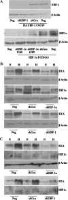
XBP-1s contributes to RTA induction by hypoxia. (A) The ability of shRNA to knock down target protein levels was demonstrated by cotransfection of HEK 293Tcells with 1 μg of target-expressing plasmid and 3 μg of relevant/irrelevant control hairpins in a pGEMT backbone. Western blots for target protein using anti-HA for HA-tagged XBP-1 detection (upper panel) and anti-HIF-1α for HIF-1α (lower panel) are shown (see Materials and Methods for plasmid descriptions). The shRNA targeting XBP-1 and the two shRNAs targeting HIF-1α are able to reduce target protein levels substantially, compared to the irrelevant control shRNA and untransduced cells (Neg). shRNA 1345 targeting HIF-1α was used for further experiments. rKSHV.219 HEK 293T cells (B) and JSC-1 cells (C) untransduced (Neg) or stably transduced with lentiviruses containing shRNA targeting XBP-1, HIF-1α, and an irrelevant control independently were cultured in normoxia (N; 21% O2) or hypoxia (H; 3% O2) for 72 h. Western blot analysis of whole-cell lysates was performed for HIF-1α, which was upregulated in all hypoxic samples except those expressing the shRNA targeting HIF-1α. The same samples probed with anti-RTA show induction of RTA in response to hypoxia, which is reduced in the presence of shRNA targeting HIF-1α or XBP-1s.
FIG. 6.
FIG. 6.
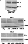
Overexpression of HIF-1α under normoxic conditions does not induce RTA. (A) Anti-HIF-1α Western blot of whole-cell lysates of JSC-1 and HBL-6 cells transduced with lentivirus expressing HIF-1α. (B) RTA Western blot of JSC-1 and HBL-6 cells untransduced (Neg) or transduced with empty vector (pIG), HIF-1α-expressing (pHIF-1αIG), and XBP-1s-expressing (pXBP-sIG) plasmids. RTA is induced only in the presence of XBP-1s expression (pXBP-sIG) under normoxic conditions.

Similar articles

Cited by

References

    1. Acosta-Alvear, D., Y. Zhou, A. Blais, M. Tsikitis, N. H. Lents, C. Arias, C. J. Lennon, Y. Kluger, and B. D. Dynlacht. 2007. XBP1 controls diverse cell type- and condition-specific transcriptional regulatory networks. Mol. Cell 2753-66. - PubMed
    1. Besnier, C., Y. Takeuchi, and G. Towers. 2002. Restriction of lentivirus in monkeys. Proc. Natl. Acad. Sci. USA 9911920-11925. - PMC - PubMed
    1. Bhende, P. M., S. J. Dickerson, X. Sun, W. H. Feng, and S. C. Kenney. 2007. X-box-binding protein 1 activates lytic Epstein-Barr virus gene expression in combination with protein kinase D. J. Virol. 817363-7370. - PMC - PubMed
    1. Cai, Q., K. Lan, S. C. Verma, H. Si, D. Lin, and E. S. Robertson. 2006. Kaposi's sarcoma-associated herpesvirus latent protein LANA interacts with HIF-1alpha to upregulate RTA expression during hypoxia: latency control under low oxygen conditions. J. Virol. 807965-7975. - PMC - PubMed
    1. Cai, Q., M. Murakami, H. Si, and E. S. Robertson. 2007. A potential alpha-helix motif in the amino terminus of LANA encoded by Kaposi's sarcoma-associated herpesvirus is critical for nuclear accumulation of HIF-1 alpha in normoxia. J. Virol. 8110413-10423. - PMC - PubMed

Publication types

MeSH terms