. 2009 Apr 14;7(4):e84.
doi: 10.1371/journal.pbio.1000084.

Sir2 paralogues cooperate to regulate virulence genes and antigenic variation in Plasmodium falciparum

Affiliations

Sir2 paralogues cooperate to regulate virulence genes and antigenic variation in Plasmodium falciparum

Christopher J Tonkin et al. PLoS Biol. .

Abstract

Cytoadherance of Plasmodium falciparum-infected erythrocytes in the brain, organs and peripheral microvasculature is linked to morbidity and mortality associated with severe malaria. Parasite-derived P. falciparum Erythrocyte Membrane Protein 1 (PfEMP1) molecules displayed on the erythrocyte surface are responsible for cytoadherance and undergo antigenic variation in the course of an infection. Antigenic variation of PfEMP1 is achieved by in situ switching and mutually exclusive transcription of the var gene family, a process that is controlled by epigenetic mechanisms. Here we report characterisation of the P. falciparum silent information regulator's A and B (PfSir2A and PfSir2B) and their involvement in mutual exclusion and silencing of the var gene repertoire. Analysis of P. falciparum parasites lacking either PfSir2A or PfSir2B shows that these NAD(+)-dependent histone deacetylases are required for silencing of different var gene subsets classified by their conserved promoter type. We also demonstrate that in the absence of either of these molecules mutually exclusive expression of var genes breaks down. We show that var gene silencing originates within the promoter and PfSir2 paralogues are involved in cis spreading of silenced chromatin into adjacent regions. Furthermore, parasites lacking PfSir2A but not PfSir2B have considerably longer telomeric repeats, demonstrating a role for this molecule in telomeric end protection. This work highlights the pivotal but distinct role for both PfSir2 paralogues in epigenetic silencing of P. falciparum virulence genes and the control of pathogenicity of malaria infection.


Conflict of interest statement

Competing interests. The authors have declared that no competing interests exist.

Figures

Figure 1
Figure 1. Identification of PfSir2B and Phylogenetic Relationships between PfSir2A and PfSir2B within Apicomplexan Parasites
(A) PfSir2B is a large gene and encodes a ∼150-kD protein. PfSir2B, like all sirtuins, contains similarity to other Sir2 molecules within the catalytic regions (black). PfSir2B has 29%/43% identity/similarity with Schizosaccharomyces pombe Sir2 (SpSir2) and 26%/51% identity/similarity with the previously identified PfSir2A. PfSir2B most closely resembles a “type IV” sirtuin, which groups together other sirtuins with diverse roles in cellular signalling such as human SIRT6, involved in telomeric chromatin maintenance [82,83] as well as SIRT7, an activator of RNA polymerase I [84]. PfSir2B has with 38%/53% identity/similarity to type IV sirtuins. (B) Phylogenetic analysis of apicomplexan Sir2 paralogues. Sir2 homologues were identified from the sequencing projects of four Plasmodium species; P. vivax [85], P. chabaudi [86], P. yoelli [86], P. berghei [86], together with Sir2 homologues from other apicomplexan parasites: T. gondii (http://toxodb.org), T. annulata [87], T. parva [87], B. bovis (www.tigr.org), and Cryptosporidium hominis [88]. Within the analysis we also included Sir2 from yeast, other related species, and unicellular protozoan parasites. Bootstraps values are indicated for important nodes, where in all cases the upper number is the bootstrap using distance, the lower is the bootstrap using maximum likelihood. “-“ sign indicates a value below 50. Two Sir2 paralogues are a common feature of apicomplexan parasites. Sir2B paralogues (red branches) show strong support for the Sir2B clade, especially within Plasmodium species. Sir2B paralogues also groups together type IV sirtuins (SIRT6 and 7) with good support. Sir2A clade (blue branches) groups together Sir2A paralogues of apicomplexan species with excellent support. Sir2A's moderate grouping with SIRT5 and Archaeoglobus fulgidus Sir2-Af1 also shows a possible functional relationship with type III sirtuins. For more in depth analysis see Text S1. P. ber, P. berghei; P. yoe, P. yoellii; P. cha, P. chabaudi; P. viv, P. vivax; P. fal, P. falciparum; T. ann, T. annulata; T. parv, T. parva; T. gondi, T. gondii; B. bov, B. bovis; A. ful, A. fulgidus; H. sap, Homo sapiens; C. hom, C. hominis; T. pse, Thalassiosira pseudonana (diatom); G. lam, Giardia lamblia; K. lac, Kluyveromyces lactis; S. cer, S. cerevisiae; L. maj, Leishmania major.
Figure 2
Figure 2. PfSir2B Gene Disruption in P. falciparum
Single crossover recombination was used to disrupt PfSir2B gene in P. falciparum. Bacterially derived plasmids were introduced containing an ∼1-kb fragment of the 5′ end of the PfSir2B gene and an hDHFR expression cassette for stable selection of transgenic parasites. Cycles of “on” and “off” drug were used to select for parasites that had integrated the plasmid by homologous integration into the PfSir2B locus. (A) Theoretical restriction mapping of the Sir2B locus, pre- and postepisome integration. Hatched line denotes bacterially derived plasmid sequence, while AmpR signifies beta lactamase expression cassette for selection for resistance on Ampicillin. The line under the 5′ end of the PfSir2B gene shows the probe (P) used for Southern blot mapping. B, BglII site. (B) Southern blot of wildtype (wt) and ΔPfSir2B disrupted parasites. Banding pattern is consistent with disruption of the Sir2B gene by multiple plasmid copies (as mapped in [A]). DNA ladder sizes are indicated on the left of the blot. (C) Northern blot showing a loss of PfSir2B transcript in ΔPfSir2B-disrupted parasites but not in ΔPfSir2A-disrupted parasites. Same probe (P) in Southern blot was used for northern blot. (D) Western blot of antisera raised against a C-terminal portion of PfSir2B. Antisera reacting with a protein in wildtype parasites contain the predicted size for PFSir2B, while this protein is absent as expected in ΔPfSir2B parasites. Cross-reactive bands indicate equal loading of lanes.
Figure 3
Figure 3. PfSir2A and PfSir2B Control the Expression of Subtelomeric Genes in P. falciparum
The expression profile of subtelomeric genes and internal var gene clusters were analysed in Sir2-disrupted lines and applied to a physical map of all 28 chromosome ends. Subtelomeric ends contain most var genes as well as the rifin gene family, which are also known to be antigenically variant. Other gene families occupy subtelomeric regions of P. falciparum chromosomes, but at this stage their biological function or whether they are antigenically variant remains elusive. As seen by the key the colour of each gene relates to the level of expression in each PfSir2-disrupted line. Black genes indicate that expression of that gene is not observed in any condition (expression signal <75th percentile). In the unusual case where PfSir2A or PfSir2B both affected the expression of a single var gene only the most dramatic change is shown. This physical map of gene expression accompanies Figure S3, which graphs the expression values of all subtelomeric genes in 3D7 and the two PfSir2-disrupted lines.
Figure 4
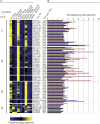
Figure 4. PfSir2 Paralogues Silence Discrete Subsets of var Genes Associated with Conserved Promoter Types That Drive Their Expression
(A) Unsupervised nonhierarchical Gaussian self-clustering analysis was performed in order to understand the relationship between var gene subtypes and the role of PfSir2 paralogues in their regulation. Genes were clustered by similarity using Pearson coefficients. Heatmap colour-scale values were assigned by ArrayMiner after further transformation of expression data (see Material and Methods for details) with +3 (yellow) being the highest expression and −3 (blue) being the lowest expression. Expression profiles of the var gene repertoire were grouped given the nine conditions examined by Affymetrix arrays (i.e., rings, trophozoites, and schizonts in 3D7 [wildtype], ΔPfSir2A, and ΔPfSir2B parasite lines). The heatmap clearly identifies three major clusters that are biologically relevant. Cluster 1 represents var genes that are expressed in standard culture conditions in 3D7 parasite isolate and are somewhat expressed in PfSir2-disrupted parasites. No correlation in promoter type is seen in var genes found is this cluster with the exception that no UpsA-var genes were found there. Var genes controlled by UpsA conserved promoter type are found within cluster 2 as are most of the internally located UpsC var genes. This finding suggests that PfSir2A plays a suppressive role in expression of var genes with an UpsA or UpsC promoter type. Cluster 3 contains almost exclusively UpsB var genes and these are de-repressed in the absence of PfSir2B. This suggests that PfSir2B is involved in the suppression of the most telomere proximal, UpsB driven var genes. Cluster 4 contains several var genes that seem to be outliers, not conforming or reaching the threshold required to fall into one of the three main clusters. Scale bar of expression goes from −3 to 3 in transformed log2 expression. (B) Raw normalised log2 expression of all var genes in ring-stage parasites aligned with their corresponding heatmap in the Gaussian self-clustering analysis. Raw normalised expression values for all var genes grouped by their promotor type as assayed by array analysis can be viewed in Figure S4. The colour of each bar refers to its strain identity: green, 3D7; red, ΔPfSir2A; blue, ΔPfSir2B.
Figure 5
Figure 5. Disruption of PfSir2 Paralogues Destroys Mutual Exclusive Expression of var Genes
In wildtype parasites selection on CSA enriches for parasites exclusively expressing var2CSA. We used this mono-allelic binding phenotype to assay if PfSir2-disrupted parasites still maintain mutual exclusive expression of var genes. (A) 2-D FACS plots showing binding of R1945 rabbit serum specifically recognises infected erythrocytes (ethidium bromide [EtBr] positive) containing var2CSA-expressing parasites. R1945 percentage positivity and negativity are separated on the basis of fluorescence levels of uninfected erythrocytes. Selection on CSA of 3D7 (3D7_CSA) greatly enhances the reactivity to R1945 serum and is comparable to the CS2 parasite line that stably expresses var2CSA. Given that ΔPfSir2A already expresses var2CSA in the absence of selection it is not surprising that CSA selection of this parasite line (ΔPfSir2A_CSA) does nothing (if anything a negative effect) to the binding of R1945 serum. ΔPfSir2A parasites show an intermediate binding of R1945 reactivity suggesting that they have lower levels of VAR2CSA on their surface. Selection of ΔPfSir2B parasites on CSA (ΔPfSir2B_CSA) also greatly enhanced R1945 binding to infected erythrocytes suggesting the ΔPfSir2B parasites are a heterogenous pool and are not confined to expressing only UpsB-driven var genes. (B) As expected qPCR of var2CSA (PFL0030c) transcript levels shows a dramatic increase upon CSA selection on 3D7 and ΔPfSir2B parasites, but not ΔPfSir2A. (C) Mutual exclusive expression of var genes is broken down in the absence of both PfSir2A and PfSir2B. As expected, 3D7_CSA mainly expresses var2CSA and while other var genes are only at a very low levels (see also Figure S5). However, when compared to 3D7_CSA, PfSir2-disrupted parasites, not only express var2CSA but also express many other var genes at much higher level than 3D7_CSA does (silenced state). This finding strongly suggests that mutual exclusive expression has broken down in these parasites. Interestingly, expression of var genes in the Sir2-disrupted lines selected on CSA also shows a bias towards var genes with certain promoter types thus confirming by another method our earlier findings.
Figure 6
Figure 6. PfSir2 Paralogues Directly Regulate var Gene Promoters and Contribute to Spreading of Transcriptional Silencing into Neighbouring Regions
(A) Plasmids bearing two expression cassettes were introduced into 3D7, ΔPfSir2A, and ΔPfSir2B parasites. The bsd cassette is driven by the housekeeping hsp86 promoter, which provides a selection mechanism for parasites stably maintaining episomes. UpsB and UpsC var promoters were placed in front of the selectable marker hdhfr and therefore var promoter activity can be monitored. All values plotted were normalised against the var promoter in its activated state. (B) PfSir2A and B paralogues differentially affect the silencing of isolated var promoters. hdhfr transcript level was measured in 3D7 parasites with silenced and activated episomally located UpsB and UpsC promoters as well as the level of hdhfr transcription in the absence of PfSir2 paralogues. Both PfSir2 paralogues can directly affect the activity of a var promoter strongly suggesting that PfSir2 paralogues do not function independently of one another on different var promoters but rather cooperate to regulate expression. In both subtelomeric and internally derived var promoters isolated from their natural context PfSir2B has a larger effect on the suppression of var promoter activity. (C) PfSir2A and PfSir2B control the spreading of silencing from var promoters. Upon activation of the downstream var promoter activity of bsd expression cassette increases suggesting that factors that normally silence var gene promoters can spread in cis to modulate expression of neighbouring regions. In the absence of both PfSir2A and PfSir2B spreading of silencing is reduced suggesting that both paralogues mediate the spreading of silencing from both internal and subtelomerically derived var gene promoters. In both cases PfSir2B has a greater influence on spreading of silencing to within the bsd cassette. qPCR was performed on three biological replicates harvested from ring-stage parasites. 95% confidence error bars derived from a calculated standard error are also displayed.
Figure 7
Figure 7. rif Genes Change Expression Timing upon Disruption of PfSir2A, but not PfSir2B
Gaussian self-clustering was used to further analyse the role of PfSir2 paralogues in rif gene expression. Expression throughout rings, trophozoite, and schizont-stage parasites show that PfSir2A can modulate the temporal expression of rif genes. Normally most rif are expressed in trophozoite stage but upon disruption of PfSir2A expression of some of this antigenically variant gene family changes to either ring- or schizont stage, or both. Interestingly the lack of PfSir2B shows a suppressive effect on rif gene expression. Unfortunately no other similarities either in promoter sequence or physical position relative to var genes could be found in any of the clusters.
Figure 8
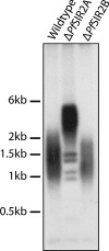
Figure 8. Sir2A, but Not PfSir2B Has a Role in Telomere Length Regulation
Genomic DNA from wildtype (3D7), ΔPfSir2A, and ΔPfSir2B was digested with four frequently cutting restriction enzymes that do not cut telomeric repeats. Samples were run on 1% agarose gel and probed with telomeric repeat-labelled with 32P. Telomere length is seen as a smear as telomere lengths vary between chromosomes and within a population telomere. 3D7 parasites have telomere lengths of between 1–2 kB. Whereas upon disruption of PfSir2A but not PfSir2B, telomere length approximately doubles in size. PfSir2B-disrupted parasites have several distinct bands of a length that correspond to nonlengthened telomeres. It is also possible that they represent telomeric repeats that have recombined into chromosome internal locations and thus are not able to lengthen.

Similar articles

Cited by

References

    1. Merrick CJ, Duraisingh MT. Heterochromatin-mediated control of virulence gene expression. Mol Microbiol. 2006;62:612–620. - PubMed
    1. Deitsch KW, Moxon ER, Wellems TE. Shared themes of antigenic variation and virulence in bacterial, protozoal, and fungal Infections. Micro Mol Biol Reviews. 1997;61:281–293. - PMC - PubMed
    1. Miller LH, Baruch DI, Marsh K, Doumbo OK. The pathogenic basis of malaria. Nature. 2002;415:673–679. - PubMed
    1. Bull PC, Lowe BS, Kortok M, Molyneux CS, Newbold CI, et al. Parasite antigens on the infected red cell surface are targets for naturally acquired immunity to malaria. Nature Med. 1998;4:358–360. - PMC - PubMed
    1. Snow RW, Guerra CA, Noor AM, Myint HY, Hay SI. The global distribution of clinical episodes of Plasmodium falciparum malaria. Nature. 2005;434:214–217. - PMC - PubMed

MeSH terms

Substances