. 2009 May 4;185(3):493-502.
doi: 10.1083/jcb.200810114. Epub 2009 Apr 27.

K63-linked ubiquitin chains as a specific signal for protein sorting into the multivesicular body pathway

Affiliations

K63-linked ubiquitin chains as a specific signal for protein sorting into the multivesicular body pathway

Elsa Lauwers et al. J Cell Biol. .

Abstract

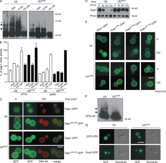
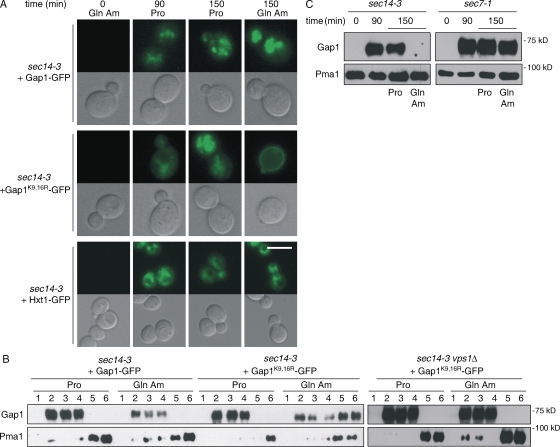
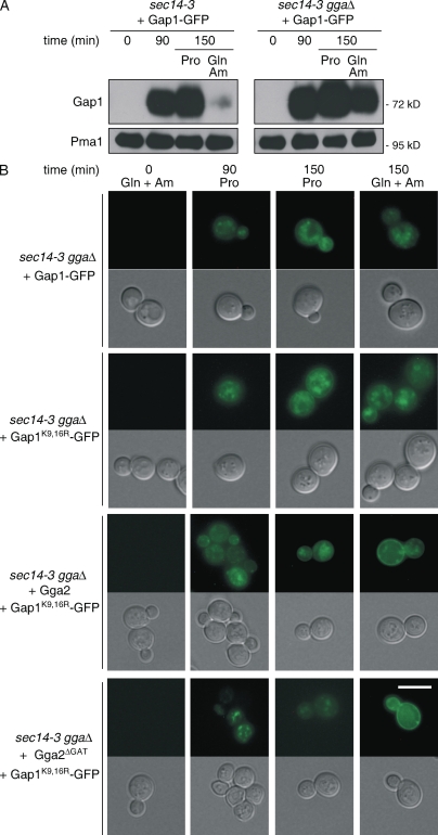
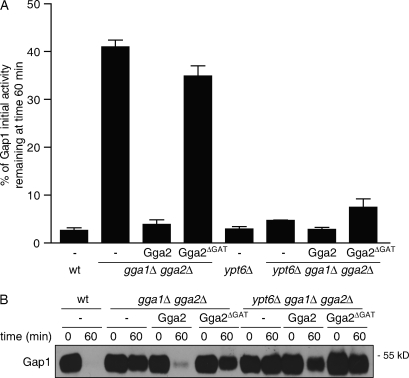
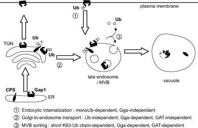
A growing number of yeast and mammalian plasma membrane proteins are reported to be modified with K63-linked ubiquitin (Ub) chains. However, the relative importance of this modification versus monoubiquitylation in endocytosis, Golgi to endosome traffic, and sorting into the multivesicular body (MVB) pathway remains unclear. In this study, we show that K63-linked ubiquitylation of the Gap1 permease is essential for its entry into the MVB pathway. Carboxypeptidase S also requires modification with a K63-Ub chain for correct MVB sorting. In contrast, monoubiquitylation of a single target lysine of Gap1 is a sufficient signal for its internalization from the cell surface, and Golgi to endosome transport of the permease requires neither its ubiquitylation nor the Ub-binding GAT (Gga and Tom1) domain of Gga (Golgi localizing, gamma-ear containing, ARF binding) adapter proteins, the latter being crucial for subsequent MVB sorting of the permease. Our data reveal that K63-linked Ub chains act as a specific signal for MVB sorting, providing further insight into the Ub code of membrane protein trafficking.


Figures

Figure 1.
Figure 1.
K63-linked ubiquitylation is required for MVB sorting of Gap1 and CPS. Cells expressing Ub or UbK63R as the sole source of Ub were grown in glucose- and proline-containing yeast nitrogen base medium. (A) gap1Δ cells were transformed with a centromeric vector carrying the GAP1 gene or its gap1K9R, gap1K16R, or gap1K9,16R allele. Cells were collected 5 min after the addition of 100 mM ammonium and were used to prepare total protein extracts, and the Gap1 ubiquitylation profile was examined by Western blotting using anti-Gap1 antibodies. Ubiquitylated forms of Gap1 corresponding to an additional mass of ∼7 kD are indicated with dots, whereas the molecular mass of the band marked with an asterisk suggests that it corresponds to a modified form of the upper ubiquitylated conjugate. In the case of cells expressing native Gap1 and Ub, more than two bands may be detected if Ub is overproduced or after a longer incubation in the presence of ammonium (Fig. S1). (B) gap1Δ or gap1Δ ypt6Δ cells were transformed with vectors as in A. Gap1 activity was measured before and 120 min after the addition of 100 mM ammonium. Graph bars represent the percentage of Gap1 initial activity remaining at time 120 min (mean of two independent experiments). Error bars represent standard deviation. (C and E) gap1Δ cells were transformed with a centromeric vector encoding Gap1-GFP, Gap1K9R-GFP, Gap1K16R-GFP, or Gap1K9,16R-GFP. Gap1-GFP localization was examined by fluorescence microscopy before and 120 min after the addition of 100 mM ammonium. The vacuolar membrane was labeled with the lipophilic marker FM4-64. (D) Total protein extracts were prepared from cells as in A and collected before and at different times after the addition of ammonium. Gap1 stability was analyzed by immunoblotting with anti-Gap1 antibodies. (F) The ubiquitylation profile of CPS was examined by Western blotting using anti-HA antibodies in total protein extracts prepared from CPS-HA3 cells. Ubiquitylated forms corresponding to an additional mass of ∼7 kD are indicated with dots. (G) Cells were transformed with a vector encoding GFP-CPS or Sna3-GFP. The localization of GFP-CPS and Sna3-GFP was examined by fluorescence microscopy. Bars, 5 µm.
Figure 2.
Figure 2.
Gap1 exit from the Golgi is Ub independent and Vps1 dependent. Cells transformed with a centromeric vector encoding Gap1-GFP or Gap1K9,16R-GFP were grown at 24°C in glucose-, glutamine-, and ammonium (Am)-containing medium to repress GAP1 gene expression. Synthesis of the permease was then induced at the restrictive temperature by transferring the cells to proline medium at 37°C. After 90 min, each culture was divided in two, glutamine and ammonium were added to one of the flasks, and the cells were incubated for 1 h more at 37°C. (A) Localization of Gap1-GFP and Gap1K9,16R-GFP in sec14-3 cells was examined at different time points by fluorescence microscopy. GFP fluorescence is shown in the top rows, and Nomarski images are shown in the bottom rows of each pair of panels. (B) Localization of Gap1-GFP and Gap1K9,16R-GFP in sec14-3 and vps1Δ sec14-3 cells at time 150 min was examined by subcellular fractionation in a sucrose gradient. (C) The level of Gap1-GFP protein was analyzed by immunoblotting in total protein extracts prepared from sec14-3 and sec7-1 cells collected at different times with anti-GFP antibodies. Bar, 5 µm.
Figure 3.
Figure 3.
The Gga proteins but not their Ub-binding GAT domain are required for Gap1 exit from the Golgi. sec14-3, gga1Δ gga2Δ, and sec14-3 gga1Δ gga2Δ cells were transformed with a centromeric vector encoding Gap1-GFP or Gap1K9,16R-GFP or the latter plasmid additionally carrying the GGA2 or gga2ΔGAT gene. Cells were treated as in Fig. 2. (A) Gap1-GFP stability was examined in total protein extracts prepared from cells collected before and at different times after the transfer to 37°C by immunoblotting with anti-GFP antibodies. (B) Gap1-GFP localization was examined by fluorescence microscopy before (not depicted) and at different times after the shift to 37°C. Am, ammonium. Bar, 5 µm.
Figure 4.
Figure 4.
The GAT domain of the Gga proteins is required for a postinternalization event during Gap1 endocytosis. (A and B) Wild-type (wt), ypt6Δ, gga1Δ gga2Δ, or ypt6Δ gga1Δ gga2Δ cells were transformed with an empty centromeric vector or with a derived plasmid carrying the GGA2 or gga2ΔGAT gene. (A) Gap1 activity was measured before and at different times after the addition of ammonium. Error bars represent standard deviation. (B) Gap1 stability was analyzed in total protein extracts prepared from the same cells as in A collected before and 60 min after the addition of ammonium.
Figure 5.
Figure 5.
Schematic model of the roles of Ub and the Gga proteins in intracellular trafficking of Gap1 and CPS. Upon addition of a favored nitrogen source or an excess of amino acids, the Gap1 permease present at the plasma membrane is K63 ubiquitylated by the Rsp5/Npi1 ligase, but monoubiquitylation of the permease on at least one lysine residue already constitutes a perfectly efficient internalization signal. K63 ubiquitylation of Gap1 is required only for further sorting into the MVB pathway. The short Ub chains are possibly recognized first by the Gga proteins (via the GAT domain) and then by Vps27 (via its Ub-interacting motif domain). If only monoubiquitylated, internalized Gap1 is partially mis-sorted to the vacuolar membrane, and the rest is recycled to the plasma membrane. Good nitrogen sources also promote K63 ubiquitylation of neosynthesized Gap1 present in the Golgi by Rsp5. Under these conditions, Gap1 exits this compartment and is sorted to the late endosome. However, this sorting step does not depend on Gap1 ubiquitylation. It requires the Gga proteins (but not their GAT domain), which likely recognize cis elements exposed by Gap1. Golgi to endosome transport of CPS is also independent of its ubiquitylation. At the late endosome level, K63-Ub chain formation is required for entry into the MVB pathway of both CPS and neosynthesized Gap1, as it is for endocytosed Gap1. This model might also be valid for many other yeast cargoes, including the Fur4, Arn1, and Sit1 permeases. All of these proteins indeed undergo Ub-independent, Gga-dependent sorting from the Golgi to the late endosome, whereas their ubiquitylation is only important for subsequent entry into the MVB pathway (Bilodeau et al., 2004; Blondel et al., 2004; Kim et al., 2007; Erpapazoglou et al., 2008). In some other cases, e.g., for the Tat2 permease and the Pma1 ATPase, cargo ubiquitylation was reported to be required for exit of the TGN (Beck et al., 1999; Helliwell et al., 2001; Umebayashi and Nakano, 2003; Pizzirusso and Chang, 2004). Yet, in these studies, vacuolar delivery was examined using experimental approaches that do not allow for distinguishing between the trafficking steps from the Golgi to the endosome and then from the endosome to the vacuole. In fact, these data are also consistent with a role of Ub only at the endosomal level, where ubiquitylation is absolutely required for MVB sorting of yeast plasma membrane proteins, whether the cargo reaches the late endosome by endocytosis or directly after exiting the Golgi (Reggiori and Pelham, 2001; Blondel et al., 2004; Stimpson et al., 2006; Kim et al., 2007; Stawiecka-Mirota et al., 2007; Erpapazoglou et al., 2008). If this ubiquitylation is defective, the cargo proteins either accumulate at the vacuolar membrane or are redirected to the plasma membrane. This direct late endosome to cell surface route taken by nonubiquitylable Gap1 variants might be equivalent to the alternative secretory pathway of S. cerevisiae involving a transit step through the late endosome (Gurunathan et al., 2002; Harsay and Schekman, 2002). Moreover, rerouting of the Arn1 and Sit1 transporters from the late endosome to the plasma membrane, without passing by the Golgi, has recently been observed under particular physiological conditions (Kim et al., 2007; Erpapazoglou et al., 2008).

Similar articles

Cited by

References

    1. Achstetter T., Franzusoff A., Field C., Schekman R. 1988. SEC7 encodes an unusual, high molecular weight protein required for membrane traffic from the yeast Golgi apparatus.J. Biol. Chem. 263:11711–11717 - PubMed
    1. Arnason T., Ellison M.J. 1994. Stress resistance in Saccharomyces cerevisiae is strongly correlated with assembly of a novel type of multiubiquitin chain.Mol. Cell. Biol. 14:7876–7883 - PMC - PubMed
    1. Babst M. 2005. A protein's final ESCRT.Traffic. 6:2–9 - PubMed
    1. Bankaitis V.A., Malehorn D.E., Emr S.D., Greene R. 1989. The Saccharomyces cerevisiae SEC14 gene encodes a cytosolic factor that is required for transport of secretory proteins from the yeast Golgi complex.J. Cell Biol. 108:1271–1281 - PMC - PubMed
    1. Barrière H., Nemes C., Du K., Lukacs G.L. 2007. Plasticity of polyubiquitin recognition as lysosomal targeting signals by the endosomal sorting machinery.Mol. Biol. Cell. 18:3952–3965 - PMC - PubMed

Publication types

MeSH terms