. 2009 Jul;29(13):3478-86.
doi: 10.1128/MCB.00013-09. Epub 2009 Apr 27.

Regulation of H3K4 trimethylation via Cps40 (Spp1) of COMPASS is monoubiquitination independent: implication for a Phe/Tyr switch by the catalytic domain of Set1

Affiliations

Regulation of H3K4 trimethylation via Cps40 (Spp1) of COMPASS is monoubiquitination independent: implication for a Phe/Tyr switch by the catalytic domain of Set1

Yoh Hei Takahashi et al. Mol Cell Biol. 2009 Jul.

Abstract

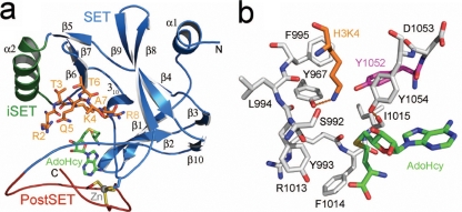
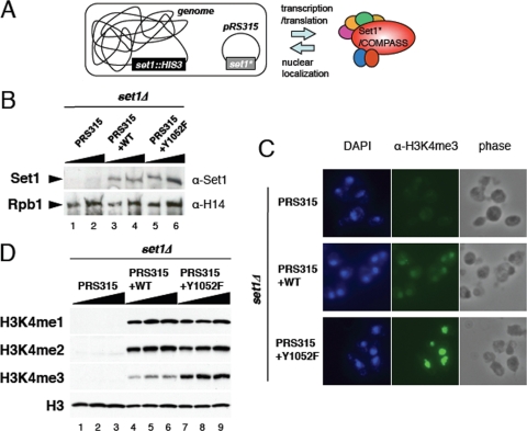
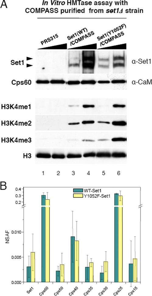
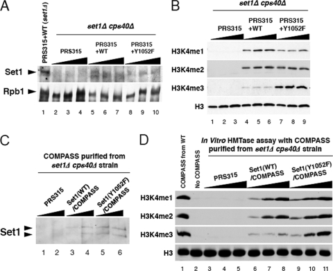
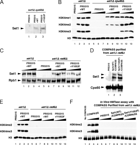
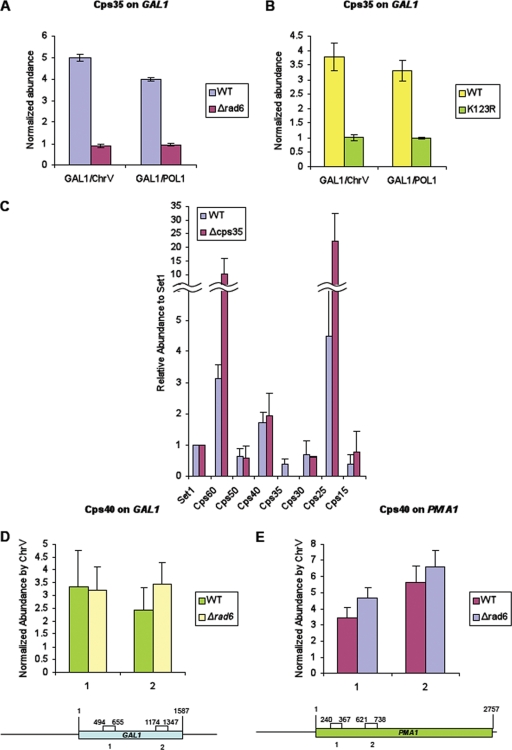
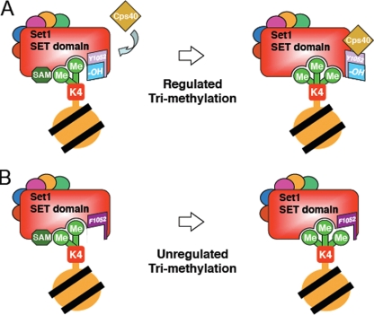
The multiprotein complex Set1/COMPASS is the founding member of the histone H3 lysine 4 (H3K4) methyltransferases, whose human homologs include the MLL and hSet1 complexes. COMPASS can mono-, di-, and trimethylate H3K4, but transitioning to di- and trimethylation requires prior H2B monoubiquitination followed by recruitment of the Cps35 (Swd2) subunit of COMPASS. Another subunit, Cps40 (Spp1), interacts directly with Set1 and is only required for transitioning to trimethylation. To investigate how the Set1 and COMPASS subunits establish the methylation states of H3K4, we generated a homology model of the catalytic domain of Saccharomyces cerevisiae yeast Set1 and identified several key residues within the Set1 catalytic pocket that are capable of regulating COMPASS's activity. We show that Tyr1052, a putative Phe/Tyr switch of Set1, plays an essential role in the regulation of H3K4 trimethylation by COMPASS and that the mutation to phenylalanine (Y1052F) suppresses the loss of Cps40 in H3K4 trimethylation levels, suggesting that Tyr1052 functions together with Cps40. However, the loss of H2B monoubiquitination is not suppressed by this mutation, while Cps40 is stably assembled in COMPASS on chromatin, demonstrating that Tyr1052- and Cps40-mediated H3K4 trimethylation takes place following and independently of H2B monoubiquitination. Our studies provide a molecular basis for the way in which H3K4 trimethylation is regulated by Tyr1052 and the Cps40 subunit of COMPASS.


Figures

FIG. 1.
FIG. 1.
Homology model of the yeast Set1 catalytic domain. (a) Ribbon representation of the secondary structure of the Set1 catalytic domain illustrating the SET domain (blue), the inserted SET motif (iSET, green), and the PostSET domain (red). A histone H3 substrate peptide spanning residues 2 to 8 (orange carbon atoms) and the product, AdoHcy (green carbons), are docked in the active site. The side chains of the four cysteines that coordinate the Zn(II) ion in the PostSET domain are also illustrated. (b) Active site of Set1. Residues comprising the enzyme's lysine binding channel are shown with gray carbon atoms, with the exception of the Phe/Tyr switch residue Tyr1052 that is highlighted by magenta carbons. AdoHcy and H3K4 are depicted as described for panel A. Hydrogen bonds are illustrated by orange dashed lines.
FIG. 2.
FIG. 2.
H3K4 methyltransferase activity of the Set1 Y1052F mutant in vivo. (A) Schematic representation of the yeast construct used for the mutational analyses of the SET domain of Set1. Yeast cells with the genomic SET1 gene replaced with the HIS3 marker are transformed with low-copy-number plasmid pRS315 harboring the SET1 coding sequence together with its own preceding promoter region, enabling an expression level equivalent to that of exogenous Set1. Point mutations were introduced into the SET1 gene carried on plasmid pRS315 (Set1*). (B) Wild-type (WT) and mutant Set1 levels in the different strains. Cell extracts prepared from logarithmically growing cells harboring SET1 plasmids were analyzed by Western blotting using polyclonal antibodies specific to Set1. H14 Western blotting detecting the large subunit of RNA Polymerase II (Rpb1) levels was used as a loading control. (C) Immunofluorescence analyses of H3K4 trimethylation by COMPASS containing wild-type or single point mutant Set1 proteins. Paraformaldehyde-fixed yeast cells were stained with DAPI and primary mouse antibodies toward trimethylated H3K4 and fluorescein-conjugated secondary antibodies. Phase-contrast images are shown in the right-hand panels. (D) Analyses of H3K4 methylation on bulk histones from cell extracts using anti-H3K4me1, anti-H3K4me2, and anti-H3K4me3-specific antibodies. Equal quantities of cell extracts from cells expressing both wild-type and mutant Set1 were subjected to SDS-PAGE, followed by Western blotting. α, anti; WT, wild-type Set1. Triangles represent increasing sample loads.
FIG. 3.
FIG. 3.
Intrinsically enhanced in vitro histone H3K4 methyltransferase activity of Set1(Y1052F) proteins within COMPASS. (A) The levels of Set1 and TAP-tagged Cps60 proteins in purified COMPASS were determined. Increasing concentrations of TAP-tagged Cps60-purified COMPASS, containing either wild-type or mutant Set1, were analyzed by Western blotting using anti-Set1 and anti-calmodulin binding peptide (CaM) antibodies. Each panel shows the results for aliquots of identical samples applied for Western blotting using different antibodies. Comparison of histone methyltransferase (HMTase) activity of wild-type and mutant Set1 within COMPASS toward histone H3K4 mono-, di-, and trimethylation (H3K4me1, -2, and -3 and H3) was performed with a histone methyltransferase assay mixture consisting of recombinant histone H3 substrate, the cofactor S-adenosylmethionine, and equal volumes of each COMPASS. Histone methyltransferase reaction products were subjected to Western analysis with the indicated antibody. Anti-H3 was used as a loading control. α, anti. (B) Subunit composition of COMPASS purified from either wild-type or Set1(Y1052F) was determined via MudPIT analysis. Error bars show standard deviations. NSAF, normalized spectral abundance factor; PRS315, plasmid pRS315; WT, wild-type Set1.
FIG. 4.
FIG. 4.
Complementation of H3K4 trimethylation in a cps40 (spp1)-deficient COMPASS both in vivo and in vitro. (A) Wild-type and Y1052F mutant Set1 protein levels in a cps40 null strain. Anti-Set1 and anti-H14 Western analyses were performed as described for Fig. 2B. (B) In vivo H3K4 methylation status in the background with cps40 deleted as revealed by Western blotting of whole-cell extracts as described for Fig. 2D but with set1 and cps40 double null cells as the genetic background. (C) Western analysis of Set1 protein levels in the TAP-purified COMPASS in wild-type and cps40 null cells. (D) In vitro H3K4 methylation catalyzed by Cps40 (Spp1)-deficient COMPASS containing wild-type or mutant Set1 proteins prepared from cps40Δ set1Δ cells harboring wild-type or mutant SET1 plasmids was assayed as described for Fig. 3A. Triangles represent increasing sample loads. PRS315, plasmid pRS315; WT, wild-type Set1.
FIG. 5.
FIG. 5.
Y1052F Set1 suppresses H3K4 di- and trimethylation defects in a cps60 (bre2) null strain, while H3K4 methylation loss caused by the loss of Rad6 is refractory to this mutation. (A) Wild-type and Y1052F mutant Set1 protein expression in a cps60 (bre2) null strain was confirmed by Western blotting with anti-Set1 antibody. (B) In vivo H3K4 methylation status in the cps60 deletion background. Assays were performed as described for Fig. 2D, except with set1 and cps60 double null cells as the genetic background. (C to F) Y1052-mediated H3K4 trimethylation regulation is independent of H2B K123 monoubiquitination via the Rad6/Bre1 pathway. (C) Equal expression of wild-type and Y1052F mutant Set1 introduced in a rad6 set1 double knockout strain as determined by Western analysis. Antibody H14, detecting Rbp1, was used as a loading control. (D) The levels of Set1 within COMPASS purified from either the wild type or rad6Δ strain carrying the vector containing either wild-type Set1 or Y1052F mutated Set1 or the vector only were examined by Western blotting using polyclonal antibodies specific to Set1. (E) Bulk H3K4 methylation levels were determined in a set1Δ strain expressing wild-type Set1 from a plasmid or a set1Δ rad6Δ strain expressing wild-type or Y1052F mutant Set1 from this vector or the vector only. (F) Wild-type or Y1052F Set1-containing COMPASS, purified from Cps60-TAP strains with RAD6 deleted, was tested for histone methyltransferase (HMTase) activity in vitro. Similar to the results of the in vivo studies described for panel E, Set1(Y1052F) cannot compensate for the loss of Rad6 in vitro. Triangles represent increasing loads of extracts. PRS315, plasmid pRS315; WT, wild-type Set1.
FIG. 6.
FIG. 6.
Cps40 (Spp1) can interact with COMPASS independently of monoubiquitinated histone H2B and Cps35 (Swd2). (A and B) ChIP of the GAL1 gene through myc-tagged Cps35 in the presence and absence of ubiquitinated H2B. Myc-Cps35 ChIP from wild-type (WT) and rad6Δ (A) and H2B(K123R) (B) strains. ChIP enrichments were normalized to the level in a ChrV nontranscribed intergenic region or the POL1 open reading frame. (C) Purification of Cps60-TAP tagged COMPASS from the wild-type (WT) and a Cps35 null strain (19). All COMPASS components except Cps35 can be detected in COMPASS purified from the cps35 null strain, indicating that Cps40 (Spp1) does not interact with COMPASS in a Cps35 (Swd2)-dependent manner. Using spectral counts, the composition of COMPASS purified from Cps35 null strain was determined (14). This histogram shows the abundances of COMPASS components purified from a cps35 null strain relative to those from the wild-type strain relative to the level of Set1. (D and E) ChIP of GAL1 (D) and PMA1 (E) genes for Cps40 in the presence and absence of monoubiquitinated H2B. Cell extracts were prepared from strains expressing nine-Myc-tagged Cps40 in the presence (wild type; WT) and absence of Rad6. (D) ChIP assay for Cps40 on the GAL1 gene was performed similarly to the ChIP assay described for Cps35 of the GAL1 gene whose results are shown in panels A and B. (E) ChIP of Cps40 on the PMA1 gene. Cells were grown in a dextrose-containing medium to an OD600 of 1.0, followed by formaldehyde-based in vivo cross-linking. Immunoprecipitations and qPCR were performed as described for panels A and B. ChIP enrichments were normalized to the levels in a ChrV nontranscribed intergenic region. Error bars show standard deviations.
FIG. 7.
FIG. 7.
Model illustrating the function of Cps40 in the regulation of histone methyltransferase activity of Set1/COMPASS. (A) Cps40 and Cps60 (yellow diamond) induce a conformational change of Set1/SET domain, pulling back Tyr1052 (light-purple trapezoid) so that its histone methyltransferase active site, encompassing the substrate of methylated lysine, becomes more open, making it easier to accommodate trimethylated lysine. Me, methyl group; yellow circle with black lines, nucleosome. (B) In the Y1052F mutant Set1, mono- and dimethylation proceed similarly to the process in the wild type. However, F1052 (dark purple), lacking the hydroxyl group (small blue trapezoid in panel A) from the phenol ring side chain, allows enough space for the methyltransferase to trimethylate the H3 peptide without the aid of Cps40. This model is consistent with our previous observation that the loss of Cps60 and Cps40 results in the loss of H3K4 trimethylation (24). SAM, S-adenosylmethionine.

Similar articles

Cited by

References

    1. Brunger, A. T., P. D. Adams, G. M. Clore, W. L. DeLano, P. Gros, R. W. Grosse-Kunstleve, J. S. Jiang, J. Kuszewski, M. Nilges, N. S. Pannu, R. J. Read, L. M. Rice, T. Simonson, and G. L. Warren. 1998. Crystallography & NMR system: a new software suite for macromolecular structure determination. Acta Crystallogr. D 54905-921. - PubMed
    1. Bupp, J. M., A. E. Martin, E. S. Stensrud, and S. L. Jaspersen. 2007. Telomere anchoring at the nuclear periphery requires the budding yeast Sad1-UNC-84 domain protein Mps3. J. Cell Biol. 179845-854. - PMC - PubMed
    1. Collins, R. E., M. Tachibana, H. Tamaru, K. M. Smith, D. Jia, X. Zhang, E. U. Selker, Y. Shinkai, and X. Cheng. 2005. In vitro and in vivo analyses of a Phe/Tyr switch controlling product specificity of histone lysine methyltransferases. J. Biol. Chem. 2805563-5570. - PMC - PubMed
    1. Couture, J. F., E. Collazo, J. S. Brunzelle, and R. C. Trievel. 2005. Structural and functional analysis of SET8, a histone H4 Lys-20 methyltransferase. Genes Dev. 191455-1465. - PMC - PubMed
    1. Couture, J. F., L. M. Dirk, J. S. Brunzelle, R. L. Houtz, and R. C. Trievel. 2008. Structural origins for the product specificity of SET domain protein methyltransferases. Proc. Natl. Acad. Sci. USA 10520659-20664. - PMC - PubMed

Publication types

MeSH terms

Associated data