

doi: 10.1101/gad.1742609.
Bmi-1 regulates the Ink4a/Arf locus to control pancreatic beta-cell proliferation
Affiliations
- PMID: 19390085
- PMCID: PMC2675870
- DOI: 10.1101/gad.1742609
Item in Clipboard
Bmi-1 regulates the Ink4a/Arf locus to control pancreatic beta-cell proliferation
Genes Dev.
.
Abstract
The molecular mechanisms that regulate the age-induced increase of p16(INK4a) expression associated with decreased beta-cell proliferation and regeneration are not well understood. We report that in aged islets, derepression of the Ink4a/Arf locus is associated with decreased Bmi-1 binding, loss of H2A ubiquitylation, increased MLL1 recruitment, and a concomitant increase in H3K4 trimethylation. During beta-cell regeneration these histone modifications are reversed resulting in reduced p16(INK4a) expression and increased proliferation. We suggest that PcG and TrxG proteins impart a combinatorial code of histone modifications on the Ink4a/Arf locus to control beta-cell proliferation during aging and regeneration.
Figures
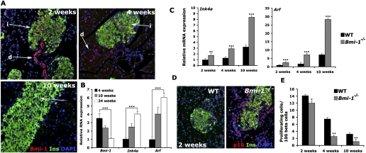
The expression of Bmi-1 declined with age and correlated with increased expression of Ink4a/Arf genes and reduced β-cell proliferation. (A) Expression pattern of Bmi-1 in the representative pancreatic sections from wild-type mice aged 2, 4, and 10 wk, respectively (20× magnification). (i) Islet cells; (d) ductal cells. (B) RNA levels of Bmi-1, Ink4a, and Arf in islets isolated from mice at different ages. (C) RNA levels of Ink4a and Arf and (D) Immunofluorescence for p16 and insulin, with DAPI, in pancreatic sections from 2-wk-old wild-type (WT) and Bmi-1−/− mice (20× magnification). (E) Quantification of proliferating β cells at 2, 4, and 10 wk in wild-type and Bmi-1−/− mice, as a percentage of Ki67+ insulin double-positive cells in islets. (*) P < 0.05; (**) P < 0.01; (***) P < 0.005.

Bmi-1−/− mice display diminished β-cell mass, hypoinsulinemia, and glucose intolerance. (A) Quantification of proliferating islet cells (left panel) and levels of Ink4a (right panel) in cultured islets isolated from 4-wk-old wild-type (WT) and Bmi-1−/− mice, transfected with Ink4a or scrambled siRNAs. (B) Growth profiles (left panel) and corresponding levels of Bmi-1 and Ink4a (right panel) in Min6 cells transfected with Bmi-1, Ink4a, Bmi-1 + Ink4a, or scrambled siRNAs. P-value symbols indicated are for comparison of Bmi-1 and Bmi-1 + Ink4a samples. (C) Pancreatic sections from 10-wk-old wild-type and Bmi-1−/− mice stained with insulin (Ins) and glucagon (Glu) with DAPI (5× magnification). (D) β-Cell mass in wild-type and Bmi-1−/− mice at 2, 4, and 10 wk of age (n = 5 for each phenotype and age). (E) GTT for wild-type and Bmi-1−/− mice at 10 wk (n = 5 for each phenotype). (F) Plasma insulin levels in 10-wk-old wild-type and Bmi-1−/− mice (fasted overnight) at 0 and 30 min, after glucose injection (n = 5 for each phenotype). (*) P < 0.05; (**) P < 0.01; (***) P < 0.005.
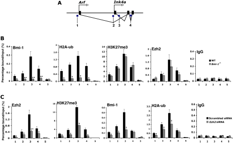
Bmi-1 plays a key role in regulation of the Ink4a/Arf locus through modulation of histone modifications. (A) Schematic representation of the Ink4a/Arf locus, with blue regions marked 1–4 indicating the amplified regions in the ChIP studies. ChIP analysis for the indicated antibodies at the Ink4a/Arf locus in islets isolated from wild-type (WT) and Bmi-1−/− mice (4 wk old) (B) and upon treatment of islets from 4-wk-old mice (C), with siRNA targeting Ezh2 or control, scrambled siRNA. Primer set 5 indicates negative control (Exon 2 of HoxC13 locus) (Cao et al. 2005). (*) P < 0.05; (**) P < 0.01; (***) P < 0.005.

Polycomb-mediated regulation of Ink4a/Arf locus during aging by histone modification (A) ChIP analysis for the indicated antibodies at the Ink4a/Arf locus with aging, with IgG as control, Young indicates 2 wk; old, 24–30 wk of age. Time points chosen to reflect very high (2 wk) and low (24–30 wk) levels of PcG proteins. (B) Coimmunostaining of ubiquitinated H2A (H2A-ub) and insulin (Ins) with DAPI, in pancreatic sections from 2-wk-old and 24-wk-old wild-type mice. (*) P < 0.05; (**) P < 0.01; (***) P < 0.005.
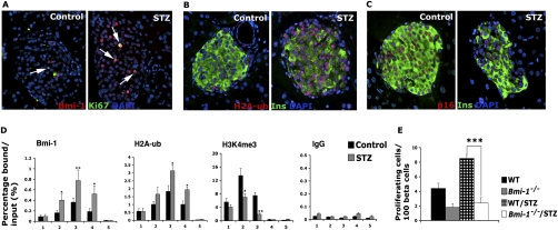
Bmi-1-dependent regulation of Ink4a/Arf locus plays a critical role in β-cell regeneration. Coimmunostaining for Bmi-1 and Ki67 (A) and coimmunostaining for H2A-ub and insulin (Ins) with DAPI (B) in pancreatic section from vehicle or STZ-treated 4-wk-old wild-type (WT) mice. Arrows indicate cells that are positive for both Bmi-1 and Ki67 (20× magnification). (C) Immunofluorescence for p16Ink4a and insulin (Ins) with DAPI on pancreatic sections from 4-wk-old mice treated with control vehicle or a single dose of STZ (20× magnification). (D) ChIP analysis for the indicated antibodies at the Ink4a/Arf locus on islets from 4-wk-old wild-type mice injected with STZ or control vehicle. Analyses A–D were performed on mice 4 d after injections. (E) Proliferation of β cells in wild-type and Bmi-1−/− mice after STZ or vehicle treatment, measured as a percentage of Ki67+ insulin double-positive islet cells. Analysis performed 15 d after STZ or control injections. n = 5 mice per treatment. (*) P < 0.05; (**) P < 0.01; (***) P < 0.005.
Similar articles
-
Polycomb protein Ezh2 regulates pancreatic beta-cell Ink4a/Arf expression and regeneration in diabetes mellitus.Genes Dev. 2009 Apr 15;23(8):975-85. doi: 10.1101/gad.1742509. Genes Dev. 2009. PMID: 19390090 Free PMC article.
-
Polycomb mediated epigenetic silencing and replication timing at the INK4a/ARF locus during senescence.PLoS One. 2009 May 20;4(5):e5622. doi: 10.1371/journal.pone.0005622. PLoS One. 2009. PMID: 19462008 Free PMC article.
-
A novel zinc finger protein Zfp277 mediates transcriptional repression of the Ink4a/arf locus through polycomb repressive complex 1.PLoS One. 2010 Aug 24;5(8):e12373. doi: 10.1371/journal.pone.0012373. PLoS One. 2010. PMID: 20808772 Free PMC article.
-
Stem cell self-renewal and cancer cell proliferation are regulated by common networks that balance the activation of proto-oncogenes and tumor suppressors.Cold Spring Harb Symp Quant Biol. 2005;70:177-85. doi: 10.1101/sqb.2005.70.057. Cold Spring Harb Symp Quant Biol. 2005. PMID: 16869752 Review.
-
Role of Ink4a/Arf locus in beta cell mass expansion under physiological and pathological conditions.J Diabetes Res. 2014;2014:873679. doi: 10.1155/2014/873679. Epub 2014 Feb 6. J Diabetes Res. 2014. PMID: 24672805 Free PMC article. Review.
Cited by
-
Clinical significance of cell cycle inhibitors in hepatocellular carcinoma.Med Mol Morphol. 2013 Dec;46(4):185-92. doi: 10.1007/s00795-013-0047-7. Epub 2013 May 3. Med Mol Morphol. 2013. PMID: 23640750 Review.
-
Bmi1 enhances tumorigenicity and cancer stem cell function in pancreatic adenocarcinoma.PLoS One. 2013;8(2):e55820. doi: 10.1371/journal.pone.0055820. Epub 2013 Feb 20. PLoS One. 2013. PMID: 23437065 Free PMC article.
-
Postnatal development, maturation and aging in the mouse cochlea and their effects on hair cell regeneration.Hear Res. 2013 Mar;297:68-83. doi: 10.1016/j.heares.2012.11.009. Epub 2012 Nov 16. Hear Res. 2013. PMID: 23164734 Free PMC article. Review.
-
Expression of linear and novel circular forms of an INK4/ARF-associated non-coding RNA correlates with atherosclerosis risk.PLoS Genet. 2010 Dec 2;6(12):e1001233. doi: 10.1371/journal.pgen.1001233. PLoS Genet. 2010. PMID: 21151960 Free PMC article.
-
Epigenetic cancer prevention mechanisms in skin cancer.AAPS J. 2013 Oct;15(4):1064-71. doi: 10.1208/s12248-013-9513-3. Epub 2013 Aug 1. AAPS J. 2013. PMID: 23904153 Free PMC article. Review.
References
-
- Bracken A.P., Kleine-Kohlbrecher D., Dietrich N., Pasini D., Gargiulo G., Beekman C., Theilgaard-Monch K., Minucci S., Porse B.T., Marine J.C., et al. The Polycomb group proteins bind throughout the INK4A–ARF locus and are disassociated in senescent cells. Genes & Dev. 2007;21:525–530. - PMC - PubMed
-
- Bruggeman S.W., Valk-Lingbeek M.E., van der Stoop P.P., Jacobs J.J., Kieboom K., Tanger E., Hulsman D., Leung C., Arsenijevic Y., Marino S., et al. Ink4a and Arf differentially affect cell proliferation and neural stem cell self-renewal in Bmi1-deficient mice. Genes & Dev. 2005;19:1438–1443. - PMC - PubMed
-
- Cao R., Tsukada Y., Zhang Y. Role of Bmi-1 and Ring1A in H2A ubiquitylation and Hox gene silencing. Mol. Cell. 2005;20:845–854. - PubMed
-
- Dor Y., Brown J., Martinez O.I., Melton D.A. Adult pancreatic β-cells are formed by self-duplication rather than stem-cell differentiation. Nature. 2004;429:41–46. - PubMed
Publication types
MeSH terms
Substances
Grants and funding
LinkOut - more resources
Full Text Sources
Medical
Molecular Biology Databases