. 2009 May 5;119(17):2357-2366.
doi: 10.1161/CIRCULATIONAHA.108.814145. Epub 2009 Apr 20.

MicroRNA-320 is involved in the regulation of cardiac ischemia/reperfusion injury by targeting heat-shock protein 20

Affiliations

MicroRNA-320 is involved in the regulation of cardiac ischemia/reperfusion injury by targeting heat-shock protein 20

Xiao-Ping Ren et al. Circulation. .

Abstract

Background: Recent studies have identified critical roles for microRNAs (miRNAs) in a variety of cellular processes, including regulation of cardiomyocyte death. However, the signature of miRNA expression and possible roles of miRNA in the ischemic heart have been less well studied.

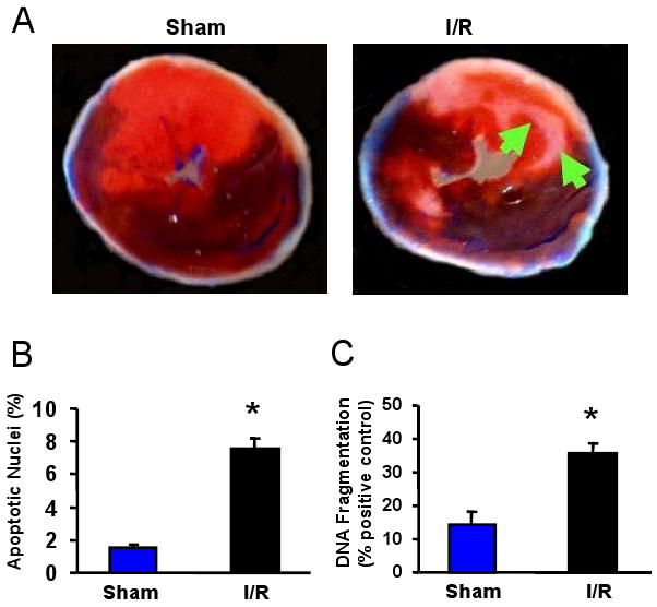
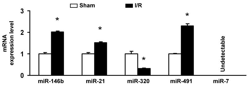
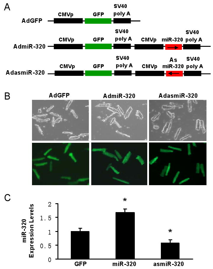
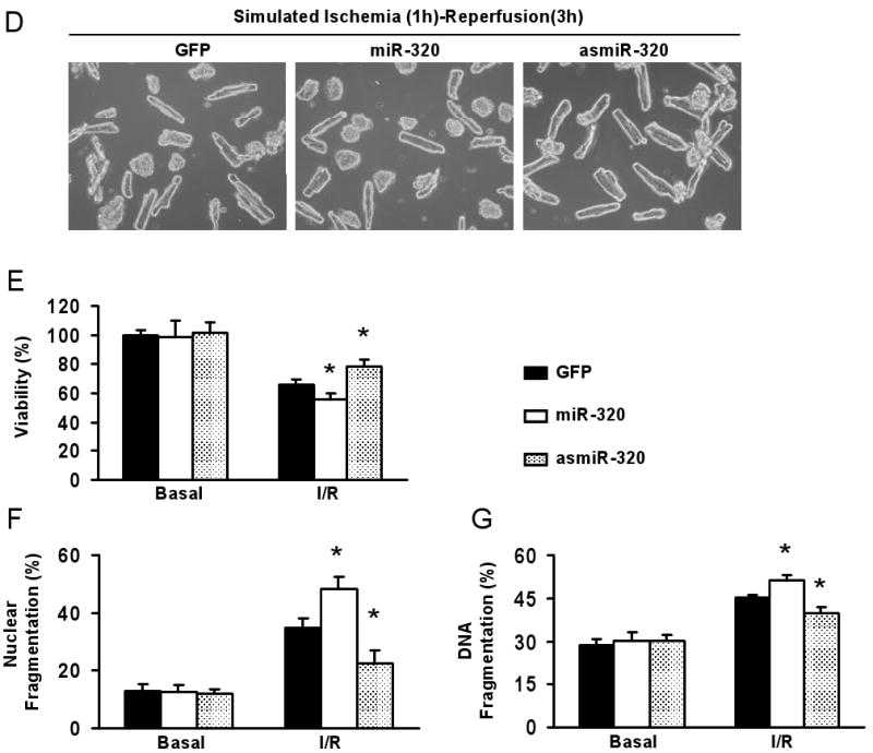
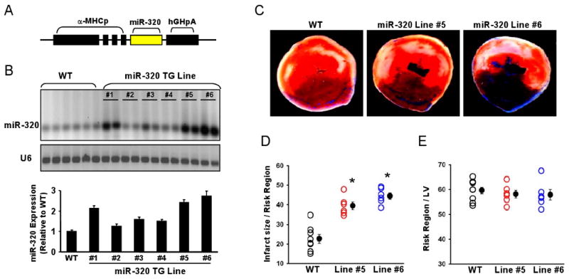
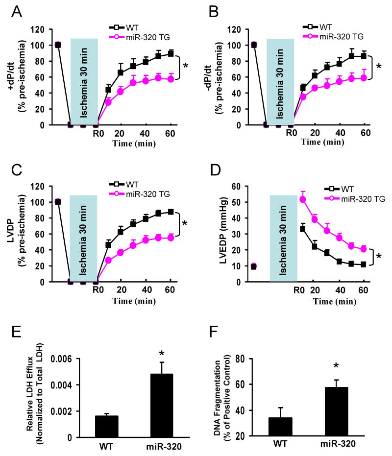
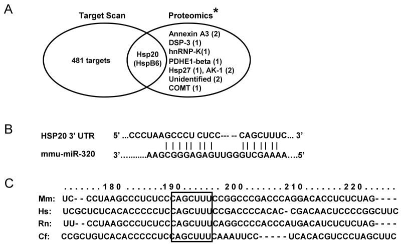
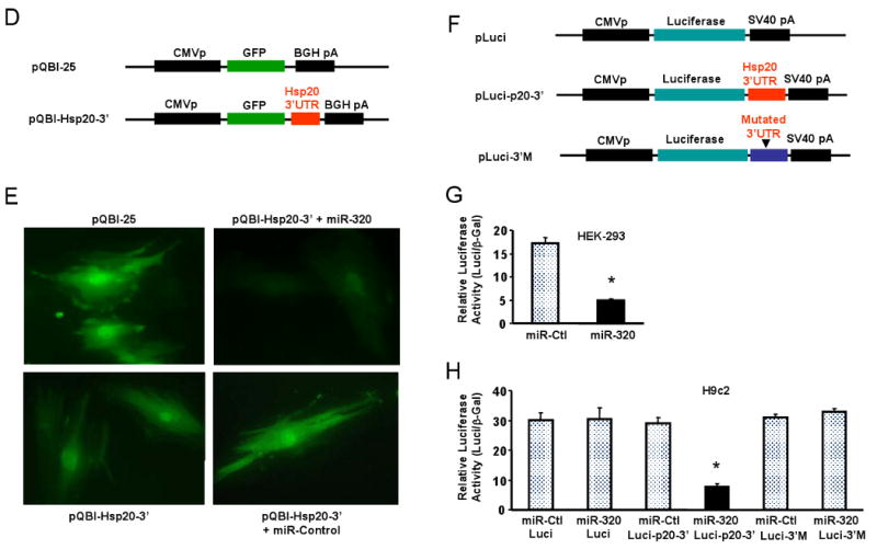
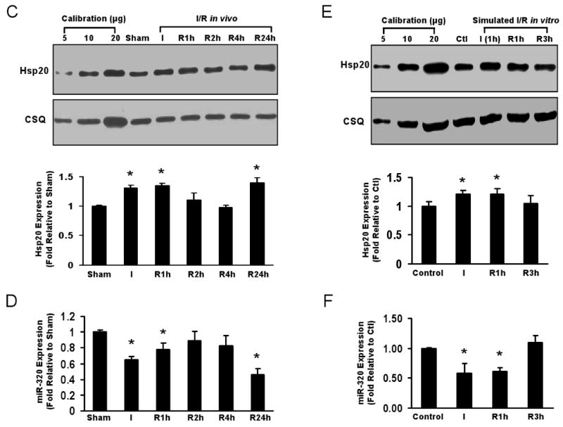
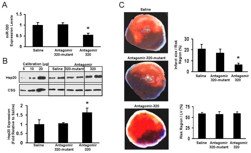
Methods and results: We performed miRNA arrays to detect the expression pattern of miRNAs in murine hearts subjected to ischemia/reperfusion (I/R) in vivo and ex vivo. Surprisingly, we found that only miR-320 expression was significantly decreased in the hearts on I/R in vivo and ex vivo. This was further confirmed by TaqMan real-time polymerase chain reaction. Gain-of-function and loss-of-function approaches were employed in cultured adult rat cardiomyocytes to investigate the functional roles of miR-320. Overexpression of miR-320 enhanced cardiomyocyte death and apoptosis, whereas knockdown was cytoprotective, on simulated I/R. Furthermore, transgenic mice with cardiac-specific overexpression of miR-320 revealed an increased extent of apoptosis and infarction size in the hearts on I/R in vivo and ex vivo relative to the wild-type controls. Conversely, in vivo treatment with antagomir-320 reduced infarction size relative to the administration of mutant antagomir-320 and saline controls. Using TargetScan software and proteomic analysis, we identified heat-shock protein 20 (Hsp20), a known cardioprotective protein, as an important candidate target for miR-320. This was validated experimentally by utilizing a luciferase/GFP reporter activity assay and examining the expression of Hsp20 on miR-320 overexpression and knockdown in cardiomyocytes.

Conclusions: Our data demonstrate that miR-320 is involved in the regulation of I/R-induced cardiac injury and dysfunction via antithetical regulation of Hsp20. Thus, miR-320 may constitute a new therapeutic target for ischemic heart diseases.


Conflict of interest statement

Conflict of Interest Disclosures: None

Figures

Figure 1
Figure 1
miRNA expression in the I/R hearts. (A-C) Occlusion of the left anterior descending coronary artery (LAD) for 30 min, followed by 24-h reperfusion (I/R), induced cardiac injury (infarction and apoptosis) in mice. (A) Infarct zone was observed in I/R hearts (white-gray zone, indicated by arrows), but not in sham samples. (B) The number of apoptotic nuclei and (C) the degree of DNA fragmentation were greatly increased in I/R hearts, compared with the shams (n=6, * P<0.01). (D) A partial heat-map of the upregulated and downregulated miRNAs (labeled by arrows). All of the microRNA array raw data is available in the Online supplemental data. (E) Dysregulated expression of miRNAs was confirmed by Taqman RT-PCR (normalized to control snoRNA412). RT-PCR primer sets for these miRNAs and control snoRNA412 were purchased from Ambion, Inc. We did not validate ambi-miR-9651 due to no available primer sets for this miRNA. (n=6, * P<0.05).
Figure 1
Figure 1
miRNA expression in the I/R hearts. (A-C) Occlusion of the left anterior descending coronary artery (LAD) for 30 min, followed by 24-h reperfusion (I/R), induced cardiac injury (infarction and apoptosis) in mice. (A) Infarct zone was observed in I/R hearts (white-gray zone, indicated by arrows), but not in sham samples. (B) The number of apoptotic nuclei and (C) the degree of DNA fragmentation were greatly increased in I/R hearts, compared with the shams (n=6, * P<0.01). (D) A partial heat-map of the upregulated and downregulated miRNAs (labeled by arrows). All of the microRNA array raw data is available in the Online supplemental data. (E) Dysregulated expression of miRNAs was confirmed by Taqman RT-PCR (normalized to control snoRNA412). RT-PCR primer sets for these miRNAs and control snoRNA412 were purchased from Ambion, Inc. We did not validate ambi-miR-9651 due to no available primer sets for this miRNA. (n=6, * P<0.05).
Figure 2
Figure 2
Overexpression of miR-320 enhanced cell death and apoptosis in cultured adult rat cardiomyocytes upon simulated ischemia/reperfusion, while knock-down was cytoprotective. (A) Diagram of recombinant adenoviral vectors. The primary miR-320 (470 bp) was PCR-amplified and inserted in the sense or antisense direction to generate AdmiR-320 or AdasmiR-320. (B) Nearly 100% cardiomyocytes were infected after 48-h adenoviral infection, as indicated by GFP fluorescence, and there were no morphological changes among GFP-, miR-320-, and asmiR-320-infected cells. (C) The levels of miR-320 were determined after 60-h infection by Taqman RT-PCR, as described in Figure 1, which confirmed the expression of mature miR-320 in AdmiR-320-infected cells, and knockdown of endogenous miR-320 in AdasmiR-320-cells. (D-G) miR-320-overexpressing cardiomyocytes were sensitive, while knockdown cells resistant to simulated ischemia/reperfusion (I/R)-induced cell death (D and E), and apoptosis (F and G), as determined by MTS incorporation (E), nuclear fragmentation by Hoechst staining (F), and cell-death-detection ELISA kit (G) (*P < 0.05 versus control AdGFP). Similar results were observed in three additional, independent experiments.
Figure 2
Figure 2
Overexpression of miR-320 enhanced cell death and apoptosis in cultured adult rat cardiomyocytes upon simulated ischemia/reperfusion, while knock-down was cytoprotective. (A) Diagram of recombinant adenoviral vectors. The primary miR-320 (470 bp) was PCR-amplified and inserted in the sense or antisense direction to generate AdmiR-320 or AdasmiR-320. (B) Nearly 100% cardiomyocytes were infected after 48-h adenoviral infection, as indicated by GFP fluorescence, and there were no morphological changes among GFP-, miR-320-, and asmiR-320-infected cells. (C) The levels of miR-320 were determined after 60-h infection by Taqman RT-PCR, as described in Figure 1, which confirmed the expression of mature miR-320 in AdmiR-320-infected cells, and knockdown of endogenous miR-320 in AdasmiR-320-cells. (D-G) miR-320-overexpressing cardiomyocytes were sensitive, while knockdown cells resistant to simulated ischemia/reperfusion (I/R)-induced cell death (D and E), and apoptosis (F and G), as determined by MTS incorporation (E), nuclear fragmentation by Hoechst staining (F), and cell-death-detection ELISA kit (G) (*P < 0.05 versus control AdGFP). Similar results were observed in three additional, independent experiments.
Figure 3
Figure 3
Overexpression of miR-320 increased I/R-induced cardiac injury in vivo. (A) Diagram of miR-320 TG construct. Transgenic mice were generated using a mouse primary miR-320 DNA under the control of the α-myosin heavy chain promoter (α-MHCp). (B) Northern blots showed that mature miR-320 was overexpressed in the transgenic hearts. Loading control was performed with an RNA probe against the small housekeeping RNA, U6 (106 nt). (C-D) miR-320 overexpression greatly increased myocardial infarct size after 30 min myocardial ischemia, via left anterior descending (LAD) coronary artery occlusion, followed by 24-h reperfusion, (E) while the region at risk was not significantly different among groups. (n=8, WT; n=6, miR-320 TG. *P<0.05 vs WT).
Figure 4
Figure 4
miR-320 overexpression depressed post-ischemic cardiac performance in isolated perfused hearts. After 30 min stabilization with KH buffer, hearts were subjected to 30 minutes of global ischemia and 1-h reperfusion. (A-C) Recovery of post-ischemic contractile function (±dP/dt) and left ventricular developed pressure (LVDP) during reperfusion was significantly depressed in TG mice; (D) Left ventricular end-diastolic pressure (LVEDP) was significantly higher in TG than that of WT hearts, (n=10, WT; n=11, miR-320 TG. *P<0.05 vs WT). (E and F) miR-320 overexpression enhanced ischemia/reperfusion-induced necrosis, measured by LDH release in the outflow of first 10-min of reperfusion; and apoptosis, measured by DNA fragmentation in the I/R hearts. The cumulative release of LDH in the outflow was normalized to the total tissue LDH content. (n=10-11 for LDH activity measurement; n=5 for DNA fragmentation measurement, *P<0.05 vs WT).
Figure 5
Figure 5
Hsp20 is a target of miR-320 in the murine heart. (A) TargetScan predicts that miR-320 has 482 targets, however, proteomic data showed that only 12 protein spots were altered in the proteome of I/R hearts (* Ref.# 22), and Hsp20 was listed in both the TargetScan results and the proteomics analysis. (B) Sequence alignment of miR-320 and 3′UTR of Hsp20. Note the complementarity at the 5′ and 3′ end of miR-320, where the crucial seed regions are located. (C) The putative miR-320-binding sites within the Hsp20 3′UTR are conserved among mammalian species (mouse, human, rat and dog). (D) Diagram of plasmid construction. A segment of Hsp20 3′ UTR was inserted downstream of the GFP-encoding sequence. (E) H9c2 cells were co-transfected with the plasmid containing the segment of Hsp20 3′ UTR and either miR-320 or a control oligoribonucleotide. Pictures were taken 48 h after transfection. (F) Diagram of plasmid construction. A segment of Hsp20 3′ UTR or a mutated segment was cloned downstream of the luciferase-encoding region. (G) Dual luciferase activity assay of HEK-293 cells co-transfected with the plasmid containing the segment of Hsp20 3′ UTR and either miR-320 or a control oligoribonucleotide showed that miR-320 inhibited luciferase activity, compared with controls. (H) Luciferase activity in H9c2 cells co-transfected with the various vectors indicated. (* P<0.05 relative to respective controls). Similar results were observed in three additional, independent experiments.
Figure 5
Figure 5
Hsp20 is a target of miR-320 in the murine heart. (A) TargetScan predicts that miR-320 has 482 targets, however, proteomic data showed that only 12 protein spots were altered in the proteome of I/R hearts (* Ref.# 22), and Hsp20 was listed in both the TargetScan results and the proteomics analysis. (B) Sequence alignment of miR-320 and 3′UTR of Hsp20. Note the complementarity at the 5′ and 3′ end of miR-320, where the crucial seed regions are located. (C) The putative miR-320-binding sites within the Hsp20 3′UTR are conserved among mammalian species (mouse, human, rat and dog). (D) Diagram of plasmid construction. A segment of Hsp20 3′ UTR was inserted downstream of the GFP-encoding sequence. (E) H9c2 cells were co-transfected with the plasmid containing the segment of Hsp20 3′ UTR and either miR-320 or a control oligoribonucleotide. Pictures were taken 48 h after transfection. (F) Diagram of plasmid construction. A segment of Hsp20 3′ UTR or a mutated segment was cloned downstream of the luciferase-encoding region. (G) Dual luciferase activity assay of HEK-293 cells co-transfected with the plasmid containing the segment of Hsp20 3′ UTR and either miR-320 or a control oligoribonucleotide showed that miR-320 inhibited luciferase activity, compared with controls. (H) Luciferase activity in H9c2 cells co-transfected with the various vectors indicated. (* P<0.05 relative to respective controls). Similar results were observed in three additional, independent experiments.
Figure 6
Figure 6
Reciprocal expression of miR-320 and Hsp20. (A) Total cellular protein from various adenoviral-infected cardiomyocytes after 60-h infection was probed using an Hsp20 antibody. Actin was used as a loading control. (B) Immunoblots for determination of Hsp20 expression in miR-320 TG hearts. * P<0.05 relative to controls, n=6-8 hearts. (C) Time course expression of Hsp20 in hearts during in vivo I/R determined by Western blots. Calsequestrin (CSQ) was used as a loading control. (D) Time course expression of miR-320 in murine hearts during I/R was determined by Taqman RT-PCR, as described in Figure 1 legend. * P<0.05 relative to shams, n=3 hearts. (E) Time course expression of Hsp20 and (F) miR-320 in ex vivo cultured adult rat cardiomyocytes subjected to simulated ischemia (1h)/reperfusion (3h). * P<0.05 relative to controls, n=3 hearts.
Figure 6
Figure 6
Reciprocal expression of miR-320 and Hsp20. (A) Total cellular protein from various adenoviral-infected cardiomyocytes after 60-h infection was probed using an Hsp20 antibody. Actin was used as a loading control. (B) Immunoblots for determination of Hsp20 expression in miR-320 TG hearts. * P<0.05 relative to controls, n=6-8 hearts. (C) Time course expression of Hsp20 in hearts during in vivo I/R determined by Western blots. Calsequestrin (CSQ) was used as a loading control. (D) Time course expression of miR-320 in murine hearts during I/R was determined by Taqman RT-PCR, as described in Figure 1 legend. * P<0.05 relative to shams, n=3 hearts. (E) Time course expression of Hsp20 and (F) miR-320 in ex vivo cultured adult rat cardiomyocytes subjected to simulated ischemia (1h)/reperfusion (3h). * P<0.05 relative to controls, n=3 hearts.
Figure 7
Figure 7
In vivo effects of antagomir-320 administration on myocardial ischemia/reperfusion. (A) The levels of miR-320 expression were determined in murine hearts 3 days after administration of antagomir-320, antagomir-320 mutant and saline control by Taqman RT-PCR, as described in Figure 1 legend. (B) Western blot and densitometric analysis of Hsp20 expression in murine hearts 3 days after treated with these anatgomirs. Calsequestrin (CSQ) was used as a loading control. (C) Antagomir-320 treatment greatly reduced myocardial infarct size after 30 min myocardial ischemia followed by24-h reperfusion, while the region at risk was not significantly different among groups. (n=7, saline; n=5, mutant antagomir-320; n=7, antagomir-320. *P<0.05 vs saline control).

Similar articles

Cited by

References

    1. Cannon RO., 3rd Mechanisms, management and future directions for reperfusion injury after acute myocardial infarction. Nat Clin Pract Cardiovasc Med. 2005;2:88–94. - PubMed
    1. Abbate A, Bussani R, Amin MS, Vetrovec GW, Baldi A. Acute myocardial infarction and heart failure: role of apoptosis. Int J Biochem Cell Biol. 2006;38:1834–1840. - PubMed
    1. Cokkinos DV, Pantos C. Myocardial protection in man--from research concept to clinical practice. Heart Fail Rev. 2007;12:345–362. - PubMed
    1. van Rooij E, Olson EN. MicroRNAs: powerful new regulators of heart disease and provocative therapeutic targets. J Clin Invest. 2007;117:2369–2376. - PMC - PubMed
    1. Latronico MV, Catalucci D, Condorelli G. Emerging role of microRNAs in cardiovascular biology. Circ Res. 2007;101:1225–1236. - PubMed

Publication types