

Acquisition of a second mutation of the Tp53 alleles immediately precedes epithelial morphological transformation in ovarian tumorigenicity
- PMID: 19375154
- PMCID: PMC2739736
- DOI: 10.1016/j.ygyno.2009.03.023
Acquisition of a second mutation of the Tp53 alleles immediately precedes epithelial morphological transformation in ovarian tumorigenicity
Abstract
Objective: Tp53 mutation is frequent and associates with malignant, high-grade ovarian cancer. However, the details about the progression of Tp53 mutation from heterozygous to homozygous, and association between genotypes and morphological transformation are not clear. We further investigated the expression and mutation of Tp53 and associated markers such as p21 and HDM2 in ovarian cancer.
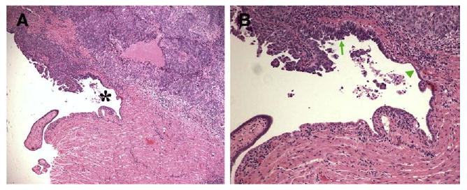
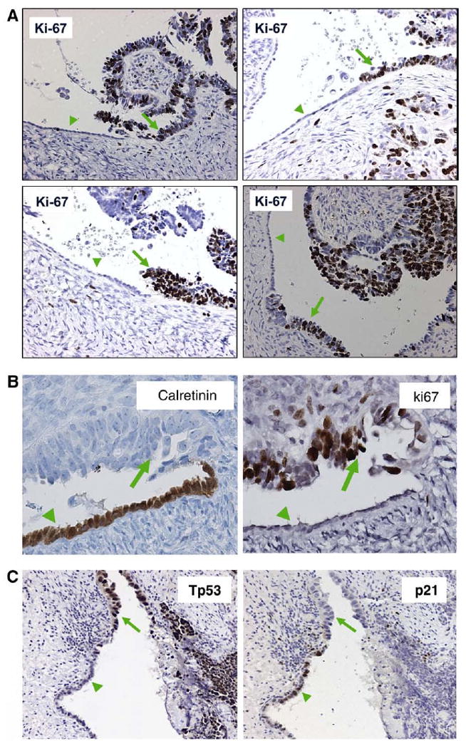
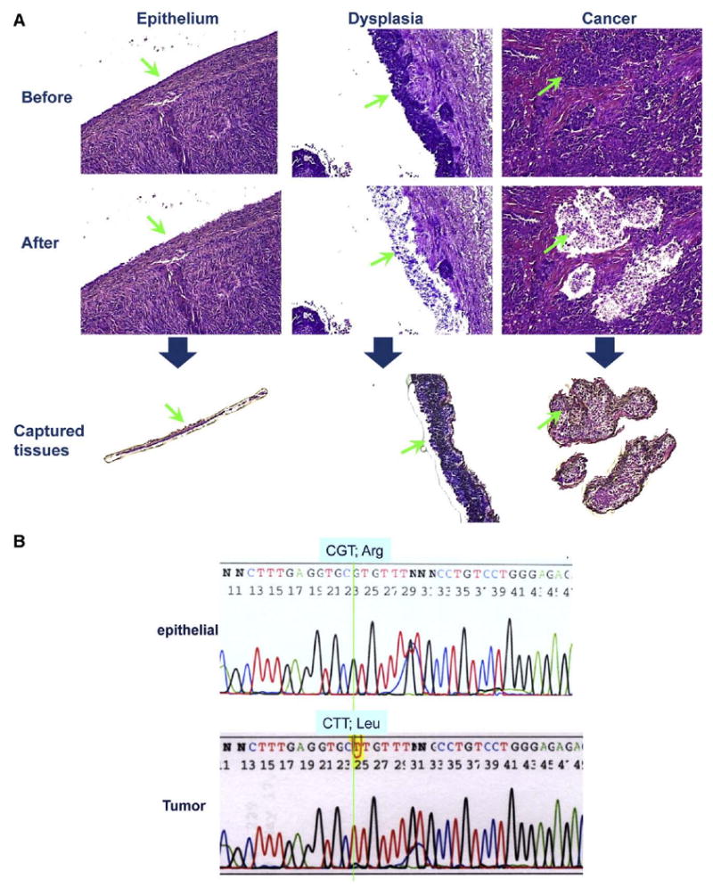
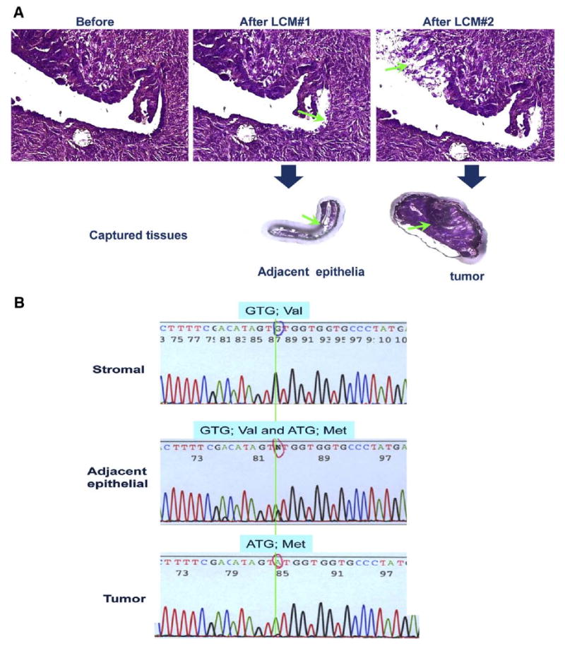
Method: Areas of contiguous ovarian surface epithelia linking morphological normal monolayer to multilayer neoplastic cells were analyzed for the correlation of Tp53 pathway alteration in relation to morphological transformation, by immunostaining and sequencing of Tp53 gene in cells from laser captured microdissection.
Results: Consistent with previous reports, Tp53 staining is positive in 78% of the tumors. The staining of p21 is positive in about 12%, and HMD2 is positive in only 1% of the tumors. In 9 out of 10 cases of p21-positive tumors, p53 is also positive. In the majority of cases of epithelial histological transitions, overexpression of Tp53 correlates with morphological transformation: Tp53 is negative in monolayered cells and positive in neoplastic lesions. Morphological transformation also closely correlates with cell proliferation as indicated by Ki-67 staining and loss of p21 expression. We detected heterozygous mutation of Tp53 in the monolayers adjacent to neoplastic cells.
Conclusions: p21 expression is an indicator of a wild type Tp53 and lack of p21 in the presence of Tp53 expression predicts an inactivated Tp53. Tp53 inactivation immediately precedes morphological transformation of the ovarian surface epithelium in most cases, and the histological transitional epithelia containing a heterozygous Tp53 mutation are thus pre-neoplastic lesions. We propose that the loss of a second allele of Tp53 leading to the loss of p21 expression, and subsequent cell proliferation, compose a sequence of events that lead to morphological transformation and instigation of ovarian epithelial tumor development.
Conflict of interest statement
Figures

Similar articles
-
Discriminating functional and non-functional p53 in human tumours by p53 and MDM2 immunohistochemistry.J Pathol. 2005 Nov;207(3):251-9. doi: 10.1002/path.1838. J Pathol. 2005. PMID: 16161005
-
Ovarian carcinoma in situ with germline BRCA1 mutation and loss of heterozygosity at BRCA1 and TP53.J Natl Cancer Inst. 2000 Jul 5;92(13):1088-91. doi: 10.1093/jnci/92.13.1088. J Natl Cancer Inst. 2000. PMID: 10880552
-
Immunohistochemical staining patterns of p53 can serve as a surrogate marker for TP53 mutations in ovarian carcinoma: an immunohistochemical and nucleotide sequencing analysis.Mod Pathol. 2011 Sep;24(9):1248-53. doi: 10.1038/modpathol.2011.85. Epub 2011 May 6. Mod Pathol. 2011. PMID: 21552211
-
TP53 Mutations in Breast and Ovarian Cancer.Cold Spring Harb Perspect Med. 2017 Jan 3;7(1):a026252. doi: 10.1101/cshperspect.a026252. Cold Spring Harb Perspect Med. 2017. PMID: 27815305 Free PMC article. Review.
-
The consequence of oncomorphic TP53 mutations in ovarian cancer.Int J Mol Sci. 2013 Sep 23;14(9):19257-75. doi: 10.3390/ijms140919257. Int J Mol Sci. 2013. PMID: 24065105 Free PMC article. Review.
Cited by
-
Poor survival with wild-type TP53 ovarian cancer?Gynecol Oncol. 2013 Sep;130(3):565-9. doi: 10.1016/j.ygyno.2013.06.016. Epub 2013 Jun 22. Gynecol Oncol. 2013. PMID: 23800698 Free PMC article.
-
Through the glass darkly: intraepithelial neoplasia, top-down differentiation, and the road to ovarian cancer.J Pathol. 2013 Dec;231(4):402-12. doi: 10.1002/path.4263. J Pathol. 2013. PMID: 24030860 Free PMC article. Review.
-
Identifying a highly-aggressive DCIS subgroup by studying intra-individual DCIS heterogeneity among invasive breast cancer patients.PLoS One. 2014 Jun 30;9(6):e100488. doi: 10.1371/journal.pone.0100488. eCollection 2014. PLoS One. 2014. PMID: 24978026 Free PMC article.
-
Molecular pathogenesis and extraovarian origin of epithelial ovarian cancer--shifting the paradigm.Hum Pathol. 2011 Jul;42(7):918-31. doi: 10.1016/j.humpath.2011.03.003. Hum Pathol. 2011. PMID: 21683865 Free PMC article. Review.
-
Nuclear envelope structural defects cause chromosomal numerical instability and aneuploidy in ovarian cancer.BMC Med. 2011 Mar 26;9:28. doi: 10.1186/1741-7015-9-28. BMC Med. 2011. PMID: 21439080 Free PMC article.
References
-
- Scully RE. Pathology of ovarian cancer precursors. J Cell Biochem Suppl. 1995;23:208–18. - PubMed
-
- Bell DA. Origins and molecular pathology of ovarian cancer. Mod Pathol. 2005;18(Suppl 2):S19–32. - PubMed
-
- Seidman JD, Russell P, Kurman RJ. Surface epithelial tumors of the ovary. In: Kurman RJ, editor. Blaustein's pathology of the female genital tract. New York: Springer Verlag; 2002. pp. 791–904.
-
- Dubeau L. The cell of origin of ovarian epithelial tumors and the ovarian surface epithelium dogma: does the emperor have no clothes? Gynecol Oncol. 1999;72:437–42. - PubMed
-
- Boyd J. Whence epithelial ovarian carcinoma? Gynecol Oncol. 2008;109:161–3. - PubMed
Publication types
MeSH terms
Substances
Grants and funding
LinkOut - more resources
Full Text Sources
Medical
Research Materials
Miscellaneous
