Comparative Study
. 2009 Apr 15;29(15):4903-10.
doi: 10.1523/JNEUROSCI.4768-08.2009.

NADPH oxidase is required for the sensory plasticity of the carotid body by chronic intermittent hypoxia

Affiliations
Comparative Study

NADPH oxidase is required for the sensory plasticity of the carotid body by chronic intermittent hypoxia

Y-J Peng et al. J Neurosci. .

Abstract

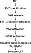
Respiratory motoneuron response to hypoxia is reflex in nature and carotid body sensory receptor constitutes the afferent limb of this reflex. Recent studies showed that repetitive exposures to hypoxia evokes long term facilitation of sensory nerve discharge (sLTF) of the carotid body in rodents exposed to chronic intermittent hypoxia (CIH). Although studies with anti-oxidants suggested the involvement of reactive oxygen species (ROS)-mediated signaling in eliciting sLTF, the source of and the mechanisms associated with ROS generation have not yet been investigated. We tested the hypothesis that ROS generated by NADPH oxidase (NOX) mediate CIH-evoked sLTF. Experiments were performed on ex vivo carotid bodies from rats and mice exposed either to 10 d of CIH or normoxia. Acute repetitive hypoxia evoked a approximately 12-fold increase in NOX activity in CIH but not in control carotid bodies, and this effect was associated with upregulation of NOX2 mRNA and protein, which was primarily localized to glomus cells of the carotid body. sLTF was prevented by NOX inhibitors and was absent in mice deficient in NOX2. NOX activation by CIH required 5-HT release and activation of 5-HT(2) receptors coupled to PKC signaling. Studies with ROS scavengers revealed that H(2)O(2) generated from O(2).(-) contributes to sLTF. Priming with H(2)O(2) elicited sLTF of carotid bodies from normoxic control rats and mice, similar to that seen in CIH-treated animals. These observations reveal a novel role for NOX-induced ROS signaling in mediating sensory plasticity of the carotid body.


Figures

Figure 1.
Figure 1.
CIH upregulates NOX enzyme activity, mRNAs in rat carotid body. A, Effect of repetitive hypoxia on NOX activity. Carotid bodies (CB) harvested from control and CIH-exposed rats were challenged with 10 episodes of repetitive hypoxia and NOX activity was determined as described in Materials and Methods. Note that acute repetitive hypoxia increases NOX activity in the CIH but not in control carotid body, and 500 μm apocynin (Apo), a NOX inhibitor, prevented this effect. The data presented are mean ± SEM, n = 3 each with normoxia (open columns) and acute repetitive hypoxia (filled columns). B, CIH upregulates NOX1, NOX2, and NOX4 mRNAs in the carotid body. Results are expressed as fold change with respect to normoxic controls. Data presented are mean ± SEM from three individual experiments performed in triplicate. Asterisks represent p < 0.01 compared with normoxia; n.s., nonsignificant. C, Localization of NOX2 and NOX4 in the rat carotid body. NOX2-like immunoreactivity (left, green color) was seen in the cytoplasm of glomus cells as evidenced by colocalization with chromagranin-A (CGA; denoted by arrows; middle). NOX4-like immunoreactivity (red color; right) is localized in the nuclei of carotid body cells. NOX4 expression can be seen in glomus cells (stained with tyrosine hydroxylase; TH; green color; denoted by arrows) as well as other cells (shown with *). Scale bar, 20 μm. CON, Control.
Figure 2.
Figure 2.
NOX inhibitors prevent sLTF of the rat carotid body. A, B, Examples of repetitive hypoxia-evoked sLTF of CIH-treated rat carotid bodies in the presence of vehicle (A) or apocynin (B). Arrows in A and B denote application of repetitive hypoxia. Superimposed action potentials of a single fiber from which the data were derived are shown in the insets. imp, Impulses. C, Average data of the effects of apocynin (500 μm), AEBFS (500 μm), and diphenyl iodinium (DPI; 3 μm) on sLTF (sensory activity averaged during 60 min postrepetitive hypoxia period). Data presented are mean ± SEM from six experiments in each group. Asterisk denotes p < 0.05 compared with CIH-exposed carotid bodies treated with vehicle.
Figure 3.
Figure 3.
Absence of sLTF of the carotid body in gp91phox/Y mice exposed to CIH. A, Examples of repetitive hypoxia-evoked sLTF of carotid bodies from CIH-treated WT (top) and gp91phox/Y (bottom) mice. Arrows denote application of repetitive hypoxia. Superimposed action potentials of a single fiber from which the data were derived are shown in the insets. imp, Impulses. B, Average data showing the magnitude of sLTF of the carotid body from WT and gp91phox/Y mice exposed to either normoxia (control, open columns) or CIH (filled columns). Data presented are mean ± SEM from WT (control and CIH, n = 7 each) and gp91phox/Y (control and CIH, n = 7 each) mice. Asterisks denote p < 0.01 compared with control carotid body.
Figure 4.
Figure 4.
Acute hypoxia facilitates 5-HT release from CIH-exposed carotid body. A, Example of 5-HT release from the carotid bodies from CIH exposed rats. Left and right represent 5-HT release during normoxia and acute hypoxia, respectively. Arrows show the elution of 5-HT during normoxia and hypoxia. B, Average data of the basal (open columns) and hypoxia-evoked (filled columns) 5-HT release from carotid bodies derived from rats reared under normoxia or CIH. Data presented are mean ± SEM from three individual experiments performed in triplicate. Asterisks denote p < 0.01 compared with basal release. Note that hypoxia had no effect on 5-HT release from carotid bodies from normoxic rats, whereas it was markedly facilitated in CIH carotid bodies and this effect was prevented by 75 μm 2-APB but not by 300 μm cadmium chloride (Cd2+).
Figure 5.
Figure 5.
Absence of sLTF of the carotid body in Pet-1−/− mice with impaired 5-HT expression in glomus cells. A, 5-HT-like immunoreactivity in glomus cells of the carotid bodies from WT and Pet-1−/− mice. Note the absence of 5-HT-like immunoreactivity in the carotid body from Pet-1−/− mice. B, Representative tracings of the carotid body sensory response to repetitive hypoxia (at arrows) from WT (top two panels) and Pet-1−/− (bottom two panels) mice exposed to either normoxia (control) or CIH. Insets represent superimposed action potentials from a single unit. C, Average data of the magnitude of sLTF (sensory activity averaged during 60 min period of postrepetitive hypoxia) presented as percentage of baseline sensory activity (i.e., before 5 episodes of repetitive hypoxia). Data represent mean ± SEM from WT (Control and CIH, n = 7 each) and Pet-1−/− (Control and CIH, n = 7 each) mice. Asterisks denote p < 0.01 compared with control. Note the absence of sLTF in Pet-1−/− mice treated with CIH.
Figure 6.
Figure 6.
NOX activation by repetitive hypoxia in the CIH-exposed rat carotid bodies requires activation of 5-HT2 receptors and PKC. A, Acute repetitive hypoxia activates PKC via 5-HT2 receptors. The level of phosphorylation of MARCKS (P-MARCKS) was monitored in the rat carotid bodies by Western blot as an index of PKC activation. Top, example of the Western blot. Effect of repetitive hypoxia on P-MARCKS expression in carotid bodies from normoxia-treated control (lane 1), CIH exposed (lane 2), CIH-treated carotid bodies in presence of 1 μm ketanserin (lane 3), or 1 μm Bis-1 (lane 4) or 100 nm PMA (positive control; lane 5). Bottom, Average data of densitometric analysis presented as percentage of control. Note that repetitive hypoxia increased P-MARCKS expression in CIH but not in control carotid bodies, and this effect was prevented by ketanserin as well as Bis-1. B, Effect of repetitive hypoxia on NOX activation in the carotid bodies from normoxia and CIH-treated carotid bodies in the absence and presence of 1 μm ketanserin or 1 μm Bis-1. Note that ketanserin and Bis-1 prevent NOX activation by repetitive hypoxia in CIH-exposed carotid bodies. Data presented in A and B represent mean ± SEM from three individual experiments performed in triplicate. Asterisks represent p < 0.01 compared with controls. Ketan, Ketanserin; CB, carotid bodies.
Figure 7.
Figure 7.
5-HT2 receptor antagonist and PKC inhibitor prevent sLTF of the carotid body. A, Examples of the effects of repetitive hypoxia (at arrows) on sensory activity of CIH-treated rat carotid bodies in the presence of 1 μm ketanserin (top) or 1 μm Bis-1 (bottom). Superimposed action potentials of a single fiber from which the data were derived are shown in the insets. Note that ketanserin and Bis-1 prevented repetitive hypoxia-evoked sLTF. imp, Impulses. B, Average data showing the absence of sLTF in presence of ketanserin or Bis-1. Data presented are mean ± SEM from CIH carotid bodies treated with vehicle (n = 7), or in presence of 1 μm ketanserin (n = 6) or 1 μm Bis-1 (n = 6). **p < 0.01 compared with CIH-exposed carotid bodies. Ketan, Ketanserin.
Figure 8.
Figure 8.
H2O2 mediates sLTF of the rat carotid bodies. A, B, Examples of CIH-treated rat carotid body sensory response to repetitive hypoxia (at arrows) in the presence of MnTMPyP (50 μm; A) or PEG-catalase (200U/ml; B). Superimposed action potentials of a single fiber from which the data were derived are shown in the insets. imp, Impulses. C, Average data of the magnitude of sLTF during the 15th, 25th, 35th, 45th, and 60th minute of postrepetitive hypoxia period in presence of vehicle (open columns, n = 6), 50 μm MnTMPyP (filled columns, n = 6), or PEG-catalase (200U/ml; gray columns, n = 6). MnTMPyP or PEG catalase was added during postrepetitive hypoxia as indicated by arrow. PEG-catalase but not MnTMPyP prevented sLTF. *p < 0.05 compared with vehicle-treated CIH carotid bodies.
Figure 9.
Figure 9.
Priming the carotid bodies with H2O2 evokes sLTF. A, Examples of the effects repetitive hypoxia (at arrows) on sensory activity of the carotid bodies from rats reared under normoxia in the presence (top) or in the absence (middle) of 500 nm H2O2. Note sLTF in the presence but not in the absence of H2O2. Continuous exposure with 500 nm H2O2 for 90 min had no effect on baseline sensory activity of the carotid body (bottom). Superimposed action potentials of a single fiber from which the data were derived are shown in the insets. B, Examples of the effects repetitive hypoxia (at arrows) on sensory activity of the carotid bodies from WT (top), gp91phox/Y (middle), and Pet-1−/− (bottom) mice in the presence of 200 nm H2O2. Note that priming with H2O2 evoked sLTF in all three mice. imp, Impulses.
Figure 10.
Figure 10.
Schematic presentation of 5-HT and NOX signaling cascade mediating sLTF of the CIH-treated carotid bodies.

Similar articles

Cited by

References

    1. Baker TL, Fuller DD, Zabka AG, Mitchell GS. Respiratory plasticity: differential actions of continuous and episodic hypoxia and hypercapnia. Respir Physiol. 2001;129:25–35. - PubMed
    1. Bánfi B, Maturana A, Jaconi S, Arnaudeau S, Laforge T, Sinha B, Ligeti E, Demaurex N, Krause KH. A mammalian H+ channel generated through alternative splicing of the NADPH oxidase homolog NOH-1. Science. 2000;287:138–142. - PubMed
    1. Bedard K, Krause KH. The NOX family of ROS-generating NADPH oxidases: physiology and pathophysiology. Physiol Rev. 2007;87:245–313. - PubMed
    1. Dinger B, He L, Chen J, Liu X, Gonzalez C, Obeso A, Sanders K, Hoidal J, Stensaas L, Fidone S. The role of NADPH oxidase in carotid body arterial chemoreceptors. Respir Physiol Neurobiol. 2007;157:45–54. - PMC - PubMed
    1. He L, Chen J, Dinger B, Sanders K, Sundar K, Hoidal J, Fidone S. Characteristics of carotid body chemosensitivity in NADPH oxidase-deficient mice. Am J Physiol Cell Physiol. 2002;282:C27–C33. - PubMed

Publication types

MeSH terms

LinkOut - more resources