. 2009 May;119(5):1335-49.
doi: 10.1172/JCI36800. Epub 2009 Apr 13.

Oxidation-specific epitopes are dominant targets of innate natural antibodies in mice and humans

Affiliations

Oxidation-specific epitopes are dominant targets of innate natural antibodies in mice and humans

Meng-Yun Chou et al. J Clin Invest. 2009 May.

Abstract

Atherosclerosis is a chronic inflammatory disease characterized by the accumulation of oxidized lipoproteins and apoptotic cells. Adaptive immune responses to various oxidation-specific epitopes play an important role in atherogenesis. However, accumulating evidence suggests that these epitopes are also recognized by innate receptors, such as scavenger receptors on macrophages, and plasma proteins, such as C-reactive protein (CRP). Here, we provide multiple lines of evidence that oxidation-specific epitopes constitute a dominant, previously unrecognized target of natural Abs (NAbs) in both mice and humans. Using reconstituted mice expressing solely IgM NAbs, we have shown that approximately 30% of all NAbs bound to model oxidation-specific epitopes, as well as to atherosclerotic lesions and apoptotic cells. Because oxidative processes are ubiquitous, we hypothesized that these epitopes exert selective pressure to expand NAbs, which in turn play an important role in mediating homeostatic functions consequent to inflammation and cell death, as demonstrated by their ability to facilitate apoptotic cell clearance. These findings provide novel insights into the functions of NAbs in mediating host homeostasis and into their roles in health and diseases, such as chronic inflammatory diseases and atherosclerosis.


Figures

Figure 1
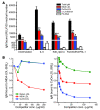
Figure 1. IgM Abs to oxidation-specific antigens are present in germ-free and conventional mice.
(A) Conventional and SPF C57BL/6 mice have similar IgM titers to oxidation-specific antigens. Plasma from 11-week-old female conventionally raised (n = 4) and SPF (n = 4) C57BL/6 mice were tested by ELISA. Values are mean and SEM. (B) MDA-LDL–specific ISCs are dominant in the spleens of conventionally raised C57BL/6 mice. Splenocytes from conventionally raised 12-week-old female C57BL/6 mice (n = 4) were tested by ELISpot assay for frequencies of ISCs as described in Methods. Values represent the number of ISCs to indicated antigen as a percentage of total ISCs (mean and SD). Data are from 1 experiment representative of 3. **P < 0.01 compared with all other antigens (1-way ANOVA with Tukey-Kramer multiple comparison test). (C) Binding curves of plasma IgM from germ-free Swiss-Webster mice to indicated antigens. Plasma samples were from 14- to 16-week old female and male mice (n = 9). Values are mean and SEM. (D) Titers of IgM Abs to oxidation-specific epitopes are present in conventional and germ-free Swiss Webster mice. Serum from 14- to 16-week-old female and male conventionally raised (n = 7), conventionalized (germ-free colonized with bacterial flora) (n = 11), and germ-free (n = 9) mice were diluted 1:400 and tested for binding to the indicated antigens. Values are mean and SEM. *P < 0.05, **P < 0.01, ***P < 0.002 compared with α1,3-dextran (1-way ANOVA with Tukey-Kramer multiple comparison test).
Figure 2
Figure 2. In vitro stimulation of B-1 cells induces increased natural IgM Ab titers to oxidation-specific antigens.
(A) Purified B-1 cells were cultured in 24-well plates in triplicate at a cell density of 1 × 10⁶ cells per well in 500 μl culture medium. Cells were stimulated with IL-5 (50 ng/ml), KdO2-Lipid A (100 ng/ml), or TLR2 agonists (a combination of Pam3CSK4 [300 ng/ml] and FSL-1 [1 μg/ml]) and incubated at 37°C for 7 days. Control B-1 cells were cultured in medium alone. Cell culture supernatants were harvested after 7 days and IgM Ab titers analyzed by ELISA at 1:45 dilution. Results were normalized to cell number recovered after 7 days. Values are mean and SEM. Data are from 1 experiment representative of 3. *P < 0.05, **P < 0.01, ***P < 0.002 compared with α1,3-dextran (repeated-measures ANOVA with Tukey-Kramer multiple comparison test). (B) Natural IgM Abs produced in vitro show specificity to MDA-LDL and CuOx-LDL. For competition immunoassay, supernatants from purified B-1 cell cultures stimulated with KdO2-Lipid A (100 ng/ml) or IL-5 (50 ng/ml) were diluted to 1:20 and incubated in the presence of the indicated concentrations of competitors (Competitor conc.) overnight. After incubation, IgM binding to MDA-LDL and CuOx-LDL was tested by ELISA. Data are the mean of triplicate determinations, expressed as ratio of IgM binding to MDA-LDL or CuOx-LDL in the presence or absence of competitor (B/B0). Data are from 1 experiment representative of 3.
Figure 3
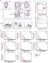
Figure 3. Characterization of Rag1–/– recipients adoptively transferred with B-1 cells.
(A) Adoptive transfer of B-1 cells into Rag1–/– mice replenishes B-1 cell population. Rag1–/– mice were injected with PBS (Rag1–/– + PBS) or with B-1 cells (Rag1–/– + B-1). Rag1–/– + PBS: Lymphocyte populations were absent in the peritoneal cavity (PEC, left). B-1 cells (IgM+CD43+) were also absent from the spleen (Spleen, left). C57BL/6: Peritoneal macrophages (CD11bhiCD5) and T cells (CD11bCD5hi) were intact (PEC, upper middle). B cells could be divided into B-1a (CD19+CD11bintCD5int), B-1b (CD19+CD11bintCD5), and B-2 cells (CD19+CD11bCD5) (PEC, lower middle). In the spleen, B-1 cells were about 2.4% of total splenocytes (Spleen, middle). Rag1–/– + B-1: B-1 cell populations were reconstituted in the peritoneal cavity (PEC, lower right) and spleen (Spleen, right), without B-2 cell or T cell contamination (PEC, right). (B) IgM Abs to oxidation-specific epitopes are present in the plasma of B-1 reconstituted Rag1–/– mice. Plasma collected after 15 weeks from Rag1–/– + B-1 (n = 8) or Rag1–/– + PBS (n = 6) and age-matched C57BL/6 mice (n = 7) were tested. Data shown are from 1 transfer experiment representative of 6. Values are mean and SEM. Numbers in the upper-right corner represent the IgM titer to each antigen. (C) Natural IgM Abs produced in vivo show specificity to MDA-LDL and CuOx-LDL. Data are the mean of triplicate determinations, expressed as the ratio of IgM binding to MDA-LDL or CuOx-LDL in the presence or absence of competitor (B/B0). Data are from 1 experiment representative of 3.
Figure 4
Figure 4. Oxidation-specific epitopes are dominant targets of NAbs.
(A) Preabsorption of plasma from Rag1–/– + B-1 mice with oxidation-specific antigens shows that oxidation-specific epitopes (OxEpitopes) are dominant targets for NAbs. Plasmas from Rag1–/– + B-1 mice were preincubated in the absence or presence of the indicated antigens (250 μg/ml total antigen) overnight and antigen-immune complexes pelleted by centrifugation. Total IgM levels were then tested by ELISA. *P < 0.05, **P < 0.01, ***P < 0.002 compared with native LDL (ANOVA with Tukey-Kramer multiple comparisons test). Data are means (and SEM) from 5 separate experiments, each using 3–7 plasma samples obtained from 5 different transfer experiments, with each sample assayed in triplicate. (B) ELISpot assay of frequencies of MDA-LDL–specific ISCs in the spleens of wild-type C57BL/6, Rag1–/– + B-1, and Rag1–/– + PBS mice. Results are from individual mice, and data are from 3 separate B-1 cell transfer experiments. Horizontal bar represents the mean for the group. P < 0.002 compared with Rag1–/– + PBS (unpaired t test). (C) B-1 cell–derived natural mAb NA-17. DNA sequences of VDJ splice sites of the VH and VL rearrangements expressed in NA-17 B-1 cell hybridoma and their relationship to the most homologous germline V, D, J gene segments. Sequence analysis of NA-17 VH rearrangement did not reveal nucleotide variation to germline genes. Sequence analysis of VL rearrangement revealed 1 nucleotide insertion between VL and JL germline gene segments.
Figure 5
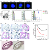
Figure 5. Natural IgM Abs recognize oxidation-specific epitopes present on apoptotic cells and atherosclerotic lesions (A) Natural IgM Abs bind to apoptotic thymocytes but not normal thymocytes.
Apoptotic thymocytes from C57BL/6 mice were incubated with plasma from Rag1–/– + B-1 or Rag1–/– + PBS mice at 1:10 dilution, NA-17 at 2.5 μg/ml, or control IgM at 5 μg/ml. Top row: Deconvolution microscopy shows that NAbs in Rag1–/– + B-1 as well as NA-17 bound to apoptotic thymocytes. Bottom row: None of the IgM bound to normal thymocytes (quadrant 1 [Q1). NAbs in plasma from Rag1–/– + B-1 bound to both early (Q2) and late apoptotic thymocytes (Q3), while NA-17 bound prominently to late apoptotic cells (Q3). Scale bar: 5 μm. 2°Ab, secondary Ab; Anti-ms-IgM-FITC, FITC-labeled anti-mouse IgM. (B) Natural IgM Abs are present in atherosclerotic lesions. Endogenous IgM Abs were detected in aortic sections from cholesterol-fed B-1 cell–reconstituted Ldlr–/–Rag1–/– mice (bottom row), but not in PBS-injected Ldlr–/–Rag1–/– mice (top row). Sections were also stained with MDA2 (5 μg/ml) for the presence of MDA epitopes. Red indicates positive staining. Original magnification, ×160. (C) NA-17 recognizes oxidation-specific epitopes present in atherosclerotic lesions. Sections of the brachiocephalic artery from cholesterol-fed Ldlr–/–Rag1–/– mice were stained with NA-17 (0.85 μg/ml) or a control natural IgM Ab, EN-2 (1.6 μg/ml). Original magnification, ×200. (D) NA-17 inhibits MDA-LDL binding to macrophages. Increasing concentrations of NA-17 or control IgM were added with a fixed amount of biotinylated MDA-LDL (Bt-MDA-LDL; 2 μg/ml) to macrophages. Data are the average of 2 experiments, expressed as the ratio of biotinylated MDA-LDL binding to macrophages in the presence or absence of IgM (B/B0).
Figure 6
Figure 6. Human umbilical cord blood contains natural IgM Abs against oxidation-specific epitopes.
(A) Left: Plasma titers of IgM in maternal and umbilical cord plasma to native LDL, KLH, and oxidation-specific antigens measured by ELISA. Right: Data are plotted as ratio of antigen-specific IgM to total IgM. ***P < 0.002 compared with maternal blood (Wilcoxon matched-pairs test and paired t test). Data shown are from 10 paired maternal-infant samples, and each sample was assayed in triplicate. Values are mean and SEM. (B) Umbilical cord IgM binds to apoptotic cells in part via binding to MDA. Apoptotic Jurkat cells, induced by UV exposure, were incubated with representative umbilical cord plasma (1:50 dilution) in the absence and presence of MDA-LDL and native LDL (1 mg/ml). Abs bound were detected by FITC-conjugated anti-human IgM. Umbilical cord IgM binding to apoptotic Jurkat cells (median fluorescence intensity [MFI], 1,917) was inhibited 45% by MDA-LDL (MFI, 1,047) while minimally affected by native LDL (MFI, 1,514).
Figure 7
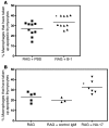
Figure 7. Natural IgM Abs against oxidation-specific epitopes facilitate apoptotic cell uptake by macrophages in vivo.
(A) Percentage of macrophages that contained fluorescently labeled apoptotic thymocytes following i.p. injection. In RAG + PBS mice, about 27% of macrophages phagocytosed apoptotic cells. The percentage was significantly increased, to 33%, in RAG + B-1 mice (*P < 0.05, unpaired t test). Horizontal bars denote means. (B) Apoptotic thymocytes were preincubated with NA-17 or a control IgM that does not bind apoptotic cells before injection into Rag1–/– mice. The phagocytic uptake was significantly different between the 3 groups (P = 0.01, 1-way ANOVA). The percentage of macrophages taking up apoptotic cells was significantly increased when the apoptotic cells were preincubated with NA-17 (RAG + NA-17), compared with control IgM (RAG + control IgM) or without preincubation (RAG) (32% vs. 20% vs. 23%; *P < 0.05, Bonferroni multiple comparison test). Horizontal bars denote means.

Similar articles

Cited by

References

    1. Binder C.J., et al. Innate and acquired immunity in atherogenesis. Nat. Med. 2002;8:1218–1226. - PubMed
    1. Hansson G.K., Libby P. The immune response in atherosclerosis: a double-edged sword. Nat. Rev. Immunol. 2006;6:508–519. - PubMed
    1. Getz G.S., Vanderlaan P.A., Reardon C.A. The immune system and murine atherosclerosis. Curr. Drug Targets. 2007;8:1297–1306. - PubMed
    1. Binder C.J., et al. The role of natural antibodies in atherogenesis. J. Lipid Res. 2005;46:1353–1363. - PubMed
    1. Boullier A., et al. Phosphocholine as a pattern recognition ligand for CD36. J. Lipid Res. 2005;46:969–976. - PubMed

Publication types

MeSH terms