. 2009 Jun;37(11):3612-24.
doi: 10.1093/nar/gkp223. Epub 2009 Apr 9.

Alternative polyadenylation variants of the RNA binding protein, HuR: abundance, role of AU-rich elements and auto-Regulation

Affiliations

Alternative polyadenylation variants of the RNA binding protein, HuR: abundance, role of AU-rich elements and auto-Regulation

Wijdan Al-Ahmadi et al. Nucleic Acids Res. 2009 Jun.

Abstract

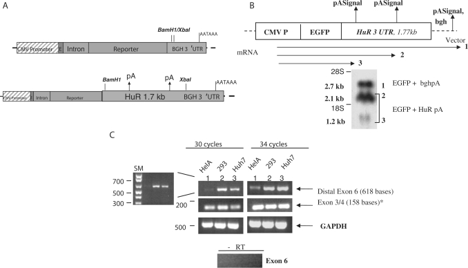
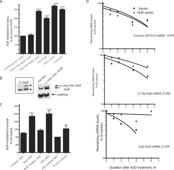
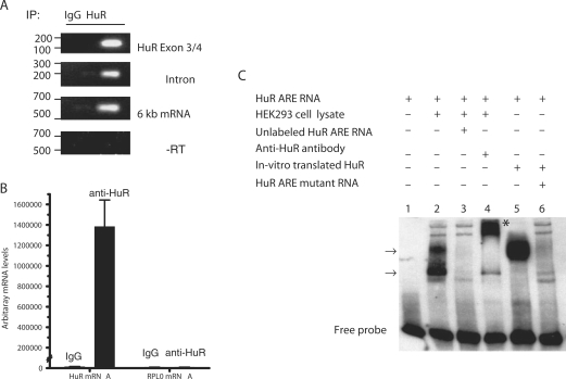
The RNA-binding protein, HuR, is involved in the stabilization of AU-rich element-containing mRNAs with products that are involved in cell-cycle progression, cell differentiation and inflammation. We show that there are multiple polyadenylation variants of HuR mRNA that differ in their abundance, using both bioinformatics and experimental approaches. A polyadenylation variant with distal poly(A) signal is a rare transcript that harbors functional AU-rich elements (ARE) in the 3'UTR. A minimal 60-nt region, but not a mutant form, fused to reporter-3'UTR constructs was able to downregulate the reporter activity. The most predominant and alternatively polyadenylated mature transcript does not contain the ARE. HuR itself binds HuR mRNA, and upregulated the activity of reporter from constructs fused with ARE-isoform and the HuR ARE. Wild-type tristetraprolin (TTP), but not the zinc finger mutant TTP, competes for HuR binding and upregulation of HuR mRNA. The study shows that the HuR gene codes for several polyadenylation variants differentially regulated by AU-rich elements, and demonstrates an auto-regulatory role of HuR.


Figures

Figure 3.
Figure 3.
Differential Involvement of AU-rich elements in HuR polyadenylation variants. (A) Schematic diagram of exon 6 of HuR that contains ARE and reporter constructs. Sequences from the EEF1A1 3′UTR (Control, construct 1), 2.7 kb mRNA 3′UTR (construct 2), 6-kb transcript 3′UTR (construct 3), uPA 3′UTR (construct 4), COX-2 3′UTR (construct 5) and IL-8 3′UTR (construct 6) were inserted in BamHI/XbaI sites as described in ‘Materials and Methods’ section. (B) Analysis of GFP reporter activity generated from GFP mRNA fused with the indicated 3′UTR. HEK293 cells were transfected with the different 3′UTR constructs in 96-well microplates. Reporter activity was assessed in 96-well black clear-bottom plates by measuring bottom fluorescence using bottom-read fluorometer. The EEF1A1 3′UTR is the control construct in which its fluorescence activity was taken as 100% and data are presented as mean ± SEM (n = 4) of % of the control. ***Denote P-values of <0.01 and <0.005, respectively (Student's t-test). Percent transfection efficiency in the control was 80% with a coefficient of variation of <6%, thus, normalization with another vector was not needed (27). (C) Analysis of GFP mRNA levels by quantitative real-time RT-PCR. HEK293 cells were transfected with the different 3′UTR constructs in 6-well plates. Total RNA was extracted and real-time quantitative RT-PCR was performed using primers and Taqman probe specific to GFP reporter sequence as described in ‘Materials and methods’ section. The fluorescence activity from control (EEF1A1) 3′UTR-fused construct was taken as 100%. Data (mean ± SEM) was presented as percentage of control. (D) A 60-nt double-stranded oligonucleotide corresponding to the ARE regions from HuR 6-kb 3′UTR, uPA 3′UTR and control stable growth hormone (GH1) 3′UTR were fused with EGFP reporter construct. Furthermore, 60-nt double-stranded oligonucleotides as mutant forms of the HuR ARE and uPA were fused with GFP reporter construct (sequences of ARE and their mutants are shown). These constructs were used for HEK293 transfection. Percentage of control 3′UTR (EEF1A1)-linked construct fluorescence activity (100%) are presented as mean ± SEM (n = 4). ***Denote P-values of <0.005 (Student t-test).
Figure 1.
Figure 1.
Bioinformatics and mRNA species assessment of HuR gene. (A) A gene structure for HuR (ELAVL1). Exons are denoted by the green boxes while the red box denotes the terminal exon that harbors the entire 3′UTR of HuR and their polyadenylation variant signals. Positions for the specific primers used in this study are shown: for the pre-mRNA (intron; blue arrows), terminal sequences in exon 6 (6-kb transcript, black arrows) and exon 3/4 (green arrows). Positions of putative pA signals leading to alternative polyadenylation are shown (vertical arrows). pA denotes polyadenylation signal. (B) EST clustering strategy view of the ESTs belonging to the HuR. The top cluster represents ∼70% of the 460 ESTs from Unigene Cluster (Hs.184492). The dark inner lines represent individual ESTs in the EST assembly. The bottom cluster represents 23% of the total ESTs in the assembly (individual ESTs are not shown). (C) Total RNA was extracted from HeLa cells and used for northern blotting with cDNA probes to HuR and β-actin mRNA. Arrows show the two different mRNA species as assessed by size markers. Lower panel showed the three mRNA variants with over-exposed autoradiography.
Figure 2.
Figure 2.
Experimental validation of the HuR polyadenylation variants. (A) Cloning of 1774 nt of 3′UTR of HuR mRNA that contains the putative two poly(A) signals into EGFP expression vector that is under the control of CMV IE promoter and stable bovine growth hormone 3′UTR/poly(A) signal. (B) HEK393 cells were transfected with the EGFP construct in (A) using Lipofectamine 2000 for 16 h. Northern blotting was performed using antisense probe to EGFP. Numbers denote the expected length of the polyadenylation variants of EGFP-HuR 3′UTR mRNAs (lanes 2 and 3) and EGFP-BGH 3′UTR transcripts (lane 1)—denote positions for the 18S (∼2 kb) and 28S rRNA (∼5 kb) bands. (C) DNase-treated total RNA extracted from three different cell lines (1, HeLa; 2, HEK293; and 3, Huh7) were used. RT-PCRs were performed for HuR gene products encoding the 6-kb transcript (distal polyadenylation variant; black-colored arrows in Figure 1A) and exon 3/4 containing forms (all polyadenylation variants, green-colored arrows in Figure 1A) using specific primers as outlined above. *These primers do not amplify intervening introns between exons 3 and 4 (7.2 kb) since RT-PCR conditions were performed with short extension times. RT-PCR control without RT to monitor genomic contamination was used with intron-specific primers.
Figure 4.
Figure 4.
Effect of HuR over-expression on HuR 3′UTR and HuR ARE. (A) HEK293 cells in 96-well plates were co-transfected with 37 ng pcDNA 3.1 empty vector or HuR-c-myc-his expressing pcDNA 3.1 vector and 25 ng of each of EGFP-3′UTR reporter vectors as shown. The reporter 3′UTR constructs were described in Figure 3A. Data mean ± SEM of quadruplicates presented as % of reporter fluorescence with control EEF1A1 3′UTR taken as 100%. Percent transfection efficiency in the control was 80% with a coefficient of variation of <6%, thus, normalization with another vector was not needed (27). (B) Representative western blotting confirming expression of the transfected c-myc-his-HuR cDNA is shown. Inset is a longer gel run for larger separation of endogenous and transfected HuR. (C) HEK293 cells in 96-well plates were co-transfected with reporter vectors and either pcDNA3.1 (control) or HuR expression pcDNA3.1. The reporter vector is EGFP fused with 3′UTR that contains a 60 nt corresponding to the ARE regions from HuR the 6-kb variant 3′UTR, uPA 3′UTR, control GH1 sequence, a 60 nt as mutant form of the HuR 6-kb ARE (Figure 3D, inset table) and another 60 nt representing U-rich control that lacks HuR binding site (17). Fluorescence data of mean ± SEM of quadruplicates were obtained. The fluorescence levels obtained from cells transfected with the pcDNA3.1 and the reporter with control GH1 3′UTR were taken as 100%. The y-axis numbers represent percentages of this control. ***Denotes P-values of <0.001 (Student t-test) when compared to control. (D) HEK293 cells were transfected with the EEF1A1 3′UTR (Control), 2.7-kb mRNA 3′UTR, 6-kb transcript 3′UTR (constructs 1–3, Figure 3A) overnight and then treated with AcD (5 μg/ml). Total RNA was extracted at the indicated time points and subjected to RT-PCR using primer pair/TaqMan probe specific to EGFP mRNA. The TaqMan primer/probe spans an intron–exon-junction sequence for efficient control of DNA contamination. Half-life mRNA determinations were obtained by plotting the changes in RPL0-normalized EGFP mRNA levels expressed as a percentage of that seen at time 0 (taken as 100%; y-axis) against AcD treatment duration (x-axis). RPL0 is a housekeeping gene. Data are from one representative experiment of at least two independent experiments. Details are given in ‘Materials and Methods’ section.
Figure 5.
Figure 5.
Binding of HuR protein to HuR mRNA and consequences of HuR over-expression. (A) The binding of endogenous HuR to endogenous target mRNA was detected by RT-PCR of RNA material obtained by precipitation with either anti-HuR antibody or goat IgG control antibody. Primers specific to the intron (pre-mRNA), distal part of exon 6 (6-kb transcript) or all mRNA (exons 3 and 4) were used in the RT-PCR reaction with a cycle number that allows at least semi-quantitative comparison as indicated in ‘Materials and Methods’ section. The positions and types/size of PCR products are shown in Figures 1A and 2C. All RNA samples were treated with DNase to eliminate genomic DNA contamination. In addition, RT-PCR (intron) was performed without RT to further monitor DNA contamination (lower panel). This protocol is performed according to a standard protocol (11,58,59). (B) Real-time RT-PCR monitoring of HuR mRNA levels following precipitation of endogenous HuR with either anti-HuR antibody or goat IgG control as described above. The real-time RT-PCR was performed with primers/TaqMan probe specific to HuR mRNA and RPL0 (background control); the TaqMan primer pair and probe span intron and exon–intron junction, respectively, in HuR mRNA to eliminate signal quantitation due to DNA contamination. Lower panel shows data in terms of % specific binding (anti-HuR/IgG). The data are one representative experiments of two with mean ± SEM of triplicates. (C) RNA-binding activity of endogenous and exogenous HuR. The HuR ARE 60-nt probe (lane 1) was incubated with 5 µg HEK293 protein lysate (lanes 2–4). Competition assay was carried in the presence (+) of 1000-fold excess of unlabeled RNA competitor (lane 3). In vitro translated HuR protein was incubated with either the HuR ARE (lane 5) or the mutant ARE (lane 6) oligonucleotide. Arrows indicate RNA–protein complexes formation in the presence of protein lysate. The supershift assay was carried out in the same manner, except that protein lysate was pre-incubated with anti-HuR antibody for 30 min at room temperature before the addition of biotinylated probe. Asterisk Indicates the super-shifted position.
Figure 6.
Figure 6.
Functional TTP-mediated antagonism of HuR autoregulation. (A) HEK293 cells were transfected with pCR3.1 vector, wild-type TTP vector or the zinc finger C124R mutant for overnight. Cell lysates were obtained, and RNA materials obtained by precipitation with either anti-HuR antibody or goat IgG control antibody were used for real-time quantitative PCR. The real-time RT-PCR was performed with primers/TaqMan probe specific to HuR mRNA and RPL0 (background control). Data show RPL0-normalized anti-HuR/PRL0-normalized anti-IgG ratios. Data are from one representative experiment (mean ± SEM of triplicate) of two. ***Denotes P-values of <0.005 one-way ANOVA test for the three group comparisons. (B) HEK293 cells in 96-well plates were co-transfected with 30-ng pCR3.1 empty vector, TTP or C124R TTP and 25 ng of each of EGFP-3′UTR reporter vectors as indicated. The reporter 3′UTR constructs are described in Figure 3A. The fluorescence levels obtained from cells transfected with the pCR3.1 and the reporter construct with control 3′UTR were taken as 100%. The y-axis numbers represent percentages of this control. ***Denotes P-values of <0.001 two-way ANOVA test when compared to the control group. (C) HEK293 cells in 96-well plates were co-transfected with 30-ng pCR3.1 empty vector, TTP or C124R TTP and 25 ng of each of EGFP-3′UTR reporter vectors as indicated. The reporter vector is EGFP fused with 3′UTR that contains a 60 nt corresponding to the ARE regions from HuR 6-kb variant 3′UTR, uPA 3′UTR, their mutant forms, and control GH1 sequence (Figure 3D, inset table). Fluorescence data of mean ± SEM of quadruplicates were obtained. The fluorescence levels obtained from cells transfected with the pCR3.1 and the reporter with control 3′UTR were taken as 100%. The y-axis numbers represent percentages of this control. **Denotes P-values of <0.01 two-way ANOVA test when compared to the control group.

Similar articles

Cited by

References

    1. Blackshear PJ. Tristetraprolin and other CCCH tandem zinc-finger proteins in the regulation of mRNA turnover. Biochem. Soc. Trans. 2002;30:945–952. - PubMed
    1. Chou CF, Mulky A, Maitra S, Lin WJ, Gherzi R, Kappes J, Chen CY. Tethering KSRP, a decay-promoting AU-rich element-binding protein, to mRNAs elicits mRNA decay. Mol. Cell Biol. 2006;26:3695–3706. - PMC - PubMed
    1. Schmidlin M, Lu M, Leuenberger SA, Stoecklin G, Mallaun M, Gross B, Gherzi R, Hess D, Hemmings BA, Moroni C. The ARE-dependent mRNA-destabilizing activity of BRF1 is regulated by protein kinase B. EMBO J. 2004;23:4760–4769. - PMC - PubMed
    1. Barreau C, Paillard L, Osborne HB. AU-rich elements and associated factors: are there unifying principles? Nucleic Acids Res. 2005;33:7138–7150. - PMC - PubMed
    1. Ma WJ, Cheng S, Campbell C, Wright A, Furneaux H. Cloning and characterization of HuR, a ubiquitously expressed Elav-like protein. J. Biol. Chem. 1996;271:8144–8151. - PubMed

Publication types