. 2009 Apr 3;137(1):60-72.
doi: 10.1016/j.cell.2009.03.018.

Acetylation targets mutant huntingtin to autophagosomes for degradation

Affiliations

Acetylation targets mutant huntingtin to autophagosomes for degradation

Hyunkyung Jeong et al. Cell. .

Abstract

Huntington's disease (HD) is an incurable neurodegenerative disease caused by neuronal accumulation of the mutant protein huntingtin. Improving clearance of the mutant protein is expected to prevent cellular dysfunction and neurodegeneration in HD. We report here that such clearance can be achieved by posttranslational modification of the mutant Huntingtin (Htt) by acetylation at lysine residue 444 (K444). Increased acetylation at K444 facilitates trafficking of mutant Htt into autophagosomes, significantly improves clearance of the mutant protein by macroautophagy, and reverses the toxic effects of mutant huntingtin in primary striatal and cortical neurons and in a transgenic C. elegans model of HD. In contrast, mutant Htt that is rendered resistant to acetylation dramatically accumulates and leads to neurodegeneration in cultured neurons and in mouse brain. These studies identify acetylation as a mechanism for removing accumulated protein in HD, and more broadly for actively targeting proteins for degradation by autophagy.


Figures

Figure 1
Figure 1. Mutant Huntingtin Is Acetylated at Lysine 444
(A) LC-MS/MS spectrum of acetylated peptide GKAcVLLGEEEALEDDSESR obtained from Htt480-68Q. The unfragmented peptide as well as a series of b-ions displayed a mass change of +42 Da, indicative of acetylation. (B) Specificity of the Ac-K444 antibody was determined in COS-7 cells transfected with Htt590-97Q or Htt590-97Q-KR, treated with TSA and NAM, and analyzed by western blotting. (C) Htt480-68Q was transfected into COS-7 cells together with indicated HATs. Levels of total and acetylated Htt are shown. β-tubulin was used as loading control. (D) CBP-HAT increases acetylation of Htt. COS-7 cells were transfected with Htt480-68Q and CBP-HAT domain, or HAT-deficient CBP-HAT-DY and Htt detected with MAB5490 and AcK444 antibodies. CBP-HAT levels were analyzed by HA antibody. (E) Htt is deacetylated by HDAC1. Neuro2a cells were transfected with Htt480-68Q along with CBP-HAT, vector, or HDACs 1–5 and levels of total and acetyl-Htt determined by western blotting. β-tubulin was used as loading control. Equal expression of all HAT and HDAC constructs was confirmed by western blotting (not shown). All data are representative of at least three independent experiments. (F) Treatment with HDAC inhibitors increased acetylation of Htt at K444. Neuro2a cells were transfected with Htt480-17Q or Htt480-68Q and treated with TSA and NAM. Htt was immunoprecipitated with anti-Htt antibody (MAB5490) and probed with AcK444 and MAB5490 antibodies. All data are representative of at least three independent experiments. (G) Full-length mutant Htt is acetylated in knock-in mouse brains. Brain samples of 140Q knock-in mice were analyzed with Htt antibody (MAB 5490; upper panel) and acetyl-Htt antibody (lower panel). Comparison of wild-type (HdhQ7/Q7), heterozygous (HdhQ7/Q140), and homozygous (HdhQ140/Q140) animals revealed acetylation of only full-length mutant Htt (lower panel). At least three independent samples were examined and a representative blot is presented. (H) Homogenates from 111Q knock-in mouse model were analyzed as in (G). The comparison of wild-type (HdhQ7/Q7) and heterozygous (HdhQ7/Q111) littermates confirmed that only mutant Htt is acetylated in vivo (lower panels). (I) Mutant huntingtin is acetylated in postmortem human HD brain. Htt was immunoprecipitated from postmortem brain tissues of presymptomatic heterozygous HD subjects (grades 0–1) and advanced HD subjects (grades 3–4) (Figure S3A) and analyzed as in (G). The upper band representing mutant huntingtin was acetylated at K444.
Figure 2
Figure 2. Acetylation-Resistant Mutant Htt Accumulates in Cultured Neurons and in Mouse Brains
(A) Rat primary cortical neurons were transduced with acetylated (lenti-Htt571-72Q) or acetylation-resistant mutant Htt (lenti-Htt571-72Q-KR) on DIV2. More than 90% of neurons expressed lenti-Htt (Ab1 antibody) (inset). Twenty-four days post-infection lenti-Htt571-72Q-KR significantly accumulated compared to lenti-Htt571-72Q. *p < 0.01 for three independent experiments. (B) Lentiviral delivery of mutant Htt in mouse cortex and striatum. Expression of lenti-Htt571-72Q and lenti-Htt571-72Q-KR Htt in contralateral brain sections was examined at 4 and 13 weeks after injection by immunostaining with EM48 antibody against mutant Htt. Serial sections from at least ten mice in each group were examined. Representative sections show predominantly cytoplasmic neuronal expression of lenti-Htt571-72Q and cytoplasmic and nuclear expression of lenti-Htt571-72Q-KR. Magnification 40× and 100×. (C) Neuronal volumes were significantly decreased in lenti-Htt571-72Q-KR compared to lenti-Htt571-72Q injected mice. Unbiased stereological analysis (Micro-BrightField) was performed 13 weeks after injection. Graphs represent means ± standard error of the mean (SEM) of five animals per group; *p < 0.01 compared to 72Q.
Figure 3
Figure 3. Acetylation of Mutant Htt at K444 Leads to Neuroprotection
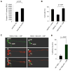
(A) Lentiviral expression of acetylation-resistant Htt (lenti-Htt571-72Q-KR) in primary corticostriatal cultures leads to increased toxicity compared to acetylated Htt (lenti-Htt571-72Q), as determined by caspase 3/7 activation. At least three independent experiments were performed; *p < 0.01. (B) Protection from Htt toxicity by CBP requires acetylation of Htt at K444. Rat primary corticostriatal neurons were transfected with Htt590-97Q or Htt590-97Q-KR together with CBP-HAT or vector, and toxicity scored for at least 150 neurons per sample. Results of three independent experiments are expressed as means + SEM (p = 0.001). (C) Acetylation of mutant Htt is protective in vivo in C. elegans. In animals expressing acetylated mutant Htt (Htt564-150Q) and CBP-HAT, the intact GFP-expressing ASH neuron (arrow, left upper panel) takes up the red fluorescent dye DiD (left middle and lower panels); no degeneration is detected. In contrast, in most animals expressing acetylation-resistant mutant Htt (Htt564-150Q-KR) and CBP-HAT, ASH neurons are GFP positive (arrow, right upper panel) and DiD negative (right middle and lower panels); neurodegeneration increases with K444R mutation. Consistent with previous studies, the ASI neurons (arrowheads) express less Htt protein, are less consistently affected by polyglutamine toxicity, and, hence, were not scored. (D) Affected ASH neurons were scored and compared for Htt564-150Q + CBP and Htt564-150Q-KR + CBP. Average percentage degenerated ± SEM is shown for multiple independent strains (p = 0.0012, see Experimental Procedures for details).
Figure 4
Figure 4. Acetylation of K444 Promotes Clearance of Mutant Htt
Neuro2a cells were transfected with Htt and CBP-HAT, treated with cycloheximide (CHX), and harvested at the indicated time points. Western blots were analyzed by densitometry and values normalized to the amount of Htt at the time of cycloheximide treatment (100%). Values represent means of at least four independent experiments + SEM; *p < 0.05; **p < 0.01 compared to Htt590-25Q (A), Htt590-97Q + CBP-HAT (B), and Htt590-97Q + CBP-HAT (C), respectively. (A) Mutant Htt590-97Q has a longer half-life than wild-type Htt590-25Q. (B) Cotransfection of CBP-HAT, but not the HAT-deficient CBP-HAT-DY, resulted in increased clearance of mutant Htt. (C) Acetylation-induced clearance of mutant Htt depends on K444. Coexpression of CBP-HAT led to increased clearance of Htt590-97Q, whereas mutation of Htt K444 to R prevented this effect of CBP-HAT. (D) Enhanced clearance of Htt590-97Q by HDAC1 knockdown requires residue K444. Efficient knockdown of HDAC1 was achieved 3 days after transfection of pSUPER-HDAC1 compared to transfection of pSUPER-luciferase in Neuro2a cells. β-tubulin was used as loading control (inset). Levels of Htt590-97Q were monitored by radiolabeled pulse-chase. Knockdown of HDAC1 led to significant clearance of Htt590-97Q (32.6% ± 4.9%), comparable to levels achieved with coexpression of CBP-HAT (20.0% ± 16.9%). Mutagenesis of K444 to R (K444R) led to a significant decrease of this clearance (73.0% ± 8.5%). Densitometry measurements are expressed as % of signal after a 1 hr chase. Three independent experiments were performed and are represented as mean + SEM. *p < 0.001 compared to Htt97Q+shHDAC1.
Figure 5
Figure 5. Acetylation of Mutant Htt Enhances Its Clearance by Autophagy
(A) Acetylation of mutant Htt at lysine 444 leads to increased recruitment of LC3 to autophagic vacuoles. RFP-Htt480-68Q and RFP-Htt480-68Q-KR were transfected into COS-7 cells together with GFP-LC3 and CFP-CBP-HAT. Live cells were sequentially scanned to detect distribution of Htt (red), LC3 (green), and CBP-HAT (blue). Scale bar, 10 μm. (a) Coexpression of RFP-Htt480-68Q with LC3 and CBP-HAT led to accumulation of LC3 puncta. (b) Cells transfected with RFP-Htt480-68Q-KR, LC3, and CBP-HAT displayed a marked decrease in the LC3 puncta. (c) LC3-G120A mutant does not form punctate structure. (d) The percentage of cells containing >5 puncta was scored in 150 cells per experiment. Values are expressed as means + SEM of three independent experiments. (B) HeLa cell line, stably expressing YFP-LC3, was transfected with Htt590-97Q or Htt590-97Q-KR plus CFP-CBP-HAT or inactive CBP-HAT-DY. ANOVA analysis of the percentage of transfected cells with Htt puncta that colocalized with LC3 puncta revealed a significant effect of the presence of CBP-HAT (p < 0.001) and the requirement of K444 (p < 0.001). (C) Accumulation of mutant acetyl-Htt after treatment with lysosomal inhibitor leupeptin (200 μM). β-tubulin was used as loading control; p < 0.001. (D) Clearance of acetylated mutant Htt is impeded by inhibition of lysosomal enzymes. Levels of Htt590-97Q, monitored by radiolabeled pulse-chase, were determined after 1, 8, 16, and 24 hr by immunoprecipitation for Htt590-97Q using an N-terminal FLAG antibody. Coexpression of CBP-HAT significantly enhanced clearance of Htt590-97Q (filled square) compared to inactive CBP-HAT-DY (open square) (p < 0.05). This clearance required K444 (open diamond) (p < 0.005) and was impeded by 200 μM leupeptin (filled diamond) (p < 0.05). Significant degradation was achieved after a chase period of 16 hr and 24 hr (*p < 0.001). Densitometry measurements are expressed as % of signal after a 1 hr chase. Representative radiograms are shown. Three independent experiments were performed and are represented as mean + SEM. ANOVA reveals a significant difference across chase time (F(3,23) = 61.779, p < 0.0001), across group (F(3,23) = 19.656, p < 0.0001), and across an interaction between chase time and group (F(9,23) = 5.479, p = 0.005). (E) Acetyl-Htt is preferentially localized to the cytoplasm. Levels of cytoplasmic and nuclear acetyl-Htt and CBP-HAT were determined by subcellular fractionation and western blotting. HDAC1 and β-tubulin were used as nuclear and cytoplasmic markers, respectively. Results are representative of three independent experiments.
Figure 6
Figure 6. Acetylated Mutant Htt Is Trafficked to Autophagosomes
(A) Purification of autophagosomes (AV). Neuro2A cells were fractionated and AV, lysosomes, and endoplasmic reticulum (ER) isolated using two distinct protocols, as described in Experimental Procedures. Cryo-EM revealed fractions enriched in multivesicular bodies that are morphologically consistent with AV (1, 2) and unilamellar structures like transport vesicles (3). (B) To determine whether LC3 was associated with membrane structures, AV fractions were incubated with an antibody against LC3 followed by iron-rich magnetic beads conjugated to Protein A. (i) Beads imaged alone. (ii) Unilamellar transport vesicles were not labeled by beads. (iii and iv) Multilamellar autophagosomal-like particles are labeled by beads. (C) Preferential trafficking of acetylated Htt to autophagosomes. Cells were fractionated and AV isolated, run on SDS-PAGE, and probed with anti-Htt antibody or LC3 antibody. Htt590-97Q was preferentially detected in the AV-enriched fractions compared to Htt590-97QKR. The cytosolic fractions predominantly contain the unbound form of LC3 (LC3-I), while the AV-enriched fractions contain the membrane-bound form of LC3 (LC3-II). Fraction “AV/ER” differs from “AV” as it is also enriched for ER markers such as calnexin (not shown). Std, protein standard. (D) Mutant Htt is detected within the AV membrane fraction. Protease protection assay was performed with 20 μg of protein from a cytosol fraction or AV fraction collected from (C), subjected to increasing amounts of proteinase K (PK) and examined by SDS-PAGE. In the cytosolic fraction, both Htt590-97Q and the LC3-I were degraded after exposure to 1 μg/ml PK. In the AV fraction, Htt590-97Q did not degrade until 25 μg/ml PK when the LC3-II was also degraded. Htt590-97Q was detected using MAB5492 (Chemicon) and LC3 with rabbit polyclonal antibody (Abcam). A representative image from three independent experiments is shown.
Figure 7
Figure 7. p62-Mediated Clearance of Mutant Htt
(A) Acetyl-Htt exists as a HMW species and accumulates upon lysosomal inhibition. Neuro2a cells were cotransfected with Htt480-68Q and CBP-HAT, in the presence of bafilomycin A1 (Baf A1, 100 nM) or DMSO. Triton-soluble lysates were analyzed by size-exclusion chromatography (SEC) and western blot using the anti-acetyl K444 antibody (bottom). Neuron-specific enolase (NSE) was used to monitor gel filtration column performance and loading. The horizontal MW marker (kDa) represents the MW obtained by SEC analysis as determined by the elution of globular protein standards. The vertical marker (kDa) represents the MW obtained by SDS-PAGE analysis of fractions. Western blots shown were analyzed by densitometric analysis from at least three independent experiments and normalized to NSE (top). Inset, the amount of monomeric Ac-Htt is shown at a smaller y axis. (B) Knockdown of p62 in inducible HN10 cell lines expressing Htt573-72Q. Values are the mean ± SEM (n = 5); *p = 0.01 for p62 shRNA compared to control (Ctrl) shRNA, normalized to α-tubulin. (C) Clearance of Ac-Htt is mediated by p62 in HN10 cells. Mutant Htt half-life was determined in untransfected (Un) or cells transfected with empty vector (Vect), CBP D-Y, CBP-HAT, control shRNA, or p62 shRNA. Half-life values of Htt573-72Q were determined as described in Experimental Procedures. The values represent the mean ± SEM (n = 5). *p < 0.01 compared to Un and Vect; *p < 0.05 compared to CBP D-Y; **p < 0.01 compared to Ctrl-shRNA.

Similar articles

Cited by

References

    1. Arrasate M, Mitra S, Schweitzer ES, Segal MR, Finkbeiner S. Inclusion body formation reduces levels of mutant huntingtin and the risk of neuronal death. Nature. 2004;431:805–810. - PubMed
    1. Bannister AJ, Kouzarides T. The CBP co-activator is a histone acetyltransferase. Nature. 1996;384:641–643. - PubMed
    1. Bates EA, Victor M, Jones AK, Shi Y, Hart AC. Differential contributions of Caenorhabditis elegans histone deacetylases to huntingtin polyglutamine toxicity. J Neurosci. 2006;26:2830–2838. - PMC - PubMed
    1. Bjorkoy G, Lamark T, Brech A, Outzen H, Perander M, Overvatn A, Stenmark H, Johansen T. p62/SQSTM1 forms protein aggregates degraded by autophagy and has a protective effect on huntingtin-induced cell death. J Cell Biol. 2005;171:603–614. - PMC - PubMed
    1. Cuervo AM. Autophagy: many paths to the same end. Mol Cell Biochem. 2004;263:55–72. - PubMed

Publication types