. 2009 Apr 3;137(1):47-59.
doi: 10.1016/j.cell.2009.01.038.

A Nurr1/CoREST pathway in microglia and astrocytes protects dopaminergic neurons from inflammation-induced death

Affiliations

A Nurr1/CoREST pathway in microglia and astrocytes protects dopaminergic neurons from inflammation-induced death

Kaoru Saijo et al. Cell. .

Abstract

Nurr1, an orphan nuclear receptor, plays an essential role in the generation and maintenance of dopaminergic neurons in the brain. Rare mutations in Nurr1 are associated with familial Parkinson's disease, but the underlying basis for this relationship has not been established. Here, we demonstrate that Nurr1 unexpectedly functions to inhibit expression of pro-inflammatory neurotoxic mediators in both microglia and astrocytes. Reduced Nurr1 expression results in exaggerated inflammatory responses in microglia that are further amplified by astrocytes, leading to the production of factors that cause death of tyrosine hydroxylase-expressing neurons. Nurr1 exerts anti-inflammatory effects by docking to NF-kappaB-p65 on target inflammatory gene promoters in a signal-dependent manner. Subsequently, Nurr1 recruits the CoREST corepressor complex, resulting in clearance of NF-kappaB-p65 and transcriptional repression. These studies suggest that Nurr1 protects against loss of dopaminergic neurons in Parkinson's disease in part by limiting the production of neurotoxic mediators by microglia and astrocytes.


Figures
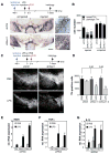
Fig. 1
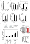
Fig. 1. Nurr1 suppresses LPS-induced inflammation and loss of TH+ neurons
A. TH-DAB staining of a representative brain section of mice injected with shCtrl- or shNurr1-lentivirus and LPS is shown at AP −3.3 mm. Regions indicated by a rectangle in the injected side of the brain are enlarged in the right panels. Scale bars: 200 μm, right panels and 50 μm, left panels. B. Quantification of TH+ cell numbers in the shNurr1 groups compared to the shCtrl-injected and the uninjected side. Bars indicate normal (black) and pathological (gray) TH+ neurons. See Supplemental Experimental Procedures for the definition of normal/pathological TH+ neurons. Asterisk, p<0.01 compared to the numbers from shCtrl-lentivirus-injected. (n=5) C. Fluorescence-TH staining of a representative brain section of mice injected with shCtrl- or shNurr1-lentivirus followed by PBS or LPS. Experimental diagram is indicated at the top. Scale bar 200 μm. D. Quantification of TH+ cell numbers in the setting of Nurr1 knockdown followed by LPS or PBS injection. Asterisk, p<0.002 compared to PBS injection (n=4). E–G. Expression of iNOS (E), TNFα (F) and IL1β (G) mRNA in Nurr1-knockdown SN 6 hours after LPS injection as determined by qPCR and normalized to HPRT expression (n=4). Error bars represent SD. Asterisk, p<0.01 compared to shCtrl/PBS-injected; **, p<0.01 compared to shCtrl/LPS-injected samples.
Fig. 2
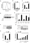
Fig. 2. Microglia initiate LPS-mediated inflammation and astrocytes propagate the production of neurotoxic factors
A. Expression of TNFα mRNA upon LPS stimulation in primary microglia, astrocytes and Neuro2A cells. B. Effect of LPS or TNFα + cycloheximide (CHX) on viability of the indicated neuronal cell lines determined using a TUNEL ELISA assay. *, p<0.01 compared to untreated sample (no Tx) (white). C. Effect of Nurr1 knockdown in Neuro2A cells on sensitivity to TNFα + CHX assessed by TUNEL ELISA assay. DE. Effect of Nurr1 knockdown inBV2 cells on LPS-induced expression of TNFα (D), iNOS (E) and IL1β (F) mRNA.*, p<0.01 compared to no stimulation (Ctrl); **, p<0.01 compared to LPS stimulation of shCtrl-BV2 cells. G. Scheme of conditioned media (CM) and cell death assay. CMs were harvested from shCtrl- and shNurr1-BV2 cells that were stimulated with LPS for 24h. Neurons or glial cells were assayed for specific markers by immunostaining and for cell death by TUNEL assay. H. Effect of CM from LPS-treated shCtrl-BV2 cells and a mixture of the CMs from shNurr1-1- and shNurr1-2-infected BV2 cells (shNurr1) on neurons and glial cells derived from in vitro differentiated neural stem cells (NSC). TUNEL assay was performed on TH-, GABA- or GFAP-positive cells derived from mouse NSC. Numbers of TUNEL-negative live cells are shown. TH-positive cells are indicated in red. I. The percentages of TUNEL-positive population are shown. *, p<0.01 and **, p<0.001 compared to no treatment (no TX). J. Effect of Nurr1 knockdown in microglia and astrocytes on the production of neurotoxic factors. Primary mouse microglia and astrocytes were infected with shCtrl- or shNurr1-lentivirus. Cells were treated with LPS for 2h and washed extensively with PBS. Cells were cultured for another 24h to generate CM. For sequential CM assay, CMs harvested from microglia were cultured with lentivirus-infected astrocytes for 24 hours. Then, CMs were harvested and tested for effect on viability of Neuro2A cells.
Fig. 3
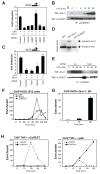
Fig. 3. Nurr1 acts as an RXR-independent, GSK3
β-dependent transrepressor for NF-κB. A. ChIP assay of Nurr1 on the TNFα-promoter in response to LPS and effect of the GSK3β-specific inhibitor SB216763 (SB21). BV2 cells were pre-incubated with DMSO or 30 μM SB21 for 1h followed by LPS stimulation for the indicated times before ChIP assay. Data are displayed as fold enrichment over control IgG. B. Repression activities of Nurr1 mutants. RAW264.7 cells were transfected with wild type Nurr1, P-box mutant (CEAA) and I-box mutant (KLL). iNOS-promoter activity in RAW264.7 cells in response to LPS was measured by luciferase-reporter assay. *, p<0.01 compared to control (Mock). C. Effect of knockdown of Ubc9 on Nurr1 repression of iNOS-promoter activity. *, p<0.01 compared to Nurr1 with control siRNA. D. Identification of the predominant SUMOylation sites of Nurr1. Flag-tag mutants of Nurr1 were transfected into Hela cells. SUMOylation assay was performed as described in Supplemental Experimental Procedure. E. Effect of reconstitution of Nurr1 shRNA-2 BV2 cells with non-targeted (NT) WT and mutant forms of Nurr1 that are not recognized by shNurr1-2. Endogenous iNOS mRNA levels are shown relative to levels in untreated BV2 cells transduced with control shRNA and mock Nurr1 lentivirus. *, p<0.01 compared to mock control cells. F. Effect of LPS stimulation on interaction of Nurr1 and p65 in BV2 cells. Lysates of BV2 cells stimulated with LPS for the indicated times were immunoprecipitated with anti-Nurr1 antibody and Western blots were developed with anti-p65 antibody. G. Effect of SB21 on Nurr1/p65 interaction. BV2 cells were incubated with SB21 at the indicated concentrations for 1h prior to stimulation with LPS. IP and Western blotting were performed as described in F. H. Effect of siRNA against GSK3β on Nurr1-mediated repression of iNOS-promoter activity. Nurr1 expression vector was transfected into RAW264.7 cells together with control siRNA or siRNA against GSK3β *, p<0.01 compared to Nurr1 with control siRNA. I. Effect of S468A mutation of p65 on Nurr1-mediated repression of iNOS-promoter activity.
Fig. 4
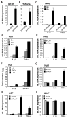
Fig. 4. CoREST repressor complex is required for Nurr1-mediated repression
A. Corepressor requirements in Nurr1-mediated repression. iNOS-luciferase and Nurr1-expression or control vector as well as siRNAs against the indicated corepressors were transfected into RAW264.7 cells and iNOS-promoter activity was assayed. *, p<0.01 compared to Nurr1 with control siRNA. B. Interaction of Nurr1 and CoREST in BV2 cells. Co-IP was performed with anti-CoREST antibody and Western blots were developed with anti-Nurr1 antibody. C. Effect of siRNAs against the indicated targets on Nurr1-mediated repression of iNOS-promoter activity. *, p<0.01, **, p<0.001 compared to Nurr1 with control siRNA. D. NLK in vitro kinase assay using GST-Nurr1 and GST-CoREST as substrates. Arrows indicate phosphorylated GST-Nurr1 and autophosphorylation of NLK. The migration position of GST-CoREST is indicated by an asterisk. GST substrates and methods are provided in Supplemental Experimental Procedures and Figure S7D. E. Effect of NLK knockdown on interaction of Nurr1 and CoREST. BV2 cells were transfected with siRNA against NLK or control siRNA. Co-IP of Nurr1 and CoREST was performed as described in B. F. Recruitment of Nurr1, CoREST and p65 to the iNOS promoter in BV2 cells shown by ChIP assay. Data represent fold enrichment of iNOS-promoter precipitated by the indicated antibodies compared to control IgG as determined by qPCR. G. ChIP analysis of Nurr1 on the iNOS promoter in the SN before and after LPS stimulation. Data are shown as averages of fold enrichment against control IgG and SD. H. Effect of Nurr1 knockdown on recruitment of CoREST (left) and p65 (right) to the TNFα promoter in BV2 cells. ChIP data are shown as fold enrichment over control IgG.
Fig. 5
Fig. 5. Nurr1 suppresses inflammatory mediators in murine astrocytes
A and B. Expression of IL1R1 (A) and p55TNFR (B) mRNA in primary astrocytes and microglia as determined by qPCR assay. *, p<0.01. C. Responses of primary mouse microglia or astrocytes to TNFα or IL1β for 6h. iNOS mRNA level was determined by qPCR. D. Regulation of Nurr1 mRNA in mouse primary astrocytes stimulated with TNFα or IL1β for the indicated times. E–I. Effect of knockdown of Nurr1 in astrocytes on induction of NO (F – measured by Greiss reaction) and mRNAs encoding neurotoxic mediators (E, G–I). Mouse primary astrocytes were infected with shCtrl- or shNurr1-lentivirus and cells were stimulated with TNFα or IL1β for 6h.
Fig. 6
Fig. 6. The CoREST complex is required for Nurr1-mediated repression in astrocytes
A. Effect of IL1β stimulation on association of Nurr1 and p65 in mouse primary astrocytes. Lysates of astrocytes stimulated with IL1β for the indicated times were immunoprecipitated with anti-Nurr1 antibody and Western blots were developed with anti-p65 antibody. B. Recruitment of Nurr1 and p65 to iNOS-promoter in mouse primary astrocyte shown by ChIP assay. Data represent fold enrichment of iNOS- promoter precipitated with the indicated antibodies compared to control IgG as determined by qPCR. C. Effect of GSK3β-specific inhibitor SB21 on recruitment of Nurr1 to iNOS-promoter. Data represent fold enrichment of iNOS-promoter precipitated with antibody against Nurr1 compared to control IgG as determined by qPCR. D. Interaction of Nurr1 and CoREST in mouse primary astrocytes. Co-IP was performed with anti-Nurr1 antibody and Western blots developed with anti-CoREST or anti-Nurr1 antibodies. E. Recruitment of Nurr1 and CoREST to iNOS-promoter in mouse primary astrocytes shown by ChIP assay. Data represent fold enrichment of iNOS-promoter precipitated with the indicated antibodies compared to control IgG as determined by qPCR. FH. Effect of knockdown of the components of CoREST-repressor complex on mRNAs encoding inflammatory mediators. Mouse primary astrocytes were infected with lentivirus carrying shRNA against CoREST, LSD1, G9a, HDAC1 or control. Cells were stimulated with IL1β for 6h and mRNA expression of iNOS (F), CSF1 (G) and Ncf1 (H) was determined by qPCR. I. Nurr1-dependent clearance of p65 from iNOS promoter. ChIP assay was performed in shNurr1- or shCtrl-astrocyte and data shown as fold enrichment over control IgG of iNOS promoter precipitated with antibody against p65.
Fig. 7
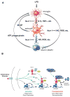
Fig. 7. Nurr1 functions to inhibit neurotoxic gene expression in microglia and astrocytes via a CoREST-dependent transrepression pathway
A. Model for communication among microglia, astrocyte and neurons. B. Model for Nurr1/CoREST-mediated repression. See main text for details.

Comment in

Similar articles

Cited by

References

    1. Aarnisalo P, Kim CH, Lee JW, Perlmann T. Defining requirements for heterodimerization between the retinoid X receptor and the orphan nuclear receptor Nurr1. J Biol Chem. 2002;277:35118–35123. - PubMed
    1. Akira S, Uematsu S, Takeuchi O. Pathogen recognition and innate immunity. Cell. 2006;124:783–801. - PubMed
    1. Ashburner BP, Westerheide SD, Baldwin AS., Jr The p65 (RelA) subunit of NF-κB interacts with the histone deacetylase (HDAC) corepressors HDAC1 and HDAC2 to negatively regulate gene expression. Mol Cell Biol. 2001;21:7065–7077. - PMC - PubMed
    1. Atkinson TJ. Toll-like receptors, transduction-effector pathways, and disease diversity: evidence of an immunobiological paradigm explaining all human illness? Int Rev Immunol. 2008;27:255–281. - PubMed
    1. Ballas N, Grunseich C, Lu DD, Speh JC, Mandel G. REST and its corepressors mediate plasticity of neuronal gene chromatin throughout neurogenesis. Cell. 2005;121:645–657. - PubMed

Publication types

MeSH terms