. 2009 Apr 17;137(2):344-55.
doi: 10.1016/j.cell.2009.01.057. Epub 2009 Apr 2.

DEX-1 and DYF-7 establish sensory dendrite length by anchoring dendritic tips during cell migration

Affiliations

DEX-1 and DYF-7 establish sensory dendrite length by anchoring dendritic tips during cell migration

Maxwell G Heiman et al. Cell. .

Abstract

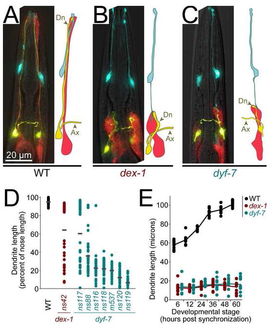
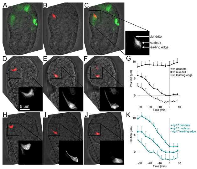
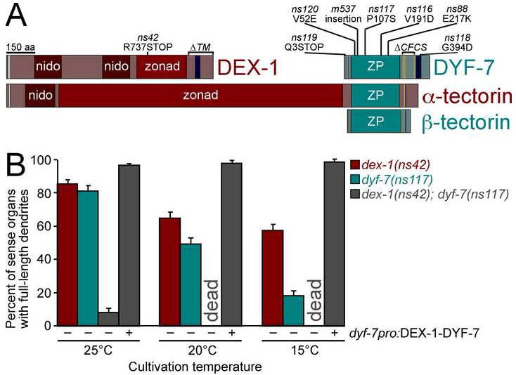
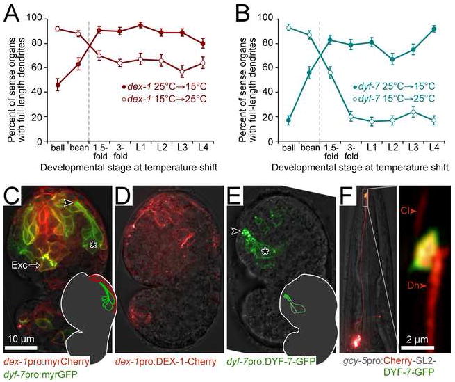
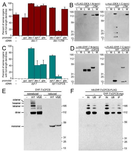
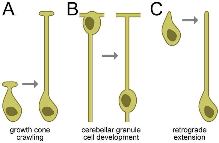
Cells are devices whose structures delimit function. For example, in the nervous system, neuronal and glial shapes dictate paths of information flow. To understand how cells acquire their shapes, we examined the formation of a sense organ in C. elegans. Using time-lapse imaging, we found that sensory dendrites form by stationary anchoring of dendritic tips during cell-body migration. A genetic screen identified DEX-1 and DYF-7, extracellular proteins required for dendritic tip anchoring, which act cooperatively at the time and place of anchoring. DEX-1 and DYF-7 contain, respectively, zonadhesin and zona pellucida domains, and DYF-7 self-associates into multimers important for anchoring. Thus, unlike other dendrites, amphid dendritic tips are positioned by DEX-1 and DYF-7 without the need for long-range guidance cues. In sequence and function, DEX-1 and DYF-7 resemble tectorins, which anchor stereocilia in the inner ear, suggesting that a sensory dendrite anchor may have evolved into part of a mechanosensor.


Figures

FIGURE 1
FIGURE 1. dex-1 and dyf-7 are required for dendrite extension
A, Wild-type; B, dex-1(ns42); and C, dyf-7(m537) animals expressing odr-1pro:YFP (AWC neurons, yellow), F16F9.3pro:mCherry (sheath glia, red), and itr-1pro:CFP (socket glia, blue). Ax, axon; Dn, dendrite. D, Animals expressing gcy-5pro:GFP (ASER neuron) with or without str-2pro:GFP (AWCL or AWCR), to mark individual dendrites, were synchronized at the first larval stage (L1) and dendrite and nose lengths measured. Each dot represents an individual (n=40 per genotype). Gray bars, means. E, Same as (D), but cohorts of L1s with short dendrites were selected by visual inspection, released from synchronization, and dendrite lengths were measured after 6 h (L1), 12 h (second larval stage, L2), 24 h (third larval stage, L3), 36 h (early fourth larval stage, L4), 48 h (late L4), and 60 h (adult). Lines connect means (n=10 per genotype at each stage).
FIGURE 2
FIGURE 2. Amphid sensory dendrite extension is coordinated
A, To compare lengths of ipsilateral and contralateral dendrites, animals bearing dex-1(ns42) or dyf-7(ns117) were made to express str-2pro:GFP (AWCL or AWCR neuron, green) and srh-142pro:RFP (ADFL and ADFR neurons, red). Closed green arrowhead, AWC dendritic tip; closed red arrowhead, ipsilateral ADF dendritic tip; open red arrowhead, contralateral ADF dendritic tip. B-C, Animals were collected as L4s and dendrite and nose lengths measured. AWC dendrite length was compared to the ipsilateral (B, closed circles) and contralateral (C, open circles) ADF dendrite lengths. Each dot represents an individual. dex-1(ns42), red symbols; dyf-7(ns117), blue symbols. Clustering of data points at corners reflects the bimodal distribution described in Fig. 1D.
FIGURE 3
FIGURE 3. Time-lapse imaging of dendrite extension
A-C, Dorsal view of a bean-stage embryo expressing the photoconvertible fluorescent protein Kaede in most sensory neurons. 20 50-msec pulses of a low-intensity 406 nm laser were used to convert Kaede from its native green-fluorescent state to its photoconverted red-fluorescent state in a single neuron. A, green, native Kaede; B, red, photoconverted Kaede; C, merged image. Box shows the vertical axis positions of the dendritic tip, nucleus, and leading edge of the photoconverted cell. D-F, Time-lapse sequence of the equivalent neuron in a different wild-type embryo. Inset is enlarged 2x. Scale bar applies to inset. D, 0 min; E, 51 min; F, 65 min. Optical stacks were collected every 4 min, and Kaede photoconversion was repeated every 16 min, as needed. G, Plot of dendrite, nucleus, and leading edge vertical axis positions over time in three wild-type embryos (Movies 1-3). Error bars, standard deviation (SD) among the individuals. A fourth wild-type embryo (Movie 4) began embryo elongation/rotation earlier and was excluded (see Supp. Fig. 2). H-J, Same as D-F, but with a dyf-7(m537) embryo. H, 0 min; I, 43 min; J, 52 min. K, Same as G, using four dyf-7(m537) embryos (Movies 4-8).
FIGURE 4
FIGURE 4. dex-1 and dyf-7 encode transmembrane proteins and interact genetically
A, Schematic diagram of DEX-1, DYF-7, α-tectorin, and β-tectorin. Nidogen (nido), zonadhesin-like (zonad) and zona pellucida (ZP) domains; consensus furin cleavage sites (CFCS, gold bars); signal sequences (gray bars); and transmembrane segments/GPI anchors (dark blue bars) are indicated. B, Animals expressing gcy-5pro:GFP and str-2pro:GFP to mark individual dendrites, and bearing the cold-sensitive mutations dex-1(ns42), dyf-7(ns117), or both were cultivated at permissive (25°C), standard (20°C), or restrictive (15°C) temperatures and dendrite lengths scored. dex-1(ns42); dyf-7(ns117) animals were scored with (+) and without (−) a dyf-7pro:DEX-1-DYF-7 fusion transgene. In each case, n≥100. For transgene experiments, three lines were scored. Error bars, standard error of the mean (SEM) except, in transgene experiments, SD among lines.
FIGURE 5
FIGURE 5. Timing and localization of DEX-1 and DYF-7 expression
Animals expressing gcy-5pro:GFP and str-2pro:GFP and bearing the cold-sensitive mutations dex-1(ns42) (A) or dyf-7(ns117) (B) were subjected to temperature shifts at the indicated stages, and dendrite lengths scored in adults. Ball, bean, 1.5-fold, and 3-fold are morphologically-defined embryonic stages; L1, L2, L3 and L4 are larval stages. Late bean stage (dashed vertical line) corresponds to Fig. 3, when dendrites grow. Error bars, SEM. C, Ventral view of wild-type embryo at the stage of dendrite growth expressing dex-1pro:myristyl-mCherry and dyf-7pro:myristyl-GFP. *, amphid neuron cell bodies. , dendrite tip. Exc, excretory cell. Inset, schematic showing position of amphid neurons (green) and neighboring dex-1-expressing cells (red). D-E, Lateral views of wild-type embryos expressing dex-1pro:DEX-1-mCherry or dyf-7pro:DYF-7-GFP. Symbols and scale as in C. Inset, schematic showing position of amphid bundle and accumulation of DYF-7-GFP at dendritic tips. F, An animal expressing gcy-5pro:mCherry-SL2-DYF-7-GFP [SL2 (splice leader 2) permits expression of two transcripts as a single operon], to simultaneously mark the cytoplasm and DYF-7-GFP in the ASER neuron. Cl, cilium. Dn, dendrite. Inset is magnified 10x.
FIGURE 6
FIGURE 6. Secretion of DEX-1 and DYF-7
A, Animals expressing gcy-5pro:GFP and str-2pro:GFP, and bearing dex-1(ns42) and transgenes with the indicated promoters (gut, pha-4pro; dex-1pro; dyf-7pro; glia, lin-26 E1 enhancer:myo-2 minimal promoter) driving dex-1 or dex-1ΔTM cDNA were cultivated at 20°C and dendrite lengths scored. The enhanced defect in dyf-7pro:DEX-1ΔTM may reflect premature nonproductive interaction of DEX-1ΔTM and DYF-7 in the secretory pathway. Each bar is the mean of three transgenic lines, n=100 per line; error bars, SD among lines. B, S2 insect cells were transfected, or not (–), with FLAG-DEX-1-myc or FLAG-DEX-1ΔTM-myc and cultured at 25°C for 2 d. Equivalent samples of cell lysate (L) and medium (M) were collected and analyzed by immunoblot. Left, anti-FLAG; right, anti-myc. Due to increased reactivity of DEX-1ΔTM C-terminal myc, these samples were diluted 1:20. *, non-specific degradation. C, same as A except animals express only gcy-5pro:GFP and bear dyf-7(m537) and transgenes with the indicated promoters driving DYF-7 or DYF-7ΔCFCS. D, same as B except cells were transfected with HA-DYF-7-FLAG or HA-DYF-7ΔCFCS-FLAG. Left, anti-HA; right, anti-FLAG. *, non-specific degradation. E, S2 cells were transfected, or not (−), with HA-DYF-7ΔCFCS-FLAG (WT) or the same construct bearing the V52E mutation (V52E). Cell lysates were collected under nonreducing (no β-mercaptoethanol (β-me), 50°C) or reducing (5% β-me, boiling) conditions and analyzed by anti-HA immunoblot. Multimers are indicated. F, S2 cells were transfected, or not (−), with HA-DYF-7ΔCFCS-FLAG alone or in conjunction with DYF-7ΔCFCS-myc. Lysates were collected under nonreducing conditions, immunoprecipated using anti-myc antibody-conjugated agarose, and dilution-normalized volumes of starting material (input, IN), unbound (UB) and immunoprecipitated (IP) fractions were analyzed by anti-HA immunoblot.
FIGURE 7
FIGURE 7. Three modes of neurite outgrowth
A, Many axons and dendrites form by a projection migrating away from a stationary cell body. B, During cerebellar granule cell development, a neurite forms in the same manner but the cell body then translocates along it. Neurite regions above the nucleus in this schematic are axons; below it are dendrites. C, Amphid sensory dendrites form by the stationary anchoring of dendritic tips during cell body migration, so the neurite is generated de novo by the migrating cell body.

Comment in

Similar articles

Cited by

References

    1. Altman J, Bayer SA. Vertical compartmentation and cellular transformations in the germinal matrices of the embryonic rat cerebral cortex. Exp Neurol. 1990;107:23–35. - PubMed
    1. Ando R, Hama H, Yamamoto-Hino M, Mizuno H, Miyawaki A. An optical marker based on the UV-induced green-to-red photoconversion of a fluorescent protein. Proc Natl Acad Sci U S A. 2002;99:12651–12656. - PMC - PubMed
    1. Bargmann CI. Chemosensation in C elegans. WormBook; 2006. pp. 1–29. - PMC - PubMed
    1. Benard CY, Boyanov A, Hall DH, Hobert O. DIG-1, a novel giant protein, non-autonomously mediates maintenance of nervous system architecture. Development. 2006;133:3329–3340. - PubMed
    1. Bi M, Hickox JR, Winfrey VP, Olson GE, Hardy DM. Processing, localization and binding activity of zonadhesin suggest a function in sperm adhesion to the zona pellucida during exocytosis of the acrosome. Biochem J. 2003;375:477–488. - PMC - PubMed

Publication types

MeSH terms

Substances

LinkOut - more resources